. 2024 Sep 11;15(9):e0069124.
doi: 10.1128/mbio.00691-24. Epub 2024 Aug 20.

Prevotella timonensis degrades the vaginal epithelial glycocalyx through high fucosidase and sialidase activities

Affiliations

Prevotella timonensis degrades the vaginal epithelial glycocalyx through high fucosidase and sialidase activities

Celia Segui-Perez et al. mBio. .

Abstract

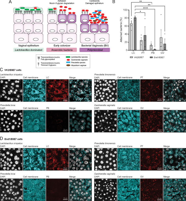
Bacterial vaginosis (BV) is a polymicrobial infection of the female reproductive tract. BV is characterized by replacement of health-associated Lactobacillus species by diverse anerobic bacteria, including the well-known Gardnerella vaginalis. Prevotella timonensis, and Prevotella bivia are anerobes that are found in a significant number of BV patients, but their contributions to the disease process remain to be determined. Defining characteristics of anerobic overgrowth in BV are adherence to the mucosal surface and the increased activity of mucin-degrading enzymes such as sialidases in vaginal secretions. We demonstrate that P. timonensis, but not P. bivia, strongly adheres to vaginal and endocervical cells to a similar level as G. vaginalis but did not elicit a comparable proinflammatory epithelial response. The P. timonensis genome uniquely encodes a large set of mucus-degrading enzymes, including four putative fucosidases and two putative sialidases, PtNanH1 and PtNanH2. Enzyme assays demonstrated that fucosidase and sialidase activities in P. timonensis cell-bound and secreted fractions were significantly higher than for other vaginal anerobes. In infection assays, P. timonensis efficiently removed fucose and α2,3- and α2,6-linked sialic acid moieties from the epithelial glycocalyx. Recombinantly expressed P. timonensis NanH1 and NanH2 cleaved α2,3 and α2,6-linked sialic acids from the epithelial surface, and sialic acid removal by P. timonensis could be blocked using inhibitors. This study demonstrates that P. timonensis has distinct virulence-related properties that include initial adhesion and a high capacity for mucin degradation at the vaginal epithelial mucosal surface. Our results underline the importance of understanding the role of different anerobic bacteria in BV.

Importance: Bacterial vaginosis (BV) is a common vaginal infection that affects a significant proportion of women and is associated with reduced fertility and increased risk of secondary infections. Gardnerella vaginalis is the most well-known BV-associated bacterium, but Prevotella species including P. timonensis and P. bivia may also play an important role. We showed that, similar to G. vaginalis, P. timonensis adhered well to the vaginal epithelium, suggesting that both bacteria could be important in the first stage of infection. Compared to the other bacteria, P. timonensis was unique in efficiently removing the protective mucin sugars that cover the vaginal epithelium. These results underscore that vaginal bacteria play different roles in the initiation and development of BV.

Keywords: Hoylesella timonensis; NanH; Prevotella bivia; Prevotella timonensis; anerobic bacteria; fucosidase; mucus; neuraminidase; vaginal microbiota; α2,3-linked sialic acids; α2,6-linked sialic acids.


Conflict of interest statement

The authors declare no conflict of interest.

Figures

Fig 1
Fig 1
Prevotella timonensis can adhere to the vaginal and endocervical epithelium. (A) Schematic representation of the different microbial communities of the vaginal epithelium in the healthy state and during the development of bacterial vaginosis. (B) Percentage of adhesion of L. crispatus (LC), P. timonensis (PT), P. bivia (PB), and G. vaginalis (GV) to VK2/E6E7 and End1/E6E7 cells assessed by quantification of colony-forming units (CFUs). The graph represents the average and SEM of at least three to four independent experiments. Statistical test: one-way ANOVA with Dunnett’s correction compared to L. crispatus in each cell line. *P < 0.05; **P < 0.01; ****P < 0.0001. (C, D) Fluorescence in situ hybridization (FISH) in combination with confocal microscopy of L. crispatus, P. timonensis, P. bivia, and G. vaginalis adhesion to (C) VK2/E6E7 and (D) End1/E6E7 cells stained for wheat germ agglutinin (WGA) and using peptide nucleotide acid (PNA) probes. For each bacterium, the corresponding PNA signal is shown in red, cell surface in cyan (WGA), and 4′,6-diamidino-2-phenylindole (DAPI) in white. White scale bars represent 20 µM.
Fig 2
Fig 2
Prevotella timonensis does not induce epithelial cytotoxicity nor is it highly inflammatory. LDH release of (A) VK2/E6E7 and (B) End1/E6E7 cells after 18 hours of infection with P. timonensis (PT), P. bivia (PB), G. vaginalis (GV), or L. crispatus (LC) at an MOI of 10 and 100. (C–F) RT-qPCR analysis of VK2/E6E7 and End1/E6E7 cell lines incubated with P. timonensis (PT), P. bivia (PB), G. vaginalis (GV), or L. crispatus (LC) at an MOI of 10 demonstrating the expression of (C) IL-1β, (D) IL-8, CCL20, and (F) CCL5. TMEM222 was used as the reference gene. As a positive control, cells were stimulated with the TLR ligand Pam3CSK4 to induce the expression of proinflammatory cytokines. The red dotted line marks significant upregulation compared to non-infected cells. The graph represents the average ± SEM of at least three independent experiments. Statistical test: one-way ANOVA with Dunnett’s correction compared to L. crispatus in each cell line. *P < 0.05; **P < 0.01; ****P < 0.0001.
Fig 3
Fig 3
Utilization of glycogen and mucins as carbon sources by BV-associated bacteria. Growth of (A) P. timonensis, (B) P. bivia, (C) G. vaginalis, and mucin-degrader Akkermansia muciniphila (D) on basal medium without carbohydrates (CMM− or MM−), basal medium supplemented with 0.5% glycogen, basal medium supplemented with 0.5% purified porcine gastric mucins (PGM), or complete medium with carbohydrate (CMM + or MM+) for up to 56 hours. The graph represents the average ± SEM of at least three independent experiments. Statistical test: two-way ANOVA with Dunnett’s correction compared to basal media without carbohydrates (CMM−). ns: not significant; *P < 0.05; **P < 0.01; ***P < 0.001; ****P < 0.0001.
Fig 4
Fig 4
High mucin degradation potential in Prevotella timonensis. (A) Abundance of predicted carbohydrate-active enzymes (CAZymes) families in the sequenced genomes of our P. timonensis, P. bivia, G. vaginalis, and A. muciniphila strains. (B) Schematic representation of a mucin glycoprotein molecule with a protein backbone and diverse O-glycan structures. Target sites for different classes of glycosyl hydrolases are depicted. (C) Number of identified O-glycan-targeting CAZymes in the genomes of the sequenced P. timonensis (PT), P. bivia (PB), G. vaginalis (GV), and A. muciniphila (AM) strains. (D–E) Domain architecture of the predicted (D) fucosidases and (E) sialidases of the designated bacteria. The displayed domains are identified by HMMER, Diamond, and Signal IP 6.0 tools and drawn to scale. (F) Fucosidase and (G) sialidase activities measured in bacterial pellets and supernatants of the different BV-associated bacteria and B. fragilis as the positive control. Abbreviations: PT (P. timonensis); PB (P. bivia); GV (G. vaginalis); BF (B. fragilis); and LC (L. crispatus). The graph represents the average ± SEM of four independent experiments. Statistical test: one-way ANOVA with Tukey’s HSD correction. ns: not significant; *P < 0.05; ** P < 0.01; *** P < 0.001; ****P < 0.0001.
Fig 5
Fig 5
Prevotella timonensis effectively removes glycans from the vaginal epithelial surface. Fluorescence confocal microscopy images of mucin O-glycan structures after incubation with P. timonensis, P. bivia, G. vaginalis, or L. crispatus at an MOI of 10 for 18 hours in anerobic conditions. Neuraminidase A and L-fucosidase were added for 3 hours as positive controls for sialidase and fucosidases activities. (A) UEA-1 (α-L fucoses), (B) MAL-II (α2,3 sialic acids), and (C) SNA (α2,6 sialic acids) stainings are shown in green, and DAPI staining is shown in white. White scale bars represent 20 µM. (D) Quantification of UEA-1, MAL-II, and SNA stainings from Fig. 5A through C. The no treatment control sample is represented as “−“ and the recombinant fucosidase or sialidase as “+.” The graph represents the average ± SEM of at least three independent experiments. Statistical test: one-way ANOVA with Dunnett’s correction and compared to untreated cells (control). *P < 0.05; **P < 0.01; ***P < 0.001; ****P < 0.0001.
Fig 6
Fig 6
P. timonensis sialidase activity at the vaginal mucosal surface can be inhibited by the sialidase inhibitors DANA and zanamivir. Fluorescence confocal microscopy images of sialic acid staining after incubation with 1 µM of recombinant P. timonensis sialidases NanH1 and NanH2 for 3 hours in anerobic conditions. (A) MAL-II (α2,3 sialic acids) and (B) SNA (α2,6 sialic acids) stainings are shown in green, and DAPI staining is shown in white. White scale bars represent 20 µM. (C) Quantification of MAL-II and SNA stainings from Fig. 6A and B. Fluorescence microscopy images of (D) MAL-II (α2,3 sialic acids) and (E) SNA (α2,6 sialic acids) after P. timonensis infection at an MOI of 10 for 18 hours in anerobic conditions in the presence/absence of 1 mM DANA or zanamivir. Lectin stainings are shown in green and DAPI in white. Statistical test: one-way ANOVA with Dunnett’s correction compared to untreated control cells. *P < 0.05; **P < 0.01. White scale bars represent 20 µM. (F) Quantification of MAL-II and SNA stainings from Fig. 6D and E. The graph represents the average ± SEM of three independent experiments.

References

    1. Cohen CR, Lingappa JR, Baeten JM, Ngayo MO, Spiegel CA, Hong T, Donnell D, Celum C, Kapiga S, Delany S, Bukusi EA. 2012. Bacterial vaginosis associated with increased risk of female-to-male HIV-1 transmission: a prospective cohort analysis among African couples. PLoS Med 9:e1001251. doi:10.1371/journal.pmed.1001251 - DOI - PMC - PubMed
    1. Gillet E, Meys JF, Verstraelen H, Bosire C, De Sutter P, Temmerman M, Broeck DV. 2011. Bacterial vaginosis is associated with uterine cervical human papillomavirus infection: a meta-analysis. BMC Infect Dis 11:10. doi:10.1186/1471-2334-11-10 - DOI - PMC - PubMed
    1. Ravel J, Moreno I, Simón C. 2021. Bacterial vaginosis and its association with infertility, endometritis, and pelvic inflammatory disease. Am J Obstet Gynecol 224:251–257. doi:10.1016/j.ajog.2020.10.019 - DOI - PubMed
    1. Dingens AS, Fairfortune TS, Reed S, Mitchell C. 2016. Bacterial vaginosis and adverse outcomes among full-term infants: a cohort study. BMC Pregnancy Childbirth 16:278. doi:10.1186/s12884-016-1073-y - DOI - PMC - PubMed
    1. Mohammadzadeh F, Dolatian M, Jorjani M, Alavi Majd H. 2015. Diagnostic value of Amsel’s clinical criteria for diagnosis of bacterial vaginosis. Glob J Health Sci 7:8–14. doi:10.5539/gjhs.v7n3p8 - DOI - PMC - PubMed

LinkOut - more resources