. 2024 Oct 3;12(10):e0036824.
doi: 10.1128/spectrum.00368-24. Epub 2024 Aug 20.

Genomic and pathogenicity analysis of two novel highly pathogenic recombinant NADC30-like PRRSV strains in China, in 2023

Affiliations

Genomic and pathogenicity analysis of two novel highly pathogenic recombinant NADC30-like PRRSV strains in China, in 2023

Hao Chang et al. Microbiol Spectr. .

Abstract

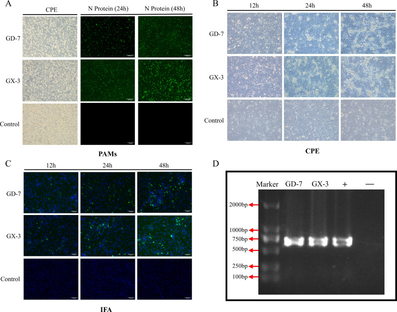
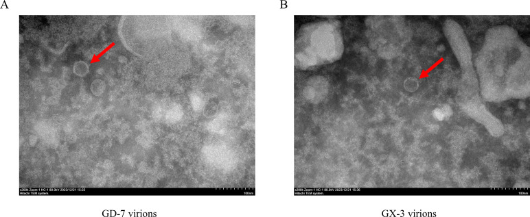
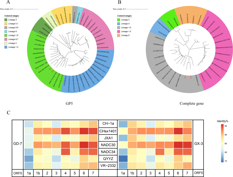
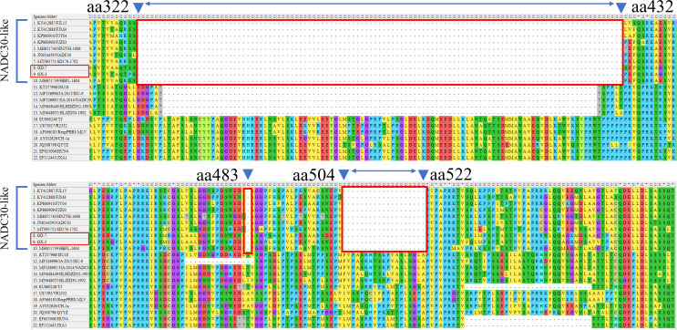
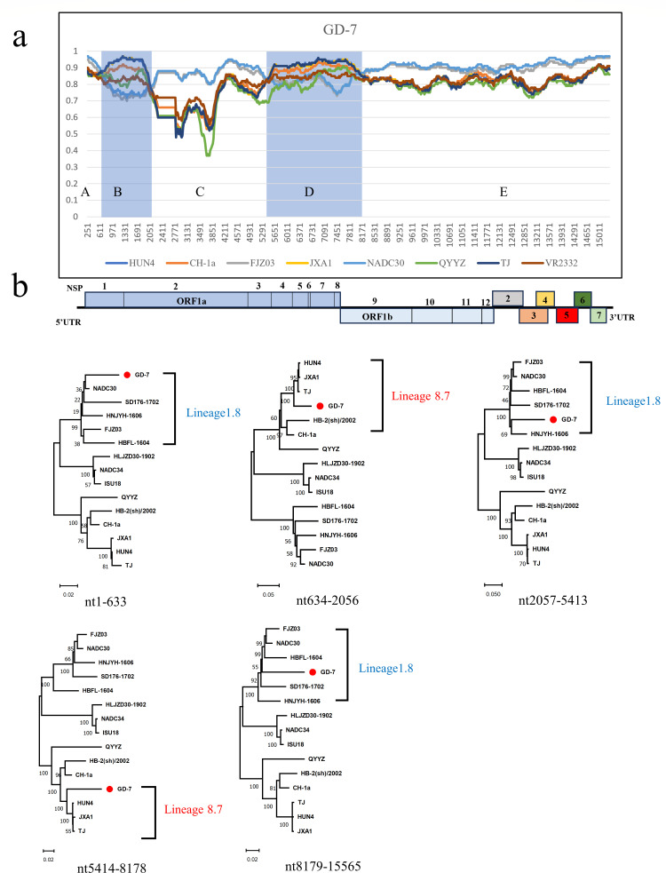
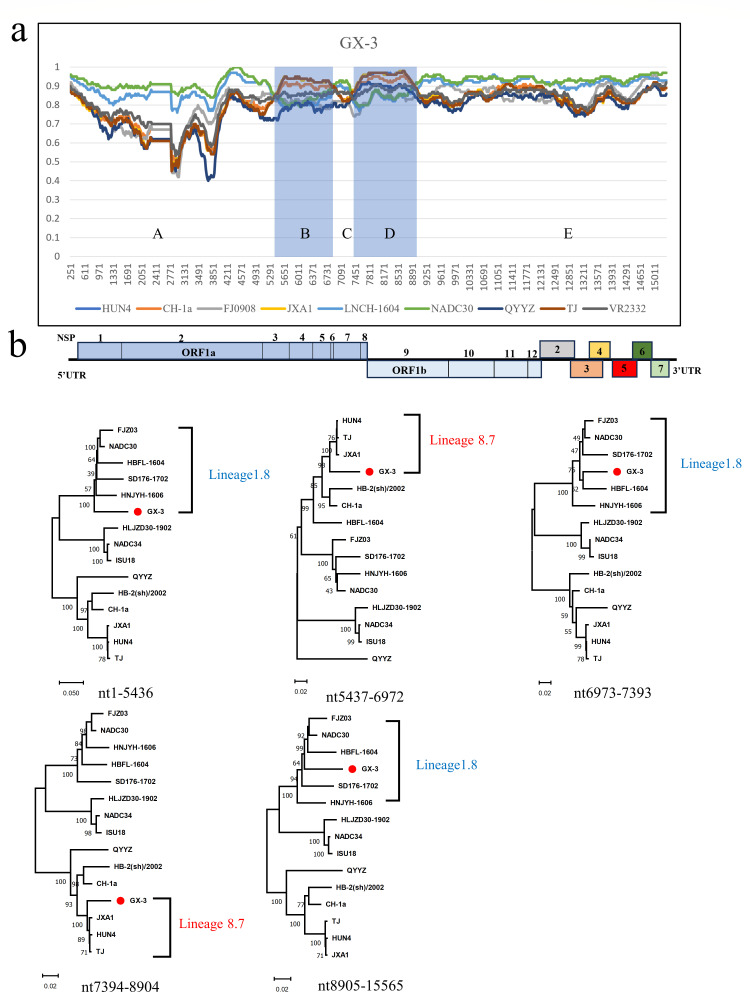
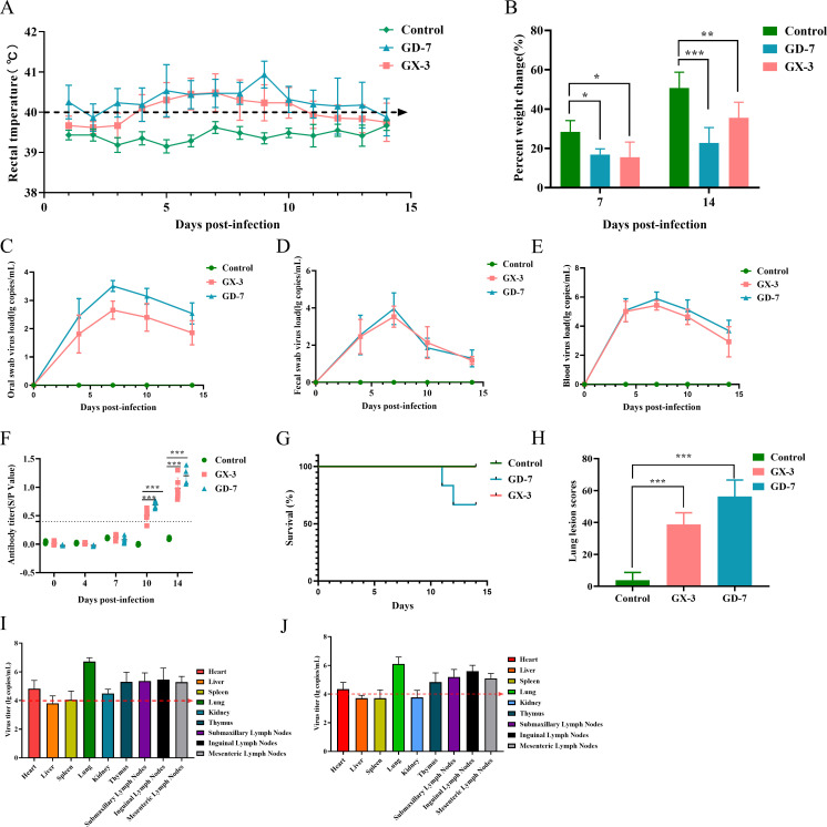
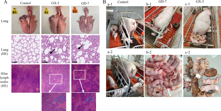
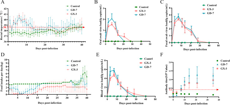
Porcine reproductive and respiratory syndrome viruses (PRRSVs) exhibit high mutability and recombination, posing challenges to their immunization and control. This study isolated two new PRRSV strains, GD-7 and GX-3, from samples collected in Guangdong and Guangxi in 2023. Whole-genome sequencing, along with phylogenetic and recombination analyses, confirmed that GD-7 and GX-3 are natural novel recombinant strains of NADC30 PRRSV. Moreover, we established a pathogenicity model for piglets and sows based on the two isolates. The results of piglet pathogenicity revealed that both GD-7 and GX-3 caused clinical symptoms such as fever, loss of appetite, depression, and slow weight gain. Moreover, we observed that the mortality rate of GD-7-inoculated group piglets was 33.3%, which was similar to that of piglets infected with other highly pathogenic PRRSV strains and exceeded the mortality rate of most NADC30-like PRRSV. In pregnant sow models, the survival rate of sows in the GD-7 group was 75%, in contrast to the GX-3 group, where no sow mortality was observed, and both strains resulted in abortion, mummified fetuses, and stillbirths. These results highlight the elevated pathogenicity of these recombinant strains in sows, with GD-7 mainly causing sows to abort, and GX-3 mainly causing sows to give birth to mummified fetuses. This study introduces two distinct clinical recombinant PRRSV strains that differ from the prevalent strains in China. This research furthers our understanding of the epidemiology of PRRSV and underscores the significance of ongoing monitoring and research in the face of evolving virus strains. Moreover, these discoveries act as early warnings, underscoring the necessity for active control and immunization against PRRSV.IMPORTANCESince the discovery of NADC30-like PRRSV in China in 2013, it has gradually become the dominant strain of PRRSV in China. NADC30-like PRRSV exhibits high recombination characteristics, constantly recombining with different strains, leading to the emergence of numerous novel strains. Of particular importance is the observation that NADC30-like PRRSV with different recombination patterns exhibits varying pathogenicity, which has a significant impact on the pig farming industry. This emphasizes the necessity of monitoring and responding to evolving PRRSV strains to develop effective immunization and control strategies. In this paper, we conducted pathogenicity studies on the isolated NADC30-like PRRSV and analyzed the differences in the genomes and pathogenicity of the different strains by recording clinical symptoms, temperature changes, detoxification tests, and changes in viremia and histopathology in infected pigs. This was done to provide a theoretical basis for the epidemiological situation and epidemic prevention and control of PRRSV.

Keywords: PRRSV; pathogenicity; phylogenetic analysis; recombination analysis.


Conflict of interest statement

The authors declare no conflict of interest.

Figures

Fig 1
Fig 1
Isolation and characterization of viruses. (A): Lesions in PAM cells after inoculation with GX-3 and GD-7 disease material and detection of PRRSVN protein by IFA. (B): Cytopathic conditions of MARC-145 at 24, 36, and 72 h post-infection (hpi). (C): FAs show the reactivity of a monoclonal antibody against PRRSV N protein to GD-7 and GX-3 strains infected at 24, 36, and 72 hpi. (D): Lesion MARC-145 cell nucleic acid was collected and detected by agarose gel electrophoresis after PCR amplification, + is the positive control, − is the blank control.
Fig 2
Fig 2
Observation of viral particles. (A) Virus particles of the GD-7 strain were observed by electron microscopy. Magnification ×200K. (B): Virus particles of the GX-3 strain observed by electron microscopy. Magnification ×200K.
Fig 3
Fig 3
Genome analysis and recombination analysis. (A) The phylogenetic tree was constructed based on the GP5 gene of GX-3 and GD-7 strains and 47 reference PRRSV strains. (B) Phylogenetic trees were constructed based on the complete genome of the GX-3 and GD-7 strains with 30 representative reference PRRSV strains. Phylogenetic trees were constructed using the distance-based neighbor-joining method with 1,000 bootstrap replicates in MEGA11. (C) Heat map constructed based on the homology of the isolated strain with other reference strains.
Fig 4
Fig 4
Amino acid deletion pattern in the NSP2 gene. The alignment of GD-7 and GX-3 is marked in red.
Fig 5
Fig 5
(a) Recombination analysis of the GD-7 strain using Simplot software, the blue-shaded region is the minor parental region and the rest is the major parental region. Below the similarity plot is a full-genome structure, with reference to CH-1a, in which the positions and boundaries of the major ORFs, nsp-encoding genes within ORF1a and ORF2b, and gaps are shown. (b) Phylogenetic trees were constructed based on GD-7 recombination intervals A, B, C, D, and E. Phylogenetic trees were constructed using the distance-based neighbor-joining method with 1,000 bootstrap replicates in MEGA11.
Fig 6
Fig 6
(a) Recombination analysis of the GX-3 strain using Simplot software, the blue-shaded region is the minor parental region and the rest is the major parental region. Below the similarity plot is a full-genome structure, with reference to CH-1a, in which the positions and boundaries of the major ORFs, nsp-encoding genes within ORF1a and ORF2b, and gaps are shown. (b) Phylogenetic trees were constructed based on GX-3 recombination intervals A, B, C, D, and E. Phylogenetic trees were constructed using the distance-based neighbor-joining method with 1,000 bootstrap replicates in MEGA11.
Fig 7
Fig 7
Pathogenicity results in piglets. (A) Body temperature change of pigs in each group after challenge. (B) Body weight gains of each group during the challenge study. (C–E): Viral load detection in blood, oral swabs, and pharyngeal swabs. (F) PRRSV-specific antibody level was detected in each group during the challenge study. (G) Ocular lesion scores of the lungs in each group. (H) Survival curves of piglets in each group. (I) The toxic load of various organs and tissues in the GD-7 group. (H) The toxic load of various organs and tissues in the GX-3 group. The significant difference is marked with the asterisk, ***P < 0.001, **P < 0.01, and *P < 0.05.
Fig 8
Fig 8
(A) Ocular lesions of the lungs and HE staining results of lung and Hilar lymphoid tissues; it shows interstitial thickening and inflammatory infiltration by black arrows; sparsely arranged lymphoid cells by blue arrow ①; and lymphocyte necrosis by blue arrow ②. (B) Clinical production results of sows in each group: a-1 and a-2 are control sows and their farrowing piglets; b-1, and b-2 are GD-7 sows and their aborting piglets; c-1 and c-2 are GX-3 sows and their farrowing mummified piglets.
Fig 9
Fig 9
Pathogenicity results in sows. (A) Body temperature change of pigs in each group after the challenge. (B and C) Viral load detection oral swabs and fecal swabs. (D) Feed intake test for each group of sows. (E) Viral load detection in blood. (F) PRRSV-specific antibody level was detected in each group during the challenge study.

Similar articles

Cited by

References

    1. Pol JM, van Dijk JE, Wensvoort G, Terpstra C. 1991. Pathological, ultrastructural, and immunohistochemical changes caused by Lelystad virus in experimentally induced infections of mystery swine disease (synonym: porcine epidemic abortion and respiratory syndrome (PEARS)). Vet Q 13:137–143. doi:10.1080/01652176.1991.9694298 - DOI - PubMed
    1. Wills RW, Zimmerman JJ, Yoon KJ, Swenson SL, McGinley MJ, Hill HT, Platt KB, Christopher-Hennings J, Nelson EA. 1997. Porcine reproductive and respiratory syndrome virus: a persistent infection. Vet Microbiol 55:231–240. doi:10.1016/s0378-1135(96)01337-5 - DOI - PubMed
    1. Khatun A, Park SY, Shabir N, Nazki S, Kang AR, Jeong CG, Seo BJ, Yang MS, Kim B, Seo YH, Kim WI. 2019. Evaluation of the inhibitory effects of (E)-1-(2-hydroxy-4,6-dimethoxyphenyl)-3-(naphthalen-1-yl)prop-2-en-1-one (DiNap), a natural product analog, on the replication of type 2 PRRSV in vitro and in vivo. Molecules 24:887. doi:10.3390/molecules24050887 - DOI - PMC - PubMed
    1. Nelsen CJ, Murtaugh MP, Faaberg KS. 1999. Porcine reproductive and respiratory syndrome virus comparison: divergent evolution on two continents. J Virol 73:270–280. doi:10.1128/JVI.73.1.270-280.1999 - DOI - PMC - PubMed
    1. Guo BQ, Chen ZS, Liu XW, Cui YZ. 1996. Isolation and identification of porcine reproductive and respiratory syndrome (PRRS) virus. Chin J Prev Vet Med 25:1–5.

MeSH terms

LinkOut - more resources