. 2024 Oct 3;12(10):e0122924.
doi: 10.1128/spectrum.01229-24. Epub 2024 Aug 20.

Orthologs of Plasmodium ICM1 are dispensable for Ca2+ mobilization in Toxoplasma gondii

Affiliations

Orthologs of Plasmodium ICM1 are dispensable for Ca2+ mobilization in Toxoplasma gondii

Gabriel Cabral et al. Microbiol Spectr. .

Abstract

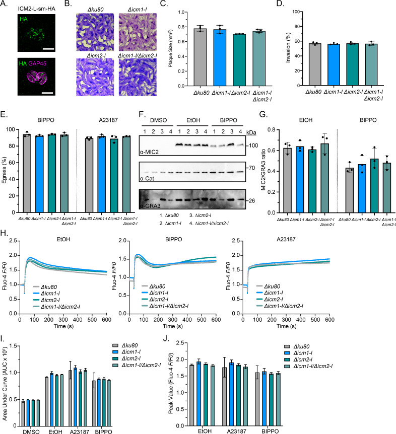
Apicomplexan parasites mobilize ionic calcium (Ca2+) from intracellular stores to promote microneme secretion and facilitate motile processes including gliding motility, invasion, and egress. Recently, a multipass transmembrane protein, ICM1, was found to be important for calcium mobilization in Plasmodium falciparum and P. berghei. Comparative genomics and phylogenetics have revealed putative ICM orthologs in Toxoplasma gondii and other apicomplexans. T. gondii possesses two ICM-like proteins, which we have named TgICM1-L (TGGT1_305470) and TgICM2-L (TGGT1_309910). TgICM1-L and TgICM2-L localized to undefined puncta within the parasite cytosol. TgICM1-L and TgICM2-L are individually dispensable in tachyzoites, suggesting a potential compensatory relationship between the two proteins may exist. Surprisingly, mutants lacking both TgICM1-L and TgICM2-L are fully viable, exhibiting no obvious defects in growth, microneme secretion, invasion, or egress. Furthermore, loss of TgICM1-L, TgICM2-L, or both does not impair the parasite's ability to mobilize Ca2+. These findings suggest that additional proteins may participate in Ca2+ mobilization or import in Apicomplexa, reducing the dependence on ICM-like proteins in T. gondii. Collectively, these results highlight similar yet distinct mechanisms of Ca2+ mobilization between T. gondii and Plasmodium.IMPORTANCECa2+ signaling plays a crucial role in governing apicomplexan motility; yet, the mechanisms underlying Ca2+ mobilization from intracellular stores in these parasites remain unclear. In Plasmodium, the necessity of ICM1 for Ca2+ mobilization raises the question of whether this mechanism is conserved in other apicomplexans. Investigation into the orthologs of Plasmodium ICM1 in T. gondii revealed a differing requirement for ICM proteins between the two parasites. This study suggests that T. gondii employs ICM-independent mechanisms to regulate Ca2+ homeostasis and mobilization. Proteins involved in Ca2+ signaling in apicomplexans represent promising targets for therapeutic development.

Keywords: Apicomplexa; Plasmodium; Toxoplasma; Toxoplasma gondii; apicomplexan; cAMP; cGMP; calcium; calcium flux; calcium signaling; cyclic GMP; motility.


Conflict of interest statement

The authors declare no conflict of interest.

Figures

Fig 1
Fig 1
Identification of ICM-Like proteins in Toxoplasma gondii. (A) Reciprocal BLASTP searches to identify orthologs of P. falciparum ICM1 in T. gondii (cutoff E < 1e-10). (B) NCBI conserved domains and TM domains identified by TOPCONS. SdaC, PotE, Aa_trans are associated with AA transport. (C) ApiICM and ApiAT sequences were aligned with MUSCLE and a consensus maximum likelihood tree was constructed with 200 bootstraps. The annotated ApiAT clades matched those published in reference (59).
Fig 2
Fig 2
Expression, localization, and conditional knockdown of TgICM1-L in tachyzoites (A) Strategy for C-terminal tagging TgICM1-L with mAID-3HA in RH TIR1-3FLAG. (B) Diagnostic PCRs from gDNA showing 3′ integration of mAID-3HA into TgICM1-L. (C) ICM1-L-mAID-3HA expression in tachyzoites is not detectable by super-resolution microscopy. Scale bar = 5 µm. (D) ICM1-L-mAID-3HA enriched by immunoprecipitation (IP) is not detectable by immunoblotting. (E) ICM1-L-mAID-3HA expression by immunoblotting following parasite treatment with IAA or the vehicle (ETOH) for 48 h was below the threshold needed to confirm conditional knockdown. (F and G) Plaque formation by 200 tachyzoites treated with IAA or the vehicle (ETOH) for 7 days. (F) Representative images. (G) Mean plaque number (N = 3, n = 9) +/-SD. n.s, not significant, unpaired two-tailed Student’s t-test. (H) IF microscopy of ICM1-L-smHA parasites confirms ICM1-L expression in tachyzoites. Scale bar = 5 µm. (I) Strategy for N-terminal tagging TgICM1-L with mAID-3HA in RH TIR1-3FLAG using FACS to select for GFP-RNP-transfected parasites. FACS gates defined with mock-transfected parasites. (J) Diagnostic PCRs from gDNA showing 5′integration of mAID-3HA into TgICM1-L. (K) mAID-3HA-ICM1-L expression in tachyzoites is not detectable by super-resolution microscopy. Scale bar = 5 µm. (L) mAID-3HA-ICM1-L enriched by IP is detectable by immunoblotting. (M) Confirmation of mAID-3HA-ICM1-L knockdown following treatment with IAA or EtOH for 48 h using IP and immunoblotting. (N and O) Plaque formation by 200 tachyzoites treated with IAA or the vehicle (ETOH) for 7 days. (N) Representative images. (O) Mean plaque number (N = 3, n = 9) ±SD. n.s, not significant, unpaired two-tailed Student’s t-test.
Fig 3
Fig 3
Deletion of TgICM1-L does not negatively impact tachyzoite fitness (A) TgICM1-L KO strategy showing diagnostic PCR positions. (B) Validation of TgICM1-L deletion by diagnostic PCR. WT = RHΔku80Δhxgprt, RHΔICM1-L. (C and D) Plaque formation by 200 RHΔku80Δhxgprt or RHΔICM1-L parasites at 7 days in HFFs. (C) Representative images. D) Mean plaque number (N = 3) ±SD. (E–G) Growth competition assay between RHΔku80Δhxgprt and RHΔICM1-L parasites. (E) Equal starting numbers of parasites were co-cultured together in HFF T-25s and passaged as needed for 29 days. gDNA samples were collected at each passage for PCR. (F) Diagnostic multiplex PCR using 50 ng of gDNA template from co-culture or standard samples. WT = internal ICM1-L 757 bp amplicon. Mut = internal HXGPRT 578 bp amplicon. (G) Percentage of RHΔku80Δhxgprt and RHΔICM1-L over 29 days based on the ratio of gDNA calculated from the standard curve for each trial. (H–J) Plaque formation by 200 RHΔku80Δhxgprt and ΔICM1-L parasites at 7 days in HFFs in the presence of increasing KCl (H), zaprinast (I), or pyrimethamine (J).
Fig 4
Fig 4
TgICM1-L and TgICM2-L are dispensable for tachyzoite growth, motility, and Ca2+ mobilization (A) IF microscopy of ICM2-L-smHA parasites confirms TgICM2-L expression in tachyzoites. Scale bar = 5 µm. (B and C) Plaque formation by tachyzoites at 7 days in HFFs. (B) Representative images. (C) Mean area number (N = 3) ±SEM. (D) Invasion assay showing the percentage of invaded tachyzoites in HFFs. (E) Egress assay showing the percentage of egressed parasite vacuoles in HFFs following treatment with BIPPO or A23187. (F–G) Microneme secretion assay of tachyzoites stimulated with DMSO, EtOH, or BIPPO. (F) Representative immunoblots of excreted secreted antigen fractions. (G) The ratio of secreted MIC2 to secreted GRA3 as quantified by immunoblotting (N = 3), no significant differences were measured. (H–J) Fluo-4 calcium reporter assay showing tachyzoite intracellular calcium changes in response to DMSO, EtOH, BIPPO, and A23187. (H) Time-resolved calcium changes in tachyzoites. Traces shown are the mean of 9 replicates per line. (I) Area under the curve (AUC) measurements. (J) Peak calcium responses. No significant differences were observed.

Similar articles

Cited by

References

    1. Janouškovec J, Paskerova GG, Miroliubova TS, Mikhailov KV, Birley T, Aleoshin VV, Simdyanov TG. 2019. Apicomplexan-like parasites are polyphyletic and widely but selectively dependent on cryptic plastid organelles. Elife 8:e49662. doi:10.7554/eLife.49662 - DOI - PMC - PubMed
    1. Cowman AF, Healer J, Marapana D, Marsh K. 2016. Malaria: Biology and Disease. Cell 167:610–624. doi:10.1016/j.cell.2016.07.055 - DOI - PubMed
    1. Venkatesan P. 2024. The 2023 WHO World malaria report. Lancet Microbe 5:e214. doi:10.1016/S2666-5247(24)00016-8 - DOI - PubMed
    1. Jalovecka M, Sojka D, Ascencio M, Schnittger L. 2019. Babesia life cycle – when phylogeny meets biology. Trends Parasitol 35:356–368. doi:10.1016/j.pt.2019.01.007 - DOI - PubMed
    1. Westblade LF, Simon MS, Mathison BA, Kirkman LA. 2017. Babesia microti: from mice to ticks to an increasing number of highly susceptible humans. J Clin Microbiol 55:2903–2912. doi:10.1128/JCM.00504-17 - DOI - PMC - PubMed

LinkOut - more resources