. 2024 Oct 3;12(10):e0066124.
doi: 10.1128/spectrum.00661-24. Epub 2024 Aug 20.

Calcium-binding protein TgpCaBP regulates calcium storage of the zoonotic parasite Toxoplasma gondii

Affiliations

Calcium-binding protein TgpCaBP regulates calcium storage of the zoonotic parasite Toxoplasma gondii

Weisong Sun et al. Microbiol Spectr. .

Abstract

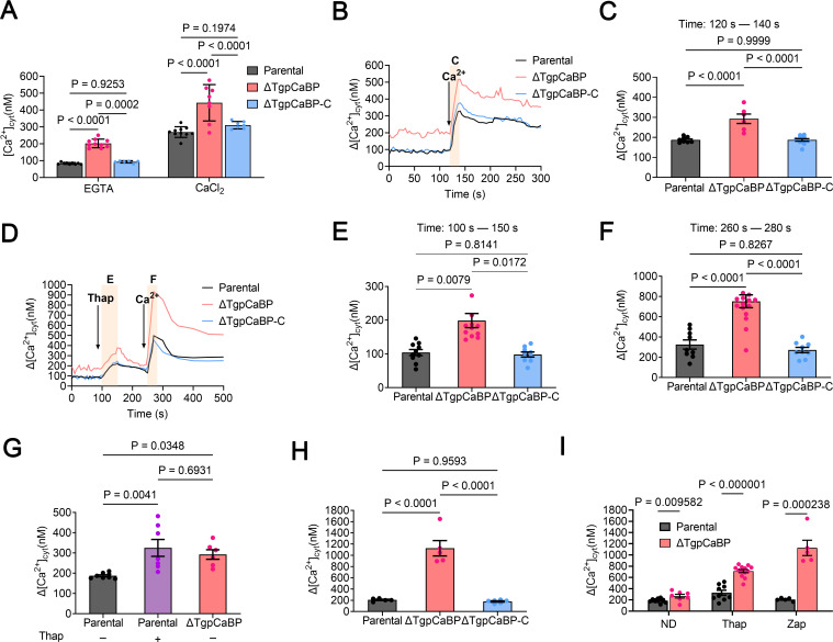
Toxoplasma gondii, the causative parasite of toxoplasmosis, is an apicomplexan parasite that infects warm-blooded mammals. The ability of the calcium-binding proteins (CBPs) to transport large amounts of Ca2+ appears to be critical for the biological activity of T. gondii. However, the functions of some members of the CBP family have not yet been deciphered. Here, we characterized a putative CBP of T. gondii, TgpCaBP (TGME49_229480), which is composed of four EF-hand motifs with Ca2+-binding capability. TgpCaBP was localized in the cytosol and ER of T. gondii, and parasites lacking the TgpCaBP gene exhibited diminished abilities in cell invasion, intracellular growth, egress, and motility. These phenomena were due to the abnormalities in intracellular Ca2+ efflux and ER Ca2+ storage, and the reduction in motility was associated with a decrease in the discharge of secretory proteins. Therefore, we propose that TgpCaBP is a Ca2+ transporter and signaling molecule involved in Ca2+ regulation and parasitization in the hosts.IMPORTANCECa2+ signaling is essential in the development of T. gondii. In this study, we identified a calcium-binding protein in T. gondii, named TgpCaBP, which actively regulates intracellular Ca2+ levels in the parasite. Deletion of the gene coding for TgpCaBP caused serious deficits in the parasite's ability to maintain a stable intracellular calcium environment, which also impaired the secretory protein discharged from the parasite, and its capacity of gliding motility, cell invasion, intracellular growth, and egress from host cells. In summary, we have identified a novel calcium-binding protein, TgpCaBP, in the zoonotic parasite T. gondii, which is a potential therapeutic target for toxoplasmosis.

Keywords: Ca2+ signaling; TgpCaBP; Toxoplasma gondii; calcium binding; parasite biology.


Conflict of interest statement

The authors declare no conflict of interest.

Figures

Fig 1
Fig 1
TgpCaBP contains a typical calcium-binding domain and is localized in the cytosol and endoplasmic reticulum of T. gondii. (A) A schematic presentation of the domain annotation of TgpCaBP of T. gondii. The predicted EF-hand domains are highlighted in orange color. An ER-targeting sequence located at the C-terminus is indicated in red color. (B) Structure modeling of TgpCaBP. Left: The structure of TgpCaBP is shown with EF-hand motifs, which consist of 12 amino acid residues (yellow) and conserved Asp residues (red). Right: Enlarged views of the EF-hand motifs of TgpCaBP showing the numbers of Asp residues. (C, D) Immunofluorescence assay (IFA) of TgpCaBP in the extracellular and intracellular tachyzoites using a rat anti-TgpCaBP antibody (1:100). TgpCaBP is localized in both cytosol and endoplasmic reticulum and co-localized with the anti-TgIPPS and TgBIP antibodies. Scale bar, 5 µm.
Fig 2
Fig 2
Calcium binding property of TgpCaBP. (A) Western blot analysis of native, HIS- and GST-tagged TgpCaBP using a rat anti-TgpCaBP antibody. (B) Calcium-dependent solubility was detected using western blotting. Parasites were lysed in 1% Triton X-100 in 5 mM EGTA or 5 mM CaCl2 and fractionated by centrifugation. TgpCaBP was detected with a rat anti-TgpCaBP antibody, a mouse anti-IMC1 antibody was used as a control for the protein in the pellet (P) and a rabbit β-Tubulin monoclonal antibody was used as a control for the protein in the supernatant (S). (C) TgpCaBP migrated faster in the presence of Ca2+. Ca2+ was replaced by Mg2+ and K+ in the control group, and EGTA was used to chelate-free calcium in the extracellular environment (Fig. S3A and B). Parasites were lysed in 1% Triton X-100 and incubated with EGTA, a combination of EGTA and CaCl2, or CaCl2. The native protein was extracted from T. gondii RH tachyzoites. (D) The expression of TgpCaBP in the parental, ΔTgpCaBP, and ΔTgpCaBP-C parasites (with two batches of proteins from each parasite line) was analyzed using western blotting with a rat anti-TgpCaBP antibody to verify the presence and absence of TgpCaBP protein in different parasite lines.
Fig 3
Fig 3
TgpCaBP was associated with intracellular growth and gliding motility of T. gondii. (A) Plaque assays were conducted for parental (P), ΔTgpCaBP (Δ), and ΔTgpCaBP-C (C) parasites. Quantification of plaque sizes from three independent biological experiments using one-way ANOVA with Tukey’s multiple comparisons. Values are means ± SEM; (n = 3). P > 0.05: not significant. Scale bar, 2 mm. (B) Red/green assays of parental, ΔTgpCaBP, and ΔTgpCaBP-C parasites calculating the invasion efficiency. The invasion capacity of the ΔTgpCaBP parasites was significantly lower than that of the parental and the gene-complemented parasites. Quantification of invasion from three independent biological experiments using one-way ANOVA with Tukey’s multiple comparisons. Values are means ± SEM; (n = 3). P > 0.05: not significant. (C) Egress assays of the parental, ΔTgpCaBP, and ΔTgpCaBP-C parasites stimulated by 3 µM A23187. The egress of the ΔTgpCaBP parasites was significantly lower than the other two parasite lines. Quantification of egress from three independent biological experiments using two-way ANOVA with Tukey’s multiple comparisons. Values are means ± SEM; (n = 3). P > 0.05: not significant. (D) Effect of A23187 on gliding of T. gondii. Indirect immunofluorescence microscopy demonstrated that the length of trails deposited during gliding with A23187 treatment in the parental and ΔTgpCaBP-C parasite was longer and more complete than the ΔTgpCaBP parasite. Trails were visualized with a mouse anti-SAG1 antibody and conjugated to Alexa 488. Scale bar, 5 µm. (E) Quantification and statistical analysis of the trail length of the parental, ΔTgpCaBP, and ΔTgpCaBP-C parasites treated with A23187 and dimethyl sulfoxide control. The motility was impaired in the ΔTgpCaBP parasites and rescued in the ΔTgpCaBP-C parasites. Values are means ± SEM; (n = 3). P > 0.05: not significant. (F) Replication assays of the parental, ΔTgpCaBP, and ΔTgpCaBP-C parasites. There was no significant difference between the replication of the ΔTgpCaBP parasites and the other two parasite lines. Quantification of replication from three independent biological experiments using two-way ANOVA with Tukey’s multiple comparisons. Values are means ± SEM; (n = 3). Ns: not significant. (G) Survival of mice challenged with 50, 500, 5 × 10³, or 5 × 10⁵ parental and ΔTgpCaBP parasites. (H) Survival of mice challenged with 50 parental, ΔTgpCaBP, and ΔTgpCaBP-C parasites. The virulence of the ΔTgpCaBP parasites decreased significantly compared to the parental parasites. The virulence was rescued in the ΔTgpCaBP-C parasites. All mice were monitored for 28 days. All data were analyzed using simple survival analysis (Kaplan-Meier), P < 0.0001.
Fig 4
Fig 4
Effect of the Ca2+ ionophore A23187 on microneme secretion of T. gondii. (A) Western blot of supernatants secreted by parasites after stimulation with A23187 at 37°C for 5 min, showing a significant difference in MIC2 secreted by the parental and the ΔTgpCaBP parasites. The pellets were monitored by a mouse anti-IMC-1 monoclonal antibody. Stimulation with 2% ethanol was used as a positive control, and DMSO was used as a solvent control. (B) Quantification and statistical analysis of MIC2 secreted by the parental and the ΔTgpCaBP parasites after treatment with 2% ethanol, DMSO, and different concentrations of A23187. Secretion of MIC2 was inhibited in the ΔTgpCaBP parasites. Values are means ± SEM; (n = 3). P > 0.05: not significant.
Fig 5
Fig 5
Role of TgpCaBP in the efflux of cytosolic Ca2+ of T. gondii. (A) Intracellular calcium concentration ([Ca2+]i) (nM) of the parental parasites, the ΔTgpCaBP and the ΔTgpCaBP-C parasites in the presence of 100 µM EGTA or 2 mM CaCl2. Values are means ± SEM; (n = 6–10). P > 0.05: not significant. (B) Measurements of dynamic cytosolic Ca2+ in the presence of Fura-2AM in tachyzoites of the parental (RH), the ΔTgpCaBP and the ΔTgpCaBP-C parasites. The buffer contains 100 µM EGTA to chelate contaminating Ca2+, and 2 mM Ca2+ was added to the suspension at 120 s. The pink box indicates the area used for the quantification presented in (C). (C) Quantification and statistical analysis of the change in cytosolic Ca2+ during the first 20 s after the addition of extracellular Ca2+. Values are means ± SEM; (n = 7–12). P > 0.05: not significant. (D) Cytosolic Ca2+ increases after adding Thap (1 µM), following Ca2+ influx resulting from adding 2 mM extracellular Ca2+ at 260 s. Thap: thapsigargin. The pink boxes indicate the area used for the quantification presented in (E) and (F). (E) Quantification and statistical analysis of the change in cytosolic Ca2+(Δ[Ca2+]cyt) at 50 s after adding Thap. Values are means ± SEM; (n = 9–12). P > 0.05: not significant. (F) Quantification and statistical analysis of the Δ[Ca2+]cyt at 20 s after adding 2 mM of Ca2+. Values are mean ± SEM; (n = 9–16). P > 0.05: not significant. (G) Quantification and statistical analysis of the Δ[Ca2+]cyt at 20 s after adding Ca2+ in the presence or absence of Thap in the parental (P) and the ΔTgpCaBP parasites. Values are means ± SEM; (n = 7–12). P > 0.05: not significant. (H) Quantification and statistical analysis of cytosolic Ca2+ increase stimulated by Zap (100 µM) in the presence of 2 mM extracellular Ca2+. Values are means ± SEM; (n = 5–6). P > 0.05: not significant. Zap: zaprinast. (I) Quantification and statistical analysis of the Δ[Ca2+]cyt during the 20 s after adding Ca2+ without additions (ND) or after adding Thap or Zap. Values are means ± SEM; (n = 5–15). P > 0.05: not significant. Two-way ANOVA with Tukey’s multiple comparison test for A and I and one-way ANOVA with Tukey’s multiple comparison tests for C, E, F, G, and H.
Fig 6
Fig 6
Schematic illustration of TgpCaBP in cytosolic Ca2+ efflux and regulatory function in ER Ca2+ storage in T. gondii. When intracellular calcium concentration is too high, TgpCaBP will participate in calcium efflux to maintain calcium balance. TgpCaBPs bind to free Ca2+ and transport them to the ER for storage. TgpCaBP participates in the regulation of the secretion of microneme proteins. The figure was designed by BioRender.

Similar articles

References

    1. Hill D, Dubey JP. 2002. Toxoplasma gondii: transmission, diagnosis and prevention. Clin Microbiol Infect 8:634–640. doi:10.1046/j.1469-0691.2002.00485.x - DOI - PubMed
    1. Robert-Gangneux F, Dardé M-L. 2012. Epidemiology of and diagnostic strategies for toxoplasmosis. Clin Microbiol Rev 25:264–296. doi:10.1128/CMR.05013-11 - DOI - PMC - PubMed
    1. Montoya JG, Rosso F. 2005. Diagnosis and management of toxoplasmosis. Clin Perinatol 32:705–726. doi:10.1016/j.clp.2005.04.011 - DOI - PubMed
    1. Gazarini ML, Thomas AP, Pozzan T, Garcia CRS. 2003. Calcium signaling in a low calcium environment. J Cell Biol 161:103–110. doi:10.1083/jcb.200212130 - DOI - PMC - PubMed
    1. Arrizabalaga G, Boothroyd JC. 2004. Role of calcium during Toxoplasma gondii invasion and egress. Int J Parasitol 34:361–368. doi:10.1016/j.ijpara.2003.11.017 - DOI - PubMed

LinkOut - more resources