. 2024 Sep;68(5):531-537.
doi: 10.1007/s10384-024-01107-w. Epub 2024 Aug 20.

Retinal vasculitis after intravitreal aflibercept 8 mg for neovascular age-related macular degeneration

Affiliations

Retinal vasculitis after intravitreal aflibercept 8 mg for neovascular age-related macular degeneration

Hidetaka Matsumoto et al. Jpn J Ophthalmol. 2024 Sep.

Abstract

Purpose: To evaluate short-term outcomes of intravitreal injection of aflibercept 8 mg for neovascular age-related macular degeneration (nAMD).

Study design: Retrospective, interventional case series.

Methods: We retrospectively studied 35 eyes of 34 consecutive patients with nAMD, assessing best-corrected visual acuity (BCVA), foveal thickness (FT), and central choroidal thickness (CCT) before and 4 weeks after the initial intravitreal dose of aflibercept 8 mg. The rate of achieving a dry macula and the incidence of intraocular inflammation (IOI) at week 4 were also determined.

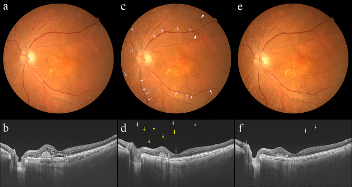
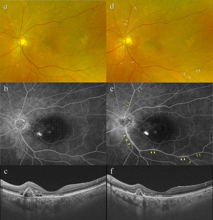
Results: BCVA showed significant improvement, with significant reductions in FT and CCT 4 weeks after the initial injection of aflibercept 8 mg (all P < 0.01), with a dry macula being achieved in 20 eyes (57.1%). However, 3 eyes (8.6%) developed non-infectious IOI associated with retinal vasculitis, an adverse event not reported previously. The IOI in these eyes was relatively mild and treated with a posterior subtenon injection of triamcinolone acetonide with or without betamethasone eye drops, resulting in amelioration of IOI without any visual loss.

Conclusions: Intravitreal aflibercept 8 mg appears to be effective for improving visual acuity and ameliorating exudative changes in eyes with nAMD. However, special attention should be given to the potential development of IOI associated with retinal vasculitis.

Keywords: Aflibercept 8 mg; Age-related macular degeneration; Anti-vascular endothelial growth factor; Intraocular inflammation; Retinal vasculitis.


Conflict of interest statement

H. Matsumoto, Payment or honoraria for lectures, presentations, speakers bureaus, manuscript writing or educational events (Novartis, Chugai, Senju), Participation on a Data Safety Monitoring Board or Advisory Board (Novartis, Chugai); J. Hoshino, Payment or honoraria for lectures, presentations, speakers bureaus, manuscript writing or educational events (Novartis, Chugai, Senju); S. Numaga, None; K. Mimura, None; Y. Asatori, None; H. Akiyama, Payment or honoraria for lectures, presentations, speakers bureaus, manuscript writing or educational events (Novartis, Chugai, Senju, Bayer, Santen, Otsuka, AMO, Pfizer, Wakamoto, Kowa, Eisai, HOYA).

Figures

Fig. 1
Fig. 1
Images of the left eye of an 82-year-old man with treatment-naïve neovascular age-related macular degeneration associated with mixed type 1 and type 2 macular neovascularization. At baseline, best-corrected visual acuity (BCVA) was 0.6 (0.22 logarithm of the minimum angle of resolution (logMAR) units). (a) Color fundus photograph shows retinal pigment epithelium (RPE) degeneration accompanied by subretinal hemorrhage and hard exudate at the macular area. The retinal vessels appear normal. (b) Optical coherence tomography (OCT) shows a shallow irregular RPE elevation and subretinal hyperreflective material, reflecting mixed type 1 and type 2 macular neovascularization, accompanied by subretinal and intraretinal fluid. The foveal thickness and central choroidal thickness are 193 μm and 214 μm, respectively. Four weeks after the initial injection of aflibercept 8 mg, BCVA of the left eye is 0.6 (0.22 logMAR units). (c) Color fundus photograph shows multiple sites of localized narrowing of the retinal vessels, especially retinal veins (arrows), and mild intraretinal hemorrhage (arrow heads). Subretinal hemorrhage and hard exudate at the macular area have decreased. (d) OCT shows several vitreous cells (arrows). Shallow irregular RPE elevation and subretinal hyperreflective material have diminished, and subretinal and intraretinal fluid have been resolved. The foveal thickness and central choroidal thickness are 145 μm and 87 μm, respectively. Aflibercept 8 mg therapy was discontinued due to the development of intraocular inflammation associated with retinal vasculitis. Two weeks after a posterior subtenon injection of triamcinolone acetonide (30 mg/0.75mL) with 0.1% betamethasone eye drops, BCVA of the left eye is 0.8 (0.10 logMAR units). (e) Color fundus photograph shows a reduction in localized narrowing of the retinal vessels, with disappearance of intraretinal hemorrhage. (f) OCT shows a reduced number of vitreous cells (arrows)
Fig. 2
Fig. 2
Images of the left eye of a 79-year-old woman with treatment-naïve neovascular age-related macular degeneration associated with polypoidal choroidal vasculopathy. At baseline, best-corrected visual acuity (BCVA) was 0.8 (0.10 logarithm of the minimum angle of resolution (logMAR) units). (a) Color fundus photograph shows retinal pigment epithelium (RPE) degeneration at the macular area. The retinal vessels appear normal. (b) Fluorescein angiography demonstrates mild leakage and window defects at the macular area. The retinal vessels appear normal. (c) Optical coherence tomography (OCT) shows a shallow irregular RPE elevation (double layer sign) and protruding RPE detachment, reflecting a branching neovascular network and polypoidal lesion, accompanied by subretinal and sub-RPE fluid. The foveal thickness and central choroidal thickness are 129 μm and 442 μm, respectively. Four weeks after initial injection of aflibercept 8 mg, BCVA of the left eye is 0.8 (0.10 logMAR units). (d) Color fundus photograph shows multiple sites of localized narrowing of the retinal vessels, especially retinal veins (arrows). (e) Fluorescein angiography reveals multiple sites of localized narrowing of the retinal vessels, especially retinal veins (arrows), and mild leakage from inferior temporal arcade retinal vein (arrow heads). (f) OCT shows that shallow irregular RPE elevation and protruding RPE detachment have diminished, and there is resolution of subretinal and sub-RPE fluid. The foveal thickness and central choroidal thickness are 118 μm and 384 μm, respectively. Aflibercept 8 mg therapy was discontinued due to the development of intraocular inflammation associated with retinal vasculitis. Subsequently, a posterior subtenon injection of triamcinolone acetonide (30 mg/0.75mL) was administered

References

    1. Yamashiro K, Oishi A, Hata M, Takahashi A, Tsujikawa A. Visual acuity outcomes of anti-VEGF treatment for neovascular age-related macular degeneration in clinical trials. Jpn J Ophthalmol. 2021;65:741–60. - PubMed
    1. Dugel PU, Koh A, Ogura Y, Jaffe GJ, Schmidt-Erfurth U, Brown DM, et al. HAWK and HARRIER: phase 3, multicenter, randomized, double-masked trials of brolucizumab for neovascular age-related macular degeneration. Ophthalmology. 2020;127:72–84. - PubMed
    1. Dugel PU, Singh RP, Koh A, Ogura Y, Weissgerber G, Gedif K, et al. HAWK and HARRIER: ninety-six-week outcomes from the phase 3 trials of brolucizumab for neovascular age-related macular degeneration. Ophthalmology. 2021;128:89–99. - PubMed
    1. Heier JS, Khanani AM, Quezada Ruiz C, Basu K, Ferrone PJ, Brittain C, et al. Efficacy, durability, and safety of intravitreal faricimab up to every 16 weeks for neovascular age-related macular degeneration (TENAYA and LUCERNE): two randomized, double-masked, phase 3, non-inferiority trials. Lancet. 2022;399:729–40. - PubMed
    1. Khanani AM, Kotecha A, Chang A, Chen SJ, Chen Y, Guymer R, et al. TENAYA and LUCERNE: two-year results from the phase 3 neovascular age-related macular degeneration trials of faricimab with treat-and-extend dosing in year 2. Ophthalmology. 2024;131:914–26. - PubMed

MeSH terms

Substances

LinkOut - more resources