Review
. 2024 Oct;26(10):1063-1076.
doi: 10.1007/s11886-024-02110-w. Epub 2024 Aug 20.

Clinical Advances in Cardiovascular Computed Tomography: From Present Applications to Promising Developments

Affiliations
Review

Clinical Advances in Cardiovascular Computed Tomography: From Present Applications to Promising Developments

Alexander Schulz et al. Curr Cardiol Rep. 2024 Oct.

Abstract

Purpose of the review: This review aims to provide a profound overview on most recent studies on the clinical significance of Cardiovascular Computed Tomography (CCT) in diagnostic and therapeutic pathways. Herby, this review helps to pave the way for a more extended but yet purposefully use in modern day cardiovascular medicine.

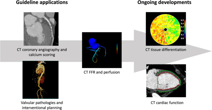
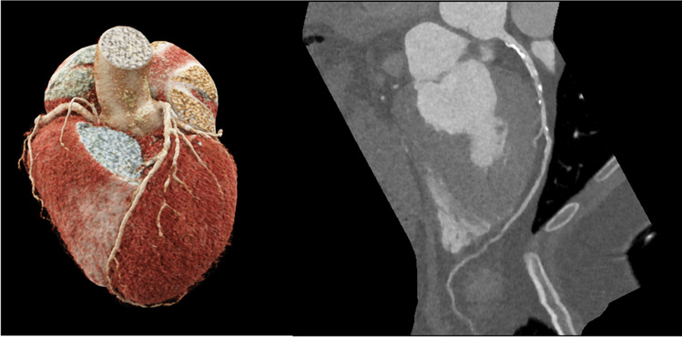
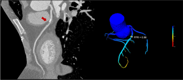
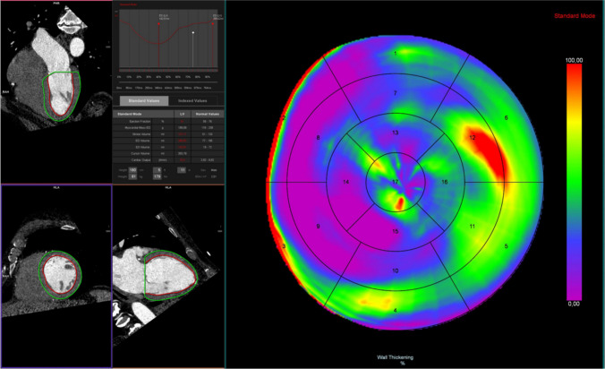
Recent findings: In recent years, new clinical applications of CCT have emerged. Major applications include the assessment of coronary artery disease and structural heart disease, with corresponding recommendations by major guidelines of international societies. While CCT already allows for a rapid and non-invasive diagnosis, technical improvements enable further in-depth assessments using novel imaging parameters with high temporal and spatial resolution. Those developments facilitate diagnostic and therapeutic decision-making as well as improved prognostication. This review determined that recent advancements in both hardware and software components of CCT allow for highly advanced examinations with little radiation exposure. This particularly strengthens its role in preventive care and coronary artery disease. The addition of functional analyses within and beyond coronary artery disease offers solutions in wide-ranging patient populations. Many techniques still require improvement and validation, however, CCT possesses potential to become a "one-stop-shop" examination.

Keywords: Cardiovascular computed tomography; Cardiovascular imaging; Coronary artery disease; Diagnostic decision-making; Structural heart disease.


Conflict of interest statement

The authors declare no competing interests.

Figures

Fig. 1
Fig. 1
Central Figure: Cardiac CT applications in guidelines and current research. Cardiac CT is widely recognized and recommended in current guidelines. Recent developments potentially enable a broader application in various diseases offering a fast and effective alternative to established diagnostic pathways. (Parts of the image have been adapted from Yamada et al. [39] © CC-BY 4.0 https://creativecommons.org/licenses/by/4.0/.)
Fig. 2
Fig. 2
CT coronary angiography. The image on the left shows a 3D model of the coronary arteries. The right image displays a radial series of the left anterior descending artery of the same patient with calcified and non-calcified plaque in the proximal segment
Fig. 3
Fig. 3
Functional testing using CT-FFR. The image on the left shows an intermediate stenosis of the left anterior descending artery by a mixed plaque. Using CT-FFR as shown on the right, it was possible to exclude haemodynamic relevance of the stenosis
Fig. 4
Fig. 4
CT-derived volumetry and functional analysis. The images on the left describe the process of CT-based volumetry using endo- and epicardial contouring. Functional assessments allow the quantification of global cardiac function as well as in-depth visualization of wall-motion abnormalities as shown by the bullet plot on the right
Fig. 5
Fig. 5
Calculation of ECV using CT. Delayed enhancement images and polar ECV maps of a patient with normal ECV (a and c) and a patient with increased ECV (b and d). ECV can be calculated by a subtraction image of a pre-contrast and a delayed enhancement post-contrast image. (By Yamada et al. [39] © CC-BY 4.0 https://creativecommons.org/licenses/by/4.0/.)
Fig. 6
Fig. 6
Transcatheter aortic valve replacement (TAVR) planning using CT. The images on the left delineate the measurement of the aortic annulus prior to TAVR in 3 different orientations of the aortic valve. The right image shows the access path via the right (A. fem. r) and left femoral artery (A. fem. l)
Fig. 7
Fig. 7
Shows the estimated optimal angiographic projection using the CT images on the right. The estimated distal landing zone shown in green. On the left panel, the actual angiographic deployment is shown taking benefit from CT-based predictions

References

    1. Toia P, et al. Technical development in cardiac CT: current standards and future improvements—a narrative review. Cardiovasc Diagn Ther. 2020;10:2018–35. - PMC - PubMed
    1. Gulati M, et al. 2021 AHA/ACC/ASE/CHEST/SAEM/SCCT/SCMR Guideline for the Evaluation and Diagnosis of Chest Pain: A Report of the American College of Cardiology/American Heart Association Joint Committee on Clinical Practice Guidelines. J Am Coll Cardiol. 2021;78:e187–285. - PubMed
    1. Lemkes JS, et al. Coronary Angiography after Cardiac Arrest without ST-Segment Elevation. N Engl J Med. 2019;380:1397–407. - PubMed
    1. Desch S, et al. Angiography after Out-of-Hospital Cardiac Arrest without ST-Segment Elevation. N Engl J Med. 2021;385:2544–53. - PubMed
    1. Zeppenfeld K, et al. 2022 ESC Guidelines for the management of patients with ventricular arrhythmias and the prevention of sudden cardiac death: Developed by the task force for the management of patients with ventricular arrhythmias and the prevention of sudden cardiac death of the European Society of Cardiology (ESC) Endorsed by the Association for European Paediatric and Congenital Cardiology (AEPC). Eur Heart J. 2022;43:3997–4126. - PubMed

MeSH terms