Review
. 2024 Sep 5;31(9):1244-1261.
doi: 10.1016/j.stem.2024.07.007. Epub 2024 Aug 19.

Hallmarks of regeneration

Affiliations
Review

Hallmarks of regeneration

Kenneth D Poss et al. Cell Stem Cell. .

Abstract

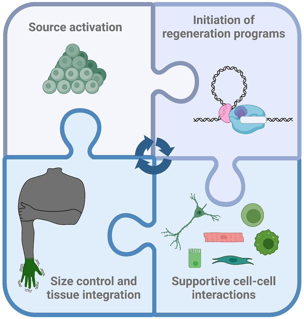
Regeneration is a heroic biological process that restores tissue architecture and function in the face of day-to-day cell loss or the aftershock of injury. Capacities and mechanisms for regeneration can vary widely among species, organs, and injury contexts. Here, we describe "hallmarks" of regeneration found in diverse settings of the animal kingdom, including activation of a cell source, initiation of regenerative programs in the source, interplay with supporting cell types, and control of tissue size and function. We discuss these hallmarks with an eye toward major challenges and applications of regenerative biology.

Keywords: regeneration.


Conflict of interest statement

Declaration of interests The authors declare no competing interests.

Figures

Fig 1.
Fig 1.
Hallmarks of regeneration.
Fig 2.
Fig 2.. Activation of cell sources.
A. Stem cells reside in a quiescence-maintaining niche before they wake to renew and differentiate upon cell loss or damage. B. Dynamic stem cell populations in the skin regenerate the epithelial barrier. C. Pluripotent stem cells populate interorgan spaces in planarian flatworms and replace differentiated cells in the process of turnover. D. Injury-induced partial dedifferentiation of cardiomyocytes leads to acquisition of embryonic features and division during zebrafish heart regeneration. E. Iris pigmented epithelial cells transdifferentiate to lens cells in amphibians, by depigmenting, forming a vesicle, and initiating production of lens crystallin proteins. F. Spared Drosophila pyloric cells endocycle in response to genetic ablation, forming a structure composed of a reduced number of large, polyploid cells with effective barrier function.
Fig 3.
Fig 3.. Initiation of regeneration programs.
A. Tissue regeneration enhancer elements (TREEs) control the expression of genes induced after injury and during regeneration. B. Dying Drosophila wing disc cells secrete Wingless (Wnt) to promote compensatory growth of the surviving tissue. Senescent cells transiently appear in the salamander limb blastema and produce mitogenic Wnt ligand. C. Skeletal muscle satellite cells in one limb respond to a contralateral limb injury by entering a Galert stage, reported to be controlled by circulating Hepatocyte growth factor activator and requiring signaling through the Hedgehog effector Gli3.
Fig 4.
Fig 4.. Interactions among supporting cell types.
A. For regeneration to supersede scarring, several cell types play a supportive role. Immune cells such as pro-regenerative macrophages, nerve cells and fibroblasts can produce cues for source cells. B. (Left) Spiny mice, but not house mice, can regenerate ear hole punches due in part to sustained high Erk signaling. (Right) Progenitors located in the mouse dermal fascia generate fibroblasts with pro-scarring, pro-inflammatory profiles that recruit immune cells, while also producing myofibroblasts that promote wound contraction. C. Antler velvet contains fibroblasts with a pro-regenerative profile that do not recruit immune cells. On the contrary, back skin has pro-scarring fibroblasts with a pro-inflammatory profile.
Fig 5.
Fig 5.. Size control and tissue integration.
A. Epigenetic memory of position is represented at least in part from the repressive histone mark H3K27me3 in fibroblasts of different limb segments. Posterior fibroblasts of the salamander blastema express the transcription factor Hand2 that initiates SHH signaling to progenitor cells located in the anterior limb. B. (Left) Wnt and Erk signaling across the body axis of planaria. After animal bissection, the Wnt signaling gradient rapidly re-scales, re-directing neoblasts to form proportionally scaled organs. (Right) Regenerated cardiomyocytes integrate with existing heart muscle in zebrafish, involving maturation and upregulation of Lrrc10 after proliferation.
Fig 6.
Fig 6.. Representation of hallmarks of regeneration in proficient and inefficient models, and potential interventions at bottlenecks.
Left: Key cellular and signaling events occur during limb regeneration in the salamander. Middle: Human limb injury leads to fibrosis without restoration of tissue patterns. Right: Manipulation of gene expression states with modern tools such as CRISPR-mediated transcriptional activation (CRISPRa), immunomodulation, and restitution of developmental genetic circuits can help unleash a regenerative response.

References

    1. Hsu YC, and Fuchs E (2022). Building and Maintaining the Skin. Cold Spring Harb Perspect Biol 14. 10.1101/cshperspect.a040840. - DOI - PMC - PubMed
    1. Hindi SM, and Millay DP (2022). All for One and One for All: Regenerating Skeletal Muscle. Cold Spring Harb Perspect Biol 14. 10.1101/cshperspect.a040824. - DOI - PMC - PubMed
    1. Biermann M, and Reya T (2022). Hematopoietic Stem Cells and Regeneration. Cold Spring Harb Perspect Biol 14. 10.1101/cshperspect.a040774. - DOI - PMC - PubMed
    1. Wagner DE, Wang IE, and Reddien PW (2011). Clonogenic neoblasts are pluripotent adult stem cells that underlie planarian regeneration. Science 332, 811–816. 10.1126/science.1203983. - DOI - PMC - PubMed
    1. Zeng A, Li H, Guo L, Gao X, McKinney S, Wang Y, Yu Z, Park J, Semerad C, Ross E, et al. (2018). Prospectively Isolated Tetraspanin(+) Neoblasts Are Adult Pluripotent Stem Cells Underlying Planaria Regeneration. Cell 173, 1593–1608 e1520. 10.1016/j.cell.2018.05.006. - DOI - PMC - PubMed

LinkOut - more resources