. 2024 Oct;31(10):1302-1317.
doi: 10.1038/s41418-024-01360-z. Epub 2024 Aug 20.

CCKBR+ cancer cells contribute to the intratumor heterogeneity of gastric cancer and confer sensitivity to FOXO inhibition

Affiliations

CCKBR+ cancer cells contribute to the intratumor heterogeneity of gastric cancer and confer sensitivity to FOXO inhibition

Zhenya Tan et al. Cell Death Differ. 2024 Oct.

Abstract

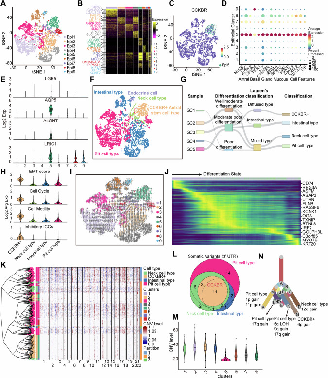
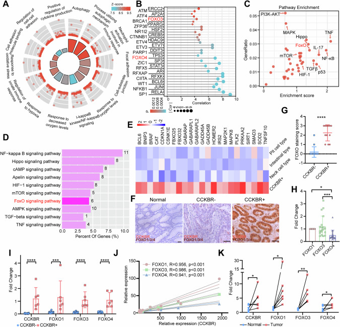
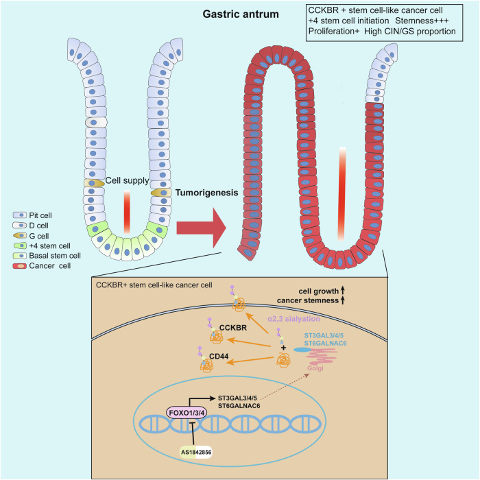
The existence of heterogeneity has plunged cancer treatment into a challenging dilemma. We profiled malignant epithelial cells from 5 gastric adenocarcinoma patients through single-cell sequencing (scRNA-seq) analysis, demonstrating the heterogeneity of gastric adenocarcinoma (GA), and identified the CCKBR+ stem cell-like cancer cells associated poorly differentiated and worse prognosis. We further conducted targeted analysis using single-cell transcriptome libraries, including 40 samples, to confirm these screening results. In addition, we revealed that FOXOs are involved in the progression and development of CCKBR+ gastric adenocarcinoma. Inhibited the expression of FOXOs and disrupting cancer cell stemness reduce the CCKBR+ GA organoid formation and impede tumor progression. Mechanically, CUT&Tag sequencing and Lectin pulldown revealed that FOXOs can activate ST3GAL3/4/5 as well as ST6GALNAC6, promoting elevated sialyation levels in CCKBR+ tumor cells. This FOXO-sialyltransferase axis contributes to the maintenance of homeostasis and the growth of CCKBR+ tumor cells. This insight provides novel perspectives for developing targeted therapeutic strategies aimed at the treating CCKBR associated gastric cancer.


Conflict of interest statement

The authors declare no competing interests.

Figures

Fig. 1
Fig. 1. Single-cell RNA sequencing (scRNA-seq) reveals the identification of CCKBR+ stem cell-like tumor cells in gastric adenocarcinoma (GA).
A t-SNE plotting of 9 tumor cell clusters in 5 GA patients. B Heatmap illustrated the expression patterns of markers within tumor cell subclusters. C t-SNE plot visualized the distribution of CCKBR expression across tumor cell clusters. D Creation of dot plots depicting the expression levels of each marker within distinct tumor cell clusters. E Violin plots were used to map the expression levels of LGR5, AQP5, A4GNT, and LRIG1 within tumor cell clusters. F t-SNE plotting of tumor cells based on the reclustering analysis using cell markers. G Sankey diagram showing the distribution of tumor cells in samples, differentiation, as well as their cell types. H Violin plots mapping the expression levels of EMT, cell cycle, cell motility, and inhibitory ICC-related genes across various types of tumor cells. I Conducting scVelo pseudotime analysis of tumor cells. The arrows indicate the potential transformation of differentiation states. J Heatmap revealing the expression characteristics of genes associated with the degree of tumor differentiation. K Phylogenetic reconstruction analysis of inferred CNVs from GC5 (left). Heatmap showing the inferred larger-scale CNVs by chromosome (right). the annotation track on the left of the heatmap indicates the inferred cell lineages and clusters, and the annotation track on the right indicates distinct CNV clusters. L The Venn diagram showing shared and unique somatic variants across tumor cell types in GC5. M Violin plots mapping CNV levels within tumor cell clusters from GC5. N Clonality trees of the single cells from GC5. The branches are delineated according to the percentage of cells in the subclone containing the corresponding CNVs. The canonical CNV events in each lesion and the predominant cell types were labeled in the clonality tree.
Fig. 2
Fig. 2. CCKBR expression is associated with low differentiation and poor prognosis in gastric adenocarcinoma.
A Immunohistochemical analysis of CCKBR expression in gastric adenocarcinomas. The experiment was repeated at least 3 times. Representative images were shown. Scale bar, 100 μm. T, tumor cell; P, paracancerous mucosa. B CCKBR-positive tumors and their corresponding paraneoplastic tissues (n = 5) were lysed. Immunoblotting of CCKBR and β-actin were performed. C Conducted statistical analysis on tumor locations and differentiation statuses based on immunohistochemical results in a cohort of 154 patients. D Kaplan-Meier survival curves showing significant differences in overall survival between samples with high and low CCKBR expression levels. P values were computed using Kaplan-Meier analysis. E Kaplan-Meier plots illustrating tumor progression differences between CCKBR high and low samples. P values were computed using Kaplan-Meier analysis. F Clinical characterization of patients with CCKBR-positive and -negative gastric antral tumors. G Immunofluorescence detected co-expression of CD44, CD133, and CCKBR in GA tumors. The experiment was repeated at least 3 times. Representative images were shown. Scale bars, 50 μm. H t-SNE plot revealing the identification of CCKBR-positive tumor cells in GSE183904 dataset across 40 patients. I Volcano plot depicting differential gene enrichment in CCKBR-positive and-negative tumor cells. J Violin plots mapping CNV levels within tumor cell types from GSE183904. ****p < 0.0001.
Fig. 3
Fig. 3. CCKBR+ tumors are inclined to form chromosomal instability (CIN) and genomically stable (GS) subtypes.
A Heatmap depicting the classification of CCKBR+ and CCKBR- tumors in TCGA STAD database; the markers of Epi5 are correlated with the expression of CCKBR. B Correlation matrix of indicated gene expression in CCKBR -positive and CCKBR-negative tumors. C Histogram depicting TCGA classification in CCKBR-negative and CCKBR-positive tumors. Chi-square test was used to determine statistical significance. D Histogram illustrating TCGA classification in tumors characterized by either abundant or deficient basal stem cell features. Chi-square test was used to determine statistical significance. E The frequency of gene mutation patterns across each sample in CCKBR-negative tumors. Different colors indicate distinct classes of mutation. F The frequency of gene mutation patterns across each sample in CCKBR+ tumors. Different colors indicate distinct classes of mutation. G A dot plot illustrating tumor-driven genes in CCKBR-negative tumors, with typical significantly driven genes labeled. H A dot plot illustrating tumor-driven genes in CCKBR+ tumors, with typical significantly driven genes labeled. I A bubble plot depicting the functional enrichment of tumor-driven genes in CCKBR-negative tumors. J A bubble plot depicting the functional enrichment of tumor-driven genes in CCKBR+ tumors. K Comparison of MSI (Microsatellite Instability) score and TMB (Tumor Mutational Burden) score in CCKBR-positive and -negative patients with simultaneous TP53 and APC mutation. L Comparison of MSI score and TMB score in CCKBR-positive and -negative patients with GLI3 or SMAD4 mutation. M Altered expression of Epi5-related markers in neoplastic and invasive gastric tissues from Tp53fl/fl;Apcwt/fl;KrasG12D/+;Cldn18-cre mice 3 months after tamoxifen induction. Data from GSE184613. Data are presented as mean ± S.D. of biological replicates. *p < 0.05; **p < 0.01; ****p < 0.0001.
Fig. 4
Fig. 4. CCKBR+ tumors displayed heightened FOXO activation.
A Circular plot illustrating the enrichment analysis of characterized genes in CCKBR-positive tumor cells. B Dot plot visualizing transcription factor enrichment in CCKBR-positive tumor cells. C Dot plot depicting signaling pathway enrichment in CCKBR-positive tumor cells. D Bar graph illustrating signaling pathway enrichment in CCKBR-positive tumor cells from the GSE183904 dataset. E Heatmap depicting the expression levels of FOXO pathway-related genes in tumor cell types. F Samples obtained from clinical gastric adenocarcinoma patients were subjected to immunohistochemical staining to investigate the localization and co-expression patterns of CCKBRs and FOXOs. Representative images were shown. Scale bars, 50 μm. G Statistical analysis of FOXO expression in CCKBR-positive (n = 12) and CCKBR-negative tumors (n = 15). H mRNA Expression levels of FOXO1, FOXO3, and FOXO4 in gastric adenocarcinoma tumors (n = 26) were determined by quantitative reverse transcription PCR (qRT-PCR). I mRNA expression levels of FOXO1, FOXO3, and FOXO4 were determined by qRT-PCR in CCKBR-positive (n = 6) and CCKBR-negative tumors (n = 20). J Point plots illustrating the correlation between the expression levels of FOXO1, FOXO3, FOXO4, and CCKBR. Pearson correlation analysis was used to determine statistical significance. K Correlation dot plots illustrating the changes in expression levels of FOXO1, FOXO3, and FOXO4 in CCKBR-positive tumors relative to paracancerous normal tissues (n = 6). Data are presented as mean ± S.D. of biological replicates. *p < 0.05; **p < 0.01; ***p < 0.001; ****p < 0.0001.
Fig. 5
Fig. 5. FOXOs maintain CCKBR-positive cancer stem cells homeostasis.
A Flow cytometry analysis of CCKBR and FOXO1/3/4 in MKN45, SGC-7901, MKN7, HGC27 and AGS cell lines. The experiment was repeated at least 3 times. Representative images were shown. B CCKBR-positive and -negative cells from MKN45 and HGC27 were sorted and cultured under conditions promoting the growth of tumorspheres. Tumorsphere formations were observed under bright field microscopy. The experiment was repeated at least 3 times. Representative images were shown. Scale bars, 100 μm. C Statistical analysis of the diameters of tumorspheres in sorted CCKBR-positive and -negative cells. DG CCKBR-positive and -negative HGC27 cells were diluted and subcutaneously injected into severely immunodeficient mice. Tumors were examined over a 33-day period (D) (n = 8 for each group). Frequency of stem cells is calculated by extreme limiting dilution analysis (E). The tumor weight (F) and tumor volume (G) were monitored in the indicated groups and at the indicated time points. H Six gastric adenocarcinoma cell lines were cultured under/without conditions favoring growth of tumorspheres (TS). The cell lysates were examined by immunoblotting as indicated. The experiment was repeated at least 3 times. I Statistical analysis of the expression of FOXOs and CCKBR in the above cell lysates. J The RNA expression levels of FOXO1, FOXO3, FOXO4, and CCKBR in six gastric adenocarcinoma cell lines forming tumorspheres. K Correlation dot plots illustrating the expression levels of FOXO1, FOXO3, FOXO4, and CCKBR. Pearson correlation analysis was used to determine statistical significance. L RT-qPCR analysis of mRNA expression levels of Epi5-related makers. MKN45 were cultured under/without conditions favoring growth of tumorspheres and then RNA was extracted. M Tumorsphere formations in MNK45 and HGC27 cells, with and without AS1842856 treatment, were observed under bright field microscopy. The experiment was repeated at least 3 times. Representative images were shown. Scale bars, 100 μm. N Statistical analysis of the diameters of tumorspheres treated with AS1842856. O Immunofluorescence staining detected the expression of CCKBR, CD133 and KI-67 in tumorspheres after treatment with AS1842856. The experiment was repeated at least 3 times. Representative images were shown. Scale bars, 20 μm. P MKN45 was transfected with shNC, shFOXO1, shFOXO3, shFOXO4, shFOXO1/3, and shFOXO1/3/4 respectively, then tumorsphere formations in MNK45 were observed under bright field microscopy. The experiment was repeated at least 3 times. Representative images were shown. Scale bars, 100 μm. Q Statistical analysis of the diameters of tumorspheres in shNC, shFOXO1, shFOXO3, shFOXO4, shFOXO1/3, and shFOXO1/3/4 group. Data are presented as mean ± S.D. of biological replicates. *p < 0.05; **p < 0.01; ***p < 0.001; ****p < 0.0001.
Fig. 6
Fig. 6. Targeting FOXO inhibits the growth of CCKBR+ tumor cells.
A CCKBR-positive and negative organoids prepared from gastric adenocarcinoma patients (n = 3 per group) were processed for H&E staining and immunofluorescence staining as indicated. Scale bars, 50 μm. B CCKBR-positive and negative organoids prepared from clinical gastric adenocarcinoma patients, with and without AS1842856 treatment, were observed under bright field microscopy (n = 3). Scale bars, 50 μm. C The cell viability of CCKBR-positive and negative organoids, with AS1842856 treatment, were analyzed (n = 3). D The cell viability of CCKBR-positive and negative organoids, with Carbenoxolone treatment, were analyzed (n = 3). E FACS examining apoptosis of CCKBR-positive organoids treated with 1 μM 5-FU and 1 μM AS1842856. F Statistical analysis of the apoptosis percentages in CCKBR-positive organoids treated with 1 μM 5-FU and 1 μM AS1842856 (AS) (n = 3 per group). GI 3 × 106 CCKBR+ MKN45 cells were injected subcutaneously into the left armpit of nude mice. Mice were intraperitoneally injected, with AS1842856 with or without 5-FU, after inoculation until the time of sacrifice. Representative tumor images (G), statistical analysis of weight (H) and volume (I) were shown (n = 5 for each group). JL CCKBR-negative AGS cells (5 × 106) were injected subcutaneously into the left armpit of nude mice. Mice were intraperitoneally injected, with AS1842856 with or without 5-FU, after inoculation until the time of sacrifice. Representative tumor images (J), statistical analysis of weight (K) and volume (L) were shown (n = 5 for each group). Data are presented as mean ± S.D. of biological replicates. ns, no significance. *p < 0.05, ***p < 0.001, ****p < 0.0001.
Fig. 7
Fig. 7. FOXOs transcriptionally regulate α2,3 sialylation, promoting the growth of CCKBR-positive tumors.
A CCKBR+ and CCKBR- tumor samples from gastric adenocarcinoma patients were performed CUT&Tag analysis. B Histogram analysis depicting the functional enrichment of genes bound by FOXOs. C Ranking the differences in promoter peaks between CCKBR+ and CCKBR- tumors based on their fold change from the CUT&Tag results. D Identification of peaks for FOXOs binding sites of ST3GAL3, ST3GAL4, ST3GAL5, and ST6GALNAC6 in both CCKBR-positive and -negative tumors. E Dot plots of correlation of FOXO1/3/4 and ST3GAL3, ST3GAL4, ST3GAL5, ST6GALNAC6. Each point represents one sample. F t-SNE plot illustrating the distribution of ST3GAL4, ST3GAL6, and ST3GALNCA6 in gastric cancer cells. G Immunohistochemical analysis of CCKBR and MAL I staining in gastric adenocarcinomas. The experiment was repeated at least 3 times. Representative images were shown. Scale bar, 50 μm. H Lectin blotting of MAL I to MKN45, MKN45 tumorspheres (MKN45-TS) and MKN45 tumorspheres treated with 1 μM AS1842856 (MKN45-TS+AS). I, J CCKBR-positive and CCKBR-negative organoids were prepared from gastric adenocarcinoma patients. CCKBR-positive organoids were treated with/without AS1842856 (AS). RT-qPCR analysis of the relative expression of STAGAL1-6 and ST6ALNAC1-6 in CCKBR-positive and CCKBR-negative organoids (I) (n = 3). The flow cytometry was used to analyze MAL I staining (J) (n = 3). K The cell viability of CCKBR-positive and CCKBR-negative organoids treated with P-3FAX-Neu5Ac was shown (n = 3). LN CCKBR-positive and -negative organoids treated with vector or AS1842856 (AS) and lysed to extract protein. MAL I pulldown and immunoblotting for CD44 and CCKBR were performed (L). Additional immunoprecipitation for CCKBR (M) or CD44 (N) and immunoblotting for MAL I was used to detection of α2,3 sialylation levels. The experiment was repeated at least 3 times. Representative images were shown. Data are presented as mean ± S.D. of biological replicates. **p < 0.01, ***p < 0.001, ****p < 0.0001.
Fig. 8
Fig. 8. CCKBR+ cancer cells exhibit stem cell-like properties in gastric adenocarcinoma.
The CCKBR+ cancer cells originate from gastric antral +4 stem cells, displaying enhanced cancer stem cell characteristics, lower differentiation, and a higher malignancy grade. This predisposes them to the formation of CIN and GS type tumors more readily. Furthermore, FOXOs play a crucial role in maintaining the homeostasis of CCKBR+ tumor cells, and inhibiting FOXO effectively suppresses their growth and cancer cell stemness. FOXOs contribute to the growth of CCKBR+ tumor cells, in part, by upregulating the level of α2,3 sialylation of CD44 and CCKBR.

References

    1. Lauren P. The two histological main types of gastric carcinoma: diffuse and so-called intestinal-type carcinoma. an attempt at a histo-clinical classification. Acta Pathol Microbiol Scand. 1965;64:31–49. - PubMed
    1. Namikawa T, Hanazaki K. Mucin phenotype of gastric cancer and clinicopathology of gastric-type differentiated adenocarcinoma. World J Gastroenterol. 2010;16:4634–9. - PMC - PubMed
    1. Imamura K, Yao K, Nimura S, Tanabe H, Kanemitsu T, Miyaoka M, et al. Characteristic endoscopic findings of gastric adenocarcinoma of fundic-gland mucosa type. Gastric Cancer. 2021;24:1307–19. - PubMed
    1. Zhang M, Hu S, Min M, Ni Y, Lu Z, Sun X, et al. Dissecting transcriptional heterogeneity in primary gastric adenocarcinoma by single cell RNA sequencing. Gut. 2021;70:464–75. - PMC - PubMed
    1. Kumar V, Ramnarayanan K, Sundar R, Padmanabhan N, Srivastava S, Koiwa M, et al. Single-cell atlas of lineage states, tumor microenvironment, and subtype-specific expression programs in gastric cancer. Cancer Discov. 2022;12:670–91. - PMC - PubMed

MeSH terms

Substances