. 2024 Aug 20;7(1):1022.
doi: 10.1038/s42003-024-06709-1.

Interleukin-33 promotes intrauterine adhesion formation in mice through the mitogen-activated protein kinase signaling pathway

Affiliations

Interleukin-33 promotes intrauterine adhesion formation in mice through the mitogen-activated protein kinase signaling pathway

Dan Liu et al. Commun Biol. .

Abstract

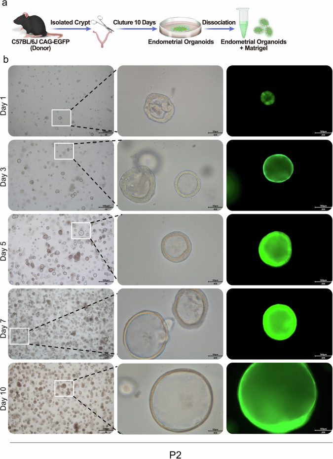
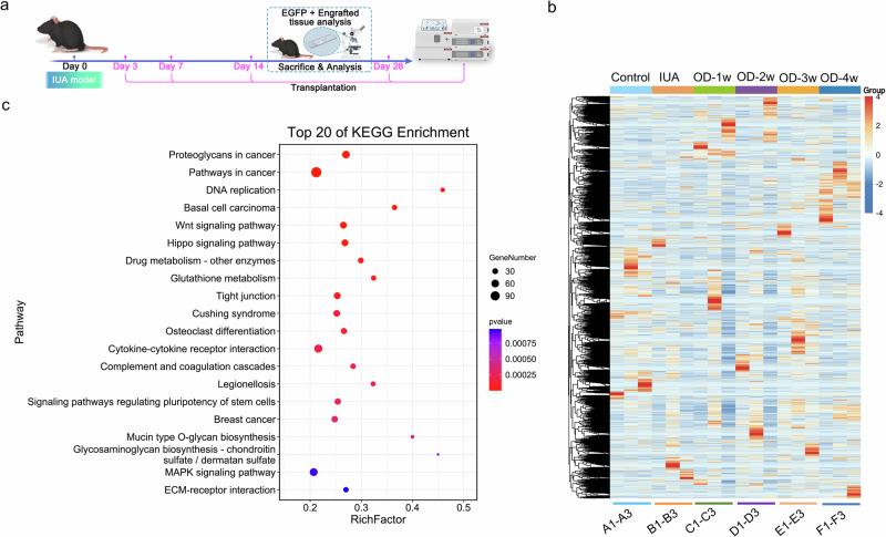
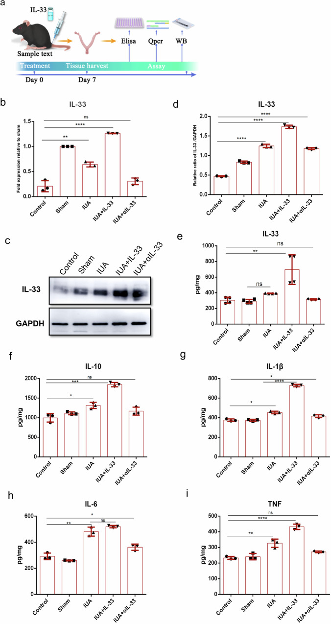
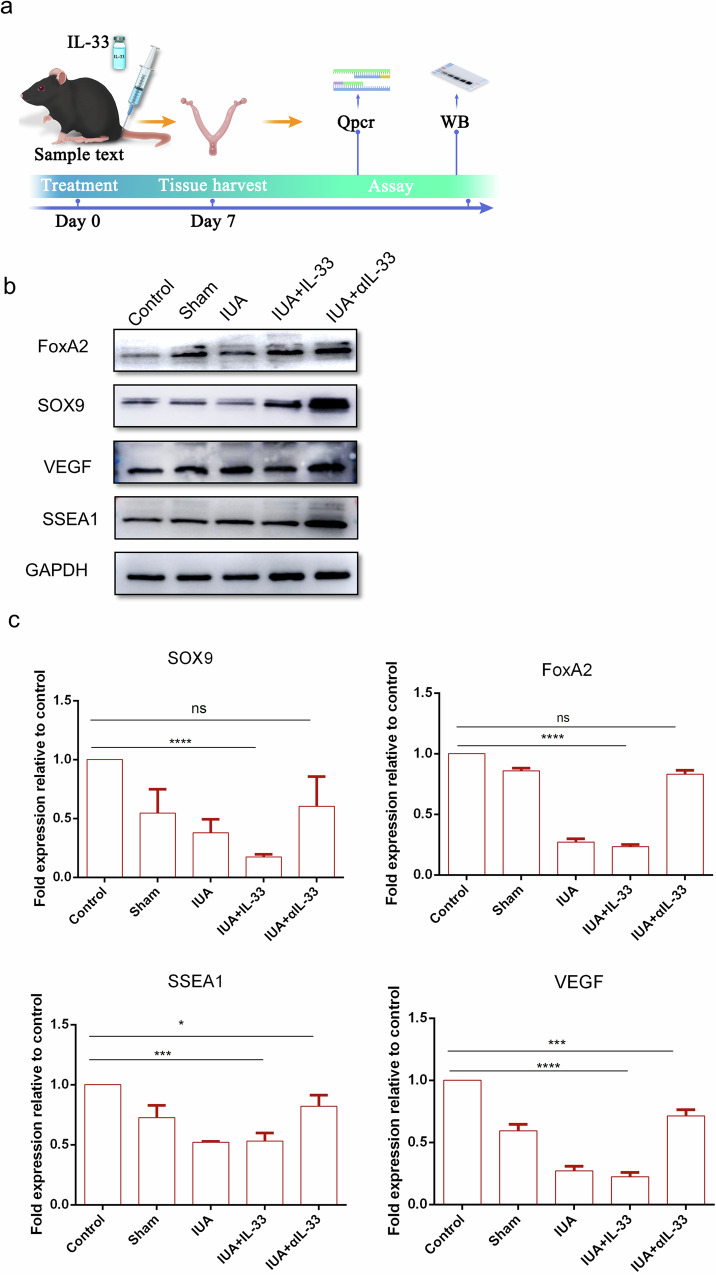
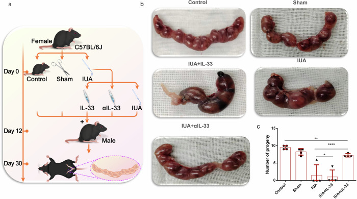
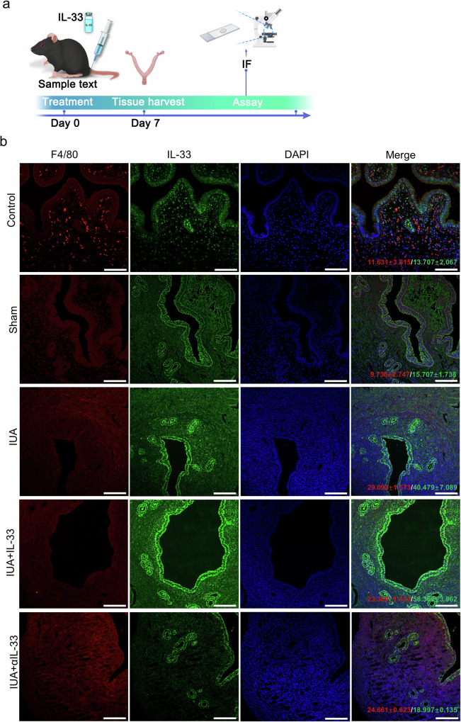
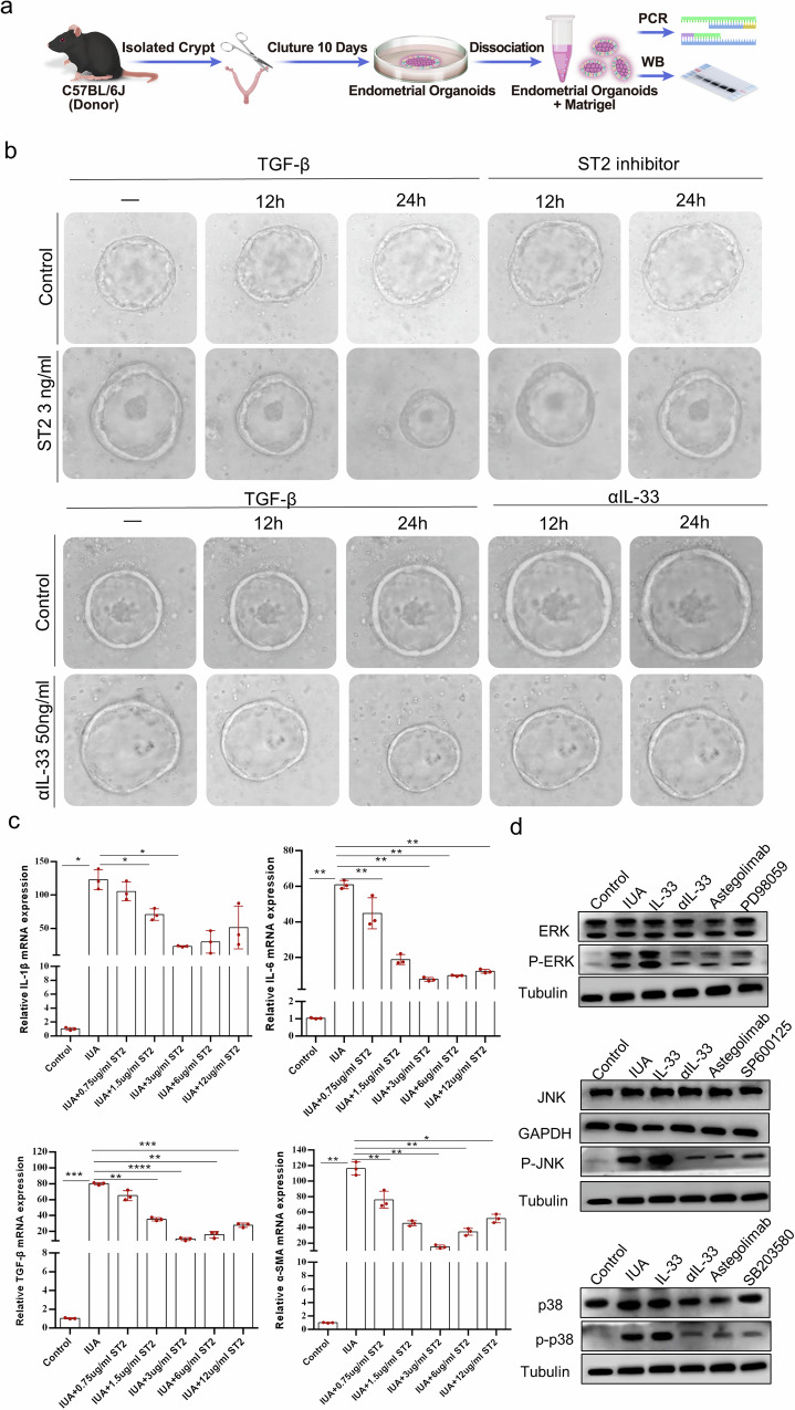
IL-33 belongs to the inflammatory factor family and is closely associated with the inflammatory response. However, its role in the development of intrauterine adhesions (IUAs) remains unclear. In this study, the role of IL-33 in the formation of IUAs after endometrial injury was identified via RNA sequencing after mouse endometrial organoids were transplanted into an IUA mouse model. Major pathological changes in the mouse uterus, consistent with the expression of fibrotic markers, such as TGF-β, were observed in response to treatment with IL-33. This finding may be attributed to activation of the phosphorylation of downstream MAPK signaling pathway components, which are activated by the release of IL-33 in macrophages. Our study provides a novel mechanism for elucidating IUA formation, suggesting a new therapeutic strategy for the prevention and clinical treatment of IUAs.


Conflict of interest statement

The authors declare no competing interests.

Figures

Fig. 1
Fig. 1. Cultivation and identification of mouse endometrial organoids.
a Schematic diagram of female C57BL/6J mouse organoid culture. b Representative brightfield images of endometrial organoids at different time points were captured under a light microscope (×4, scale bar, 500 μm; ×40, scale bar, 50 μm; ×20, scale bar, 100 μm).
Fig. 2
Fig. 2. RNA sequencing (RNA-seq) analysis of changes in the microenvironment of the uterine cavity of mice.
a Schematic diagram of the RNA-seq data of female C57BL/6 J mice. b Heatmap depicting all differentially expressed genes identified by RNA-seq analysis of the endometria from endometrial organoid-transplanted mice and normal mice. The colors range from blue (indicating low expression) to red (indicating high expression). OD organoid. c KEGG analysis of genes involved in the IL-33-related module (highlighted in blue). The size of the nodes represents the gene count, and the color of the nodes reflects the statistical significance represented as [−log10 (p value)]. The data are from one experiment with three biological replicates per group (n = 3).
Fig. 3
Fig. 3. Expression level of IL-33 in the mouse uterus.
a Schematic diagram of experiments carried out in mice. The mice were randomly assigned to the following groups (30 in each group): the control group; the sham operation group (sham), in which the mice underwent laparotomy without any treatment; the IUA model group (IUA), in which mice underwent induction of previously described mechanical damage; the rmIL-33-treated group (IUA + IL-33), in which mice experienced intrauterine injection of rmIL-33 (4 µg) on day 0 on both sides of the uterus during the mechanical scratching process; the αIL-33-treated group (IUA + αIL-33), in which the mice received an intrauterine injection of αIL-33 (10 µg) on both sides of the uterus on day 0 during the mechanical scratching process. The mice from each group were killed on day 7. bd qPCR and Western blotting were employed to analyze the mRNA and protein levels of IL-33 in the endometrium. The full-length blots/gels are presented in Supplementary Fig. 2 (ei). The protein levels of IL-33 and fibrogenic cytokines in the uterus were determined via ELISAs. The data are from one experiment with three independent experiments with three mice per group (n = 3). The values are the means ± SDs. *p < 0.05, **p < 0.01, ***p < 0.001, ns denotes p > 0.05 (unpaired Student’s t test).
Fig. 4
Fig. 4. Pathological features of IUAs in the IUA mouse model.
a Schematic diagram of experiments carried out in mice. The mice were randomly assigned to the following groups (30 in each group): the control group; the sham operation group (sham), in which the mice underwent laparotomy without any treatment; the IUA model group (IUA), in which the mechanical damage of the mice was induced as previously described; the rmIL-33-treated group (IUA + IL-33), in which the mice received an intrauterine injection of rmIL-33 (4 µg) on both sides of the uterus on day 0 during the mechanical scratching process; and the αIL-33-treated group (IUA + αIL-33), in which the mice received an intrauterine injection of αIL-33 (10 µg) on both sides of the uterus on day 0 during the mechanical scratching process. The mice from each group were killed on day 7. b HE staining was performed to determine changes in the glands in each group at different stages of treatment. The capillaries of the IUA model group and the IUA+rmIL-33 group were disrupted and congested, and inflammatory cells infiltrated the stroma, as shown via HE staining. c Masson’s trichrome staining was used to analyze changes in fibrosis in the different groups. d Immunohistochemical staining showing positive expression of TGF-β. The data are from one experiment with three independent experiments with three mice per group (n = 3) (×4, scale bar, 500 μm; ×20, scale bar, 100 μm). The results of the statistical analysis of the quantitative data are presented in Supplementary Fig. 1. The values are the means ± SDs. *p < 0.05, **p < 0.01, ***p < 0.001, ns denotes p > 0.05 (unpaired Student’s t test).
Fig. 5
Fig. 5. Increase in the expression of endometrial stem cell markers under αIL-33 treatment.
a Schematic diagram of experiments carried out in mice. The mice were randomly assigned to the following groups (30 in each group): the control group; the sham operation group (sham), in which the mice underwent laparotomy without any treatment; the IUA model group (IUA), in which the mice underwent induction of previously described mechanical damage; the rmIL-33-treated group (IUA + IL-33), in which the mice experienced intrauterine injection of IL-33 (4 µg) on both sides of the uterus during the mechanical scratching process; and the αIL-33-treated group (IUA + αIL-33), in which the mice experienced intrauterine injection of αIL-33 (10 µg) on both sides of the uterus during the mechanical scratching process. The mice from each group were killed on day 7. b Western blotting was conducted to determine the protein levels of the stem cell markers in each group. The full-length blots/gels are presented, and statistical analysis of the quantitative results is presented in Supplementary Fig. 2. c qRT‒PCR was used to measure the mRNA expression levels of SOX9, SSEA1, VEGF, and FoxA2 in the endometrial tissues of each group. The data are from one experiment with three independent experiments with three mice per group (n = 3). The values are the means ± SDs. *p < 0.05, **p < 0.01, ***p < 0.001, ns denotes p > 0.05 (unpaired Student’s t test).
Fig. 6
Fig. 6. The pregnancy rate changed after treatment with αIL-33.
a Schematic diagram of the experiments carried out in mice. b Representative images of embryos in the uterine cavity in each group. c Quantitative analysis of the pregnancy rate in each group. The data are from three independent experiments with three mice per group (n = 3). The values are the means ± SDs. *p < 0.05, **p < 0.01, ***p < 0.001, ns denotes p > 0.05 (unpaired Student’s t test).
Fig. 7
Fig. 7. Macrophages are the source of IL-33 production.
a Schematic diagram of experiments in the mouse uterus. b IF staining to determine the protein levels of IL-33 and F4/80 in endometrial tissues (×40, scale bar, 50 μm). The data are from one experiment with three independent experiments with three mice per group (n = 3). The values are the means ± SDs. *p < 0.05, **p < 0.01, ***p < 0.001, ns denotes p > 0.05 (unpaired Student’s t test).
Fig. 8
Fig. 8. IL-33 activates the formation of IUAs via the MAPK signaling pathway.
a Schematic diagram of mouse organoid culture and experiments carried out in mice. b Representative images of endometrial organoids treated with ST2 inhibitors at different time points were captured under a light microscope. The results of the gradient concentration experiment for ST2 are presented in Supplementary Fig. 4. c qRT‒PCR analysis of the mRNA expression levels of IL6, IL-1β, TGF-β, and α-SMA in endometrial organoids. d The protein expression levels of JNK, ERK, and p38 were measured via Western blotting after endometrial organoids were treated with the JNK inhibitor SP600125, the ERK/MEK1 inhibitor PD98059, or the p38 inhibitor SB203580 before rmIL-33 was added at 100 ng/ml. Full-length blots/gels are presented, and the results of the statistical analysis of the quantitative results are presented in Supplementary Fig. 5. The data are from one experiment with three independent experiments with three mice per group (n = 3). The values are the means ± SDs. *p < 0.05, **p < 0.01, ***p < 0.001, ns denotes p > 0.05 (unpaired Student’s t test).

Similar articles

Cited by

References

    1. Yu, Y. et al. Clinical features of the predilection and severer sites of intrauterine adhesions. Zhong Nan Da Xue Xue Bao Yi Xue Ban.47, 1568–1574 (2022). - PMC - PubMed
    1. Hooker, A. B. et al. Systematic review and meta-analysis of intrauterine adhesions after miscarriage: prevalence, risk factors and long-term reproductive outcome. Hum. Reprod.20, 262–278 (2014). - PubMed
    1. Xin, L. et al. A collagen scaffold loaded with human umbilical cord-derived mesenchymal stem cells facilitates endometrial regeneration and restores fertility. Acta Biomater.92, 160–171 (2019). 10.1016/j.actbio.2019.05.012 - DOI - PubMed
    1. Gürgan, T. et al. Systematic and standardized hysteroscopic endometrial injury for treatment of recurrent implantation failure. Reprod. Biomed. Online39, 477–483 (2019). 10.1016/j.rbmo.2019.02.014 - DOI - PubMed
    1. Kim, J., Koo, B. K. & Knoblich, J. A. Human organoids: model systems for human biology and medicine. Nat. Rev. Mol. Cell Biol.21, 571–584 (2020). 10.1038/s41580-020-0259-3 - DOI - PMC - PubMed

Publication types

MeSH terms

LinkOut - more resources