. 2024 Aug 20;43(1):234.
doi: 10.1186/s13046-024-03150-4.

Repurposed AT9283 triggers anti-tumoral effects by targeting MKK3 oncogenic functions in Colorectal Cancer

Affiliations

Repurposed AT9283 triggers anti-tumoral effects by targeting MKK3 oncogenic functions in Colorectal Cancer

Valentina Piastra et al. J Exp Clin Cancer Res. .

Abstract

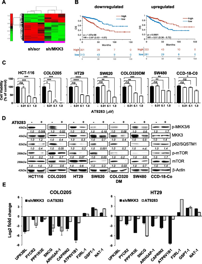
Background: Colorectal cancer (CRC) is the third most common type of cancer and the second leading cause of cancer-related deaths worldwide, with a survival rate near to 10% when diagnosed at an advanced stage. Hence, the identification of new molecular targets to design more selective and efficient therapies is urgently required. The Mitogen activated protein kinase kinase 3 (MKK3) is a dual-specificity threonine/tyrosine protein kinase that, activated in response to cellular stress and inflammatory stimuli, regulates a plethora of biological processes. Previous studies revealed novel MKK3 roles in supporting tumor malignancy, as its depletion induces autophagy and cell death in cancer lines of different tumor types, including CRC. Therefore, MKK3 may represent an interesting new therapeutic target in advanced CRC, however selective MKK3 inhibitors are currently not available.

Methods: The study involved transcriptomic based drug repurposing approach and confirmatory assays with CRC lines, primary colonocytes and a subset of CRC patient-derived organoids (PDO). Investigations in vitro and in vivo were addressed.

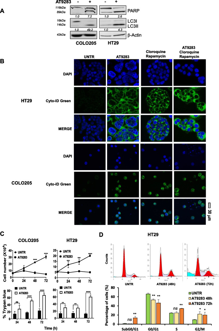
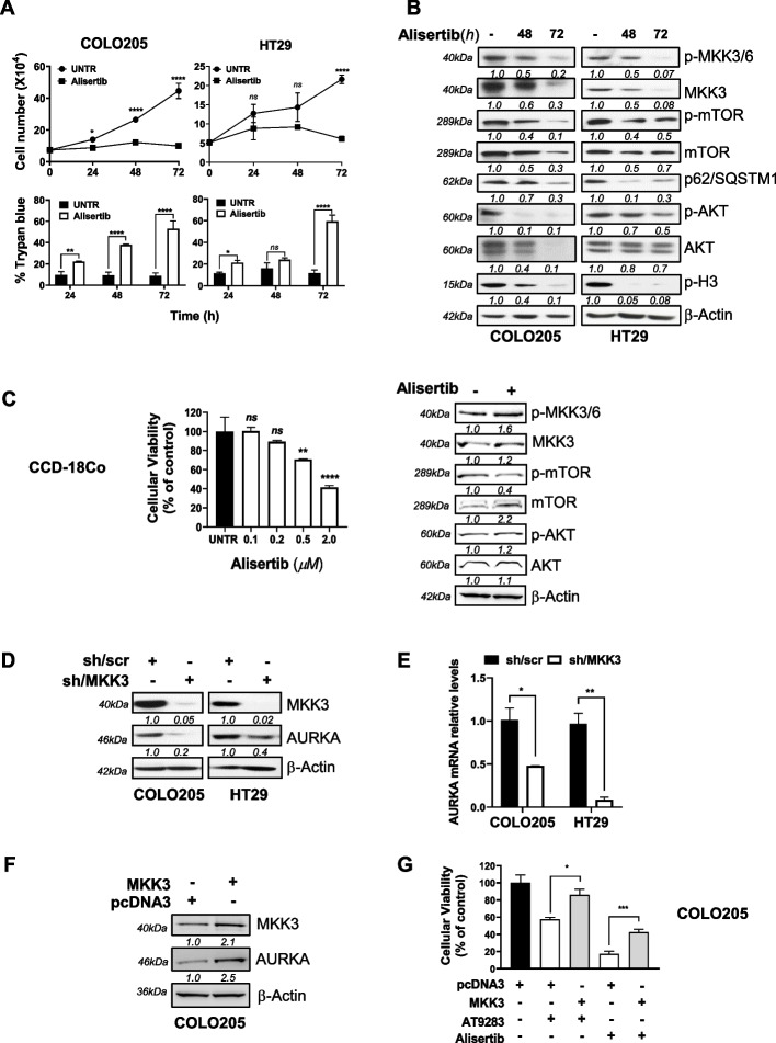
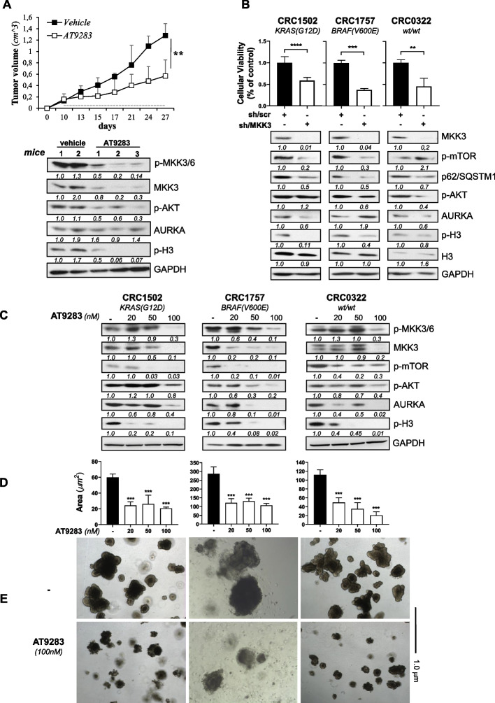
Results: The repurposing approach identified the multitargeted kinase inhibitor AT9283 as a putative compound with MKK3 depletion-mimicking activities. Indeed, AT9283 drops phospho- and total-MKK3 protein levels in tested CRC models. Likely the MKK3 silencing, AT9283 treatment: i) inhibited cell proliferation promoting autophagy and cell death in tested CRC lines and PDOs; ii) resulted well-tolerated by CCD-18Co colonocytes; iii) reduced cancer cell motility inhibiting CRC cell migration and invasion; iv) inhibited COLO205 xenograft tumor growth. Mechanistically, AT9283 abrogated MKK3 protein levels mainly through the inhibition of aurora kinase A (AURKA), impacting on MKK3/AURKA protein-protein interaction and protein stability therefore uncovering the relevance of MKK3/AURKA crosstalk in sustaining CRC malignancy in vitro and in vivo.

Conclusion: Overall, we demonstrated that the anti-tumoral effects triggered by AT9283 treatment recapitulated the MKK3 depletion effects in all tested CRC models in vitro and in vivo, suggesting that AT9283 is a repurposed drug. According to its good tolerance when tested with primary colonocytes (CCD-18CO), AT9283 is a promising drug for the development of novel therapeutic strategies to target MKK3 oncogenic functions in late-stage and metastatic CRC patients.

Keywords: Aurora kinase A; Colorectal cancer (CRC); Drug repurposing; MKK3/p38MAPK; Target therapy.


Conflict of interest statement

All authors disclose any competing financial interests or personal relationships that could have appeared to influence the work reported in this paper.

Figures

Fig. 1
Fig. 1
Drug repurposing to identify putative MKDMA drugs. A Hierarchical clustering analyses of DEGs accurately distinguished between sh-/scr and sh/MKK3 conditions; B Overall survival (OS) data from the TCGA COADREAD cohort of downregulated (n. 52) and upregulated (n. 16) genes in the sh/MKK3 subline compared with the control (sh/scr) subline. Patients were categorized into high- and low-signal intensity groups based on positive and negative z scores, respectively. The log-rank test was used to evaluate differences between survival curves. A multivariate Cox hazard regression model was utilized with adjustments for T, N, M, stage, and MSI status. C AT9283 effects on cell proliferation with reported CRC lines and primary colonocytes (CCD-18CO) by MTT assay 72 h later. Outputs were quantified with respect to untreated cells (-) set to 1.0 and eported as mean ± SD. Representative results of three independent experiments in technical triplicates are reported. Significance was assessed with ordinary one-way ANOVA followed by Dunnett’s post-hoc multiple comparisons test: ns, not significant; *p < 0.05; **p < 0.01; ***p < 0.001. D AT9283 IC50s treatments: HCT116 and HT29 (50 nM), COLO205 (15 nM), SW620 (100 nM), COLO320DM (885 nM), SW480 (1.6 μM), and CCD-18CO (4 μM) and protein lysates generated 72 h later were analysed by WB with the indicated antibodies. More relevant bands from the same filter at the same exposure length are reported. E RNAs was isolated from: I) sh/MKK3 and sh/scr sublines treated 144 h with DOX (1 μg/ml); II) AT9283 treated COLO205 (15 nM) and HT29 (100 nM) cells 48 h. Expression of subset of DEGs was assessed by qPCR. Results were normalized to GAPDH housekeeping gene and quantified with respect to relative controls (untreated; sh/scr) set to 1.0, outcomes were log2-transformed and reported mean ± SD for each DEG. Representative results of three independent experiments in technical triplicates are reported. Significance was assessed with an unpaired Student’s t test: p < 0.0001
Fig. 2
Fig. 2
AT9283 induces autophagy and cell death in the tested CRC cells. A COLO205 and HT29 cells treated for 72 h with AT9283 at a relative IC50 of 15 nM or 100 nM, respectively, were collected, and protein lysates were analysed by western blotting with the indicated antibodies. More relevant bands from the same filter at the same exposure length are reported. B CRC cells were treated with AT9283 at the relative IC50, and 72 h later, the cells were stained for autophagolysosome structures and analysed via confocal microscopy (40X magnification). Representative images from three independent experiments with similar results are shown. C CRC cells were treated with AT9283 at the relative IC50, and viable and trypan blue-positive cells were quantified at different time points. The representative results of three independent experiments performed in technical triplicates are reported as the means ± SDs. Significance was assessed with ordinary one-way ANOVA followed by Dunnett’s post hoc multiple comparisons test: ns, not significant; *p < 0.05; **p < 0.01; ***p < 0.001; ****p < 0.0001. D HT29 cells were left untreated (UNTR) or treated with AT9283 at the IC50 (100 nM) for 72 h and analysed by flow cytometry. Representative data from three independent experiments with similar results are reported
Fig. 3
Fig. 3
AT9283 triggers anti-tumor effects mainly inhibiting AURKA activity in CRC. A Alisertib IC50 effects on COLO205 (65 nM) and HT29 (100 nM) cells proliferation and survival. Representative results of three independent experiments performed in technical triplicates are reported as means ± SDs. Significance was assessed with ordinary one-way ANOVA followed by Dunnett’s post hoc multiple comparisons test: ns, not significant; *p < 0.05; **p < 0.01; ****p < 0.0001. B Protein lysates, from 48 and 72 h alisertib IC50 pre-treated cells, were analysed by WB with the indicated antibodies. More relevant bands from the same filter at the same exposure length are reported; C (left panel). MTT assays with colonocytes treated 72 h with alisertib at indicated doses. Results, quantified to untreated cells (UNTR), are reported as the mean ± SD. Representative data of three independent experiments in technical triplicates are reported. Significance was assessed with ordinary one-way ANOVA followed by Dunnett’s post hoc multiple comparisons test: ns, not significant; **p < 0.01; ****p < 0.0001; (right panel). Protein lysates from colonocytes pre-treated 72 h with alisertib IC50 (2.0 μM), were analysed by WB with the indicated antibodies. More relevant bands from the same filter at the same exposure length are reported. D Protein lysates from COLO205 and HT29-sh/scr and -sh/MKK3 sublines pre-treated 96 h with DOX (1 μg/mL) were analysed by WB with the indicated antibodies. More relevant bands from the same filter at the same exposure length are reported. E Cells were treated as in D and RNAs analysed by qPCR. Data were normalized to GAPDH, quantified with respect to sh/scr set to 1.0, and reported as the mean ± S.D. Representative results of three independent experiments (biological replicates) are reported. Significance was assessed with unpaired Student’s t test: *p < 0.05; **p < 0.01; F Protein lysates from transiently transfected (48 h) COLO205-empty (pcDNA3) or HA-tagged MKK3- (pDNA3HA-MKK3) were analysed by WB with the indicated antibodies or (G) treated with AT9283 (15 nM) or alisertib (60 nM). Effects on cell proliferation assessed 72 h later by MTT. The results are reported as the mean ± SD of three independent experiments with similar results. Significance was assessed with two-tailed unpaired Student’s t tests: *p < 0.05, ***p < 0.001
Fig. 4
Fig. 4
AT9283 abrogates MKK3/AURKA nuclear co-localization in CRC cells. A Protein lysates from COLO205 and HT29 cells, left untreated or treated for 72 h with AT9283 at the IC50, were immune-precipitated either with the anti-pMKK3 or anti-AURKA antibody, and immuno-complexes were revealed by western blot analysis with the indicated antibodies. More relevant bands from the same filter at the same exposure length are reported; B Cells were treated or left untreated, and immunofluorescence was performed with the indicated antibodies. Immunostained cells were analysed at 40X magnification under a confocal microscope. Representative data from three independent experiments with similar results are reported; C Quantifications of the intensities of each fluorophore in the nucleus (N) and cytoplasm (C) were performed with ImageJ softwar. Results are reported as the mean ± S.D. Significance was assessed with two-tailed unpaired Student’s t tests: *p < 0.05, ***p < 0.001, ****p < 0.0001. UNTR = untreated
Fig. 5
Fig. 5
AT9283 mimics the MKK3 depletion effects hampering CRC cell motility. A, B COLO205 and HT29-sh/scr, -sh/MKK3 sublines were treated for 120 h with DOX (1 μg/mL) (A), whereas parental cells were treated for 72 h with AT9283 at the IC50 (B), and protein lysates were analysed via western blotting with the indicated antibodies. More relevant bands from the same filter at the same exposure length are reported; C (left panels). COLO205 and HT29-sh/scr-sh/MKK3 sublines pre-treated for 96 h with DOX (1 μg/mL) and parental cells left untreated or pre-treated for 72 h with AT9283 at the IC50 were plated on cell inserts and stained with crystal violet 24 h later. Images were acquired with an optical microscope at 20X magnification. CT, relative control (untreated or sh/scr). Representative images of three independent experiments are shown. C (right panels). Quantification of the acquired images of the stained cells was performed with ImageJ software. The representative results of three independent experiments, quantified with respect to relative controls (set to 1.0), are reported as the mean ± SD. Significance was assessed with ordinary one-way ANOVA followed by Dunnett’s post hoc multiple comparisons test: ****p < 0.0001. D Wounds were generated at 80% confluence from COLO205- and HT29-sh/scr and -sh/MKK3 sublines pre-treated for 96 h with DOX (1 μg/mL) and from parental cells left untreated or pre-treated for 72 h with AT9283 at the IC50. Twenty-four hours later, the wounds were analysed under an optical microscope (20X magnification). Representative images of three independent experiments are shown (left panels). Wounds were quantified with ImageJ software on acquired images and the percentage of confluent wounds was estimated. The representative results of three independent experiments are reported as the mean ± SD (right panel). Significance was assessed with ordinary one-way ANOVA followed by Dunnett’s post hoc multiple comparisons test: ns, not significant; ***p < 0.001; ****p < 0.0001
Fig. 6
Fig. 6
AT9283 recapitulated the effects of MKK3 depletion in preclinical models and in CRC patient-derived organoids. A COLO205 xenografted cells (6 mice/group) were treated daily with either AT9283 (15 mg/kg) or vehicle solution. Upper panel: Tumour growth effects are reported as the means ± S.D. Significance was analysed using two-way ANOVA: **p < 0.01. Lower panel: Protein lysates from explanted tumors were analysed by WB with the indicated antibodies. More relevant bands from the same filter at the same exposure length are reported. B Engineered -sh/scr, and -sh/MKK3 PDOs were pre-treated 144 h with DOX (1 μg/mL) and effects on cell proliferation assessed by MTT assay (upper panels). Results were quantified with respect to the control set to 1.0 and reported as the mean ± SD. Representative results of three independent experiments are reported. Significance was assessed with two-tailed unpaired Student’s t test: ns, not significant; **p < 0.01; ***p < 0.001, ****p < 0.0001. Lower panels. Protein lysates from engineered sh/scr and sh/MKK3 PDOs, pre-treated 144 h with DOX (1 μg/mL), were analysed by WB with indicated antibodies. More relevant bands from the same filter at the same exposure length are reported. C Protein lysates from PDOs pre-treated 144 h with AT9283, were analysed by WB with the indicated antibodies. More relevant bands from the same filter at the same exposure length are reported. D, E PDOs pre-treated as in C, were analysed at microscope (4X magnification) and relative area (μm2) quantified with ImageJ software on collected images. The representative results of three independent experiments are reported as the mean ± SD. Significance was assessed with ordinary one-way ANOVA followed by Dunnett’s post hoc multiple comparisons test: ***p < 0.001; E Representative images of PDOs left untreated or treated with AT9283 at the highest dosage tested

References

    1. Bray F, Laversanne M, Sung H, Ferlay J, Siegel RL, Soerjomataram I, Jemal A. Global cancer statistics 2022: GLOBOCAN estimates of incidence and mortality worldwide for 36 cancers in 185 countries. CA Cancer J Clin. 2024;74(3):229–63 PMID: 38572751. 10.3322/caac.21834 - DOI - PubMed
    1. Patel SG, Karlitz JJ, Yen T, Lieu CH, Boland CR. The rising tide of early-onset colorectal cancer: a comprehensive review of epidemiology, clinical features, biology, risk factors, prevention, and early detection. Lancet Gastroenterol Hepatol. 2022;7(3):262–74 PMID: 35090605. 10.1016/S2468-1253(21)00426-X - DOI - PubMed
    1. Dariya B, Aliya S, Merchant N, Alam A, Nagaraju GP. Colorectal cancer biology, diagnosis, and therapeutic approaches. Crit Rev Oncog. 2020;25(2):71–94 PMID: 33389859. 10.1615/CritRevOncog.2020035067 - DOI - PubMed
    1. Lee YT, Tan YJ, Oon CE. Molecular targeted therapy: treating cancer with specificity. Eur J Pharmacol. 2018;834:188–96 PMID: 30031797. 10.1016/j.ejphar.2018.07.034 - DOI - PubMed
    1. Bossi G. MKK3 as oncotarget. Aging (Albany NY). 2016;8(1):1–2 PMID: 26805700. 10.18632/aging.100878 - DOI - PMC - PubMed

MeSH terms