. 2024 Aug 21;43(1):236.
doi: 10.1186/s13046-024-03143-3.

The DNA repair pathway as a therapeutic target to synergize with trastuzumab deruxtecan in HER2-targeted antibody-drug conjugate-resistant HER2-overexpressing breast cancer

Affiliations

The DNA repair pathway as a therapeutic target to synergize with trastuzumab deruxtecan in HER2-targeted antibody-drug conjugate-resistant HER2-overexpressing breast cancer

Jangsoon Lee et al. J Exp Clin Cancer Res. .

Abstract

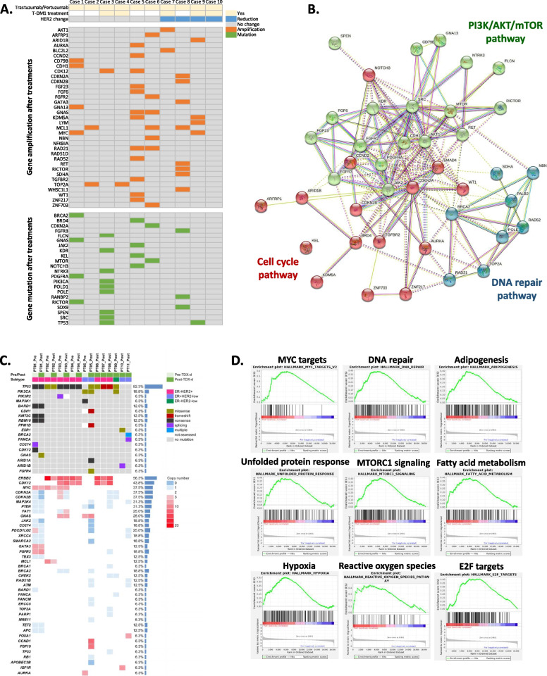
Background: Anti-HER2 therapies, including the HER2 antibody-drug conjugates (ADCs) trastuzumab emtansine (T-DM1) and trastuzumab deruxtecan (T-DXd), have led to improved survival outcomes in patients with HER2-overexpressing (HER2+) metastatic breast cancer. However, intrinsic or acquired resistance to anti-HER2-based therapies remains a clinical challenge in these patients, as there is no standard of care following disease progression. The purpose of this study was to elucidate the mechanisms of resistance to T-DM1 and T-DXd in HER2+ BC patients and preclinical models and identify targets whose inhibition enhances the antitumor activity of T-DXd in HER2-directed ADC-resistant HER2+ breast cancer in vitro and in vivo.

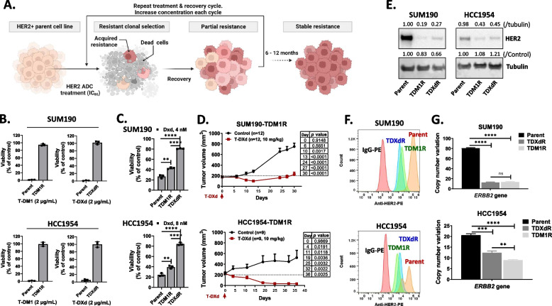
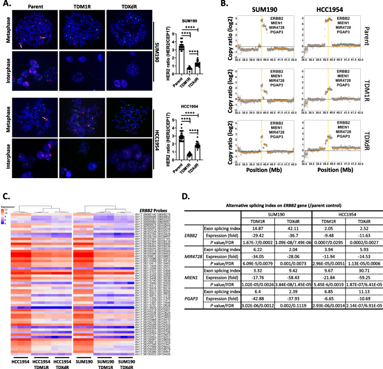
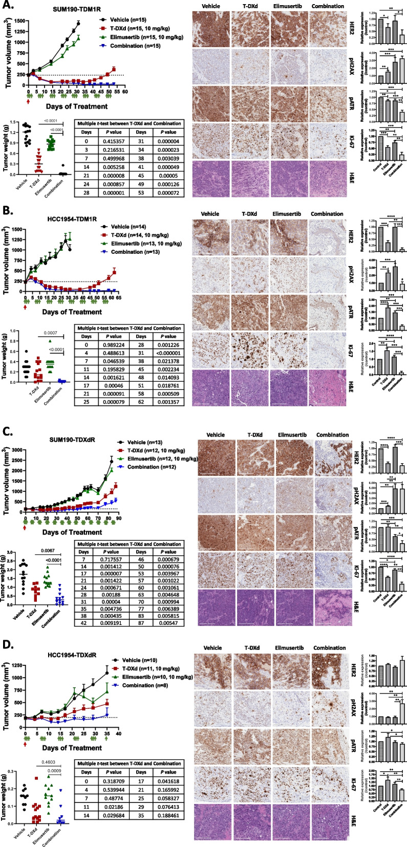
Methods: Targeted DNA and whole transcriptome sequencing were performed in breast cancer patient tissue samples to investigate genetic aberrations that arose after anti-HER2 therapy. We generated T-DM1 and T-DXd-resistant HER2+ breast cancer cell lines. To elucidate their resistance mechanisms and to identify potential synergistic kinase targets for enhancing the efficacy of T-DXd, we used fluorescence in situ hybridization, droplet digital PCR, Western blotting, whole-genome sequencing, cDNA microarray, and synthetic lethal kinome RNA interference screening. In addition, cell viability, colony formation, and xenograft assays were used to determine the synergistic antitumor effect of T-DXd combinations.

Results: We found reduced HER2 expression in patients and amplified DNA repair-related genes in patients after anti-HER2 therapy. Reduced ERBB2 gene amplification in HER2-directed ADC-resistant HER2+ breast cancer cell lines was through DNA damage and epigenetic mechanisms. In HER2-directed ADC-resistant HER2+ breast cancer cell lines, our non-biased RNA interference screening identified the DNA repair pathway as a potential target within the canonical pathways to enhance the efficacy of T-DXd. We validated that the combination of T-DXd with ataxia telangiectasia and Rad3-related inhibitor, elimusertib, led to significant breast cancer cell death in vitro (P < 0.01) and in vivo (P < 0.01) compared to single agents.

Conclusions: The DNA repair pathways contribute to HER2-directed ADC resistance. Our data justify exploring the combination treatment of T-DXd with DNA repair-targeting drugs to treat HER2-directed ADC-resistant HER2+ breast cancer in clinical trials.

Keywords: DNA damage repair pathway; HER2 antibody–drug conjugates; HER2+ breast cancer; HER2-directed ADC resistance; T-DXd.


Conflict of interest statement

Naoto T. Ueno and Jangsoon Lee have contracted research with Daiichi Sankyo. Seock-Ah Im reports advisory role for AstraZeneca, Daiichi-Sankyo, Eisai, Hanmi, Idience, Lilly, MSD, Novartis, Pfizer, Bertis, and Roche and has received research grants through her institution from AstraZeneca, Boryung Pharm, Daiichi-Sankyo, Daewoong Pharm, Eisai, Pfizer, and Roche. All other authors declare no competing interests.

Figures

Fig. 1
Fig. 1
DNA repair pathways are activated after HER2-targetd drug treatment in patients with HER2+ BC. A Targeted WGS was performed on 10 paired patient samples after treatment with trastuzumab/pertuzumab or T-DM1. The table shows gene amplification or variation after treatment. B The gene list was analyzed using STRING software (version 11.0) to show similar categories by functions of genes. In the context of the STRING analysis, k-means clustering was applied to identify groups of genes with similar behavior. Each color indicates co-regulated gene modules related to specific canonical signaling pathways. C Targeted DNA sequencing was performed to identify gene alteration profiling from five pairs before and after T-DXd treatment and six after T-DXd treatment in BC patients tissue samples. Genomic DNA was collected from FFPE tissue samples. D Gene expression analysis in three pairs before and after T-DXd treatment in HER2+ BC patients tissue samples. Total RNA was collected from FFPE tissue slides, and an RNA-seq analysis was conducted
Fig. 2
Fig. 2
Anti-HER2 antibody–drug conjugate (HER2-directed ADC)–resistant HER2+ BC cell line generation. A TDM1R and TDXdR cell lines generated by continuous treatment/recovery cycle with HER2-directed ADC. SUM190 (1 million) and HCC1954 (500,000) cells were added to the 100-mm culture dish. The next day, cells were treated with T-DM1 or T-DXd at the 80% inhibitory concentration (IC80) for 3–5 days and then replaced with fresh complete media until cells recovered at a normal growth rate. This treatment/recovery cycle was repeated for about 6–12 months. B Clonogenic assay. Parent TDM1R and TDXdR cell lines were treated with 2 µg/ml of T-DM1 or T-DXd for 14 days, and viability was measured by an SRB staining assay. Experiments were repeated three times independently. Data were collected from three biological replicates. C Antiproliferation effect of T-DXd payload and DXd in parent and TDM1R and TDXdR cell lines. Cells were treated with DXd for 14 days, and viability was measured by the SRB staining assay. Data were collected from three biological replicates. D T-DXd significantly reduces tumor growth in SUM190-TDM1R (n = 12 per group) and HCC1954-TDM1R (n = 9 per group) xenograft models. A multiple t-test comparison was used to compare tumor size between the control and treatment groups. E TDM1R and TDXdR cell lines showed reduced HER2 expression. The ImageJ program was used to measure intensity. Western blotting. F FACS analysis. TDM1R and TDXdR cell lines showed reduced cell-surface HER2 expression. Cells were maintained without drug for 7 days and collected to measure HER2 expression on the cell surface with anti-HER2-PE. Three biological replicates showed similar results. G Droplet digital PCR assay. CNV indicates copy number variation. TDM1R and TDXdR cell lines showed a reduced ERBB2 gene copy number. Each box shows mean with standard deviation; **, P < 0.01; ***, P < 0.001, ****, P < 0.0001, n.s. not significant. Data were collected from three biological replicates
Fig. 3
Fig. 3
HER2-directed ADC-resistant HER2+ BC cell lines showed reduced ERBB2 gene amplification. A Fluorescence in situ hybridization analysis. The red color indicates the amplification of ERBB2 gene, and the green color indicates the centromere on chromosome 17 (CEP17). A total of 25 individual cells were evaluated for ERBB2 gene amplification by measuring the HER2/CEP17 ratio from each cell line. Each box shows mean ± s.d.; ****, P < 0.0001. Three biological replicated experiments showed similar results. B Whole-genome sequencing data analysis. ERBB2, MIEN1, MIR4728, and PGAP3 gene copy numbers were reduced on chromosome 17 in TDM1R and TDXdR cell lines. C Transcriptome analysis of ERBB2 gene. All ERBB2 probes were pulled out from Affymetrix Clariom D Human microarray data and clustered by differential expression. Expression indicates log2. Data were collected from three biological replicates. D Alternative splicing was increased in ERBB2, MIEN1, MIR4728, and PGAP3 genes on chromosome 17 compared to in parent cells. Transcriptome Analysis Console (TAC, Affymetrix, Inc) software used for the Affymetrix Clariom D Human microarray database to compare differential gene splicing between HER2-ADC-resistant cells and their parent cells. Data were collected from three biological replicates
Fig. 4
Fig. 4
A gene expression analysis and synthetic lethal kinome library high-throughput RNAi screening revealed that the DNA repair pathway is a target for enhancing the efficacy of T-DXd in TDM1R and TDXdR cell lines. A Functional gene-set enrichment analysis using Affymetrix Clariom D Human Transcriptome array data. B Illustration of synthetic lethal kinome library high-throughput RNAi screening. C and D STRING interaction analysis of the top 50 target genes from kinome library high-throughput RNAi screening. K-means clustering was applied to identify groups of genes with similar behavior. Each color indicated co-regulated gene modules related to a specific canonical signaling pathway. SUM190-TDM1R (C), SUM190-TDXdR (D). E Bliss independence dose–response assay. Cells were treated with T-DXd and elimusertib for 5 days, and viability was measured using SRB staining. The data shown are representative of three independent experiments with similar results—the table indicates viability, and the Bliss synergy score was evaluated and visualized using Synergyfinderplus software (right, www.synergyfinderplus.org). F Clonogenic assay. Cells were treated with T-DXd and/or elimusertib for 14 days, and cell viability was measured by SRB staining. Data are presented as mean ± standard deviation. Two-tailed unpaired Student’s t-test. Experiments were repeated in triplicate. G. Western blotting. Cells were treated with T-DXd (1 µg/ml) and/or elimusertib (100 nM) for 48 h, and whole-cell lysates were collected for immunoblotting. Protein expression was normalized with actin level in control cells from each TDM1R and TDXdR cell line using ImageJ software. The data shown are representative of three independent experiments with similar results
Fig. 5
Fig. 5
Combination treatment with T-DXd and elimusertib enhanced the antitumor effect compared with monotherapy in TDM1R and TDXdR HER2+ BC in vitro and in vivo. A-D Xenograft assay using SUM190-TDM1R (A), HCC1954-TDM1R (B), SUM190-TDXdR (C), and HCC1954-TDXdR (D). Cells were injected into the mammary fat pad of nude mice, and treatments were started when tumors were an average of 200—250 mm3. T-DXd (10 mg/kg) was administered one time on Day 0 via tail-vein injection. Elimusertib (10 mg/kg) was administered via oral gavage twice a day (6-h intervals) for 3 consecutive days per week. Data are presented as mean ± standard deviation. Left, tumor growth and tumor weight (endpoint) measurements. Table shows multiple t-tests between T-DXd and combination on each measurement date. Right, IHC images of expression levels of HER2, pH2AX, pATR, and Ki-67 in xenograft tumor tissues. Multiple t-test comparison tests were used for tumor growth. Table shows t-tests between T-DXd and a combination of elimusertib and T-DXd on each measurement date. A two-tailed unpaired Student’s t-test was used for tumor weight comparison. Scale bars = 200 µm. IHC intensity was evaluated using the ImageJ program. Each box shows the mean with standard deviation. * P < 0.05, ** P < 0.01, *** P < 0.001, **** P < 0.0001. The data shown are representative of three tumor samples per group with similar results

References

    1. Burstein HJ. The distinctive nature of HER2-positive breast cancers. N Engl J Med. 2005;353(16):1652–4. - PubMed
    1. Moasser MM. The oncogene HER2: its signaling and transforming functions and its role in human cancer pathogenesis. Oncogene. 2007;26(45):6469–87. - PMC - PubMed
    1. Payne SJ, Bowen RL, Jones JL, Wells CA. Predictive markers in breast cancer–the present. Histopathology. 2008;52(1):82–90. - PubMed
    1. Arteaga CL, Sliwkowski MX, Osborne CK, Perez EA, Puglisi F, Gianni L. Treatment of HER2-positive breast cancer: current status and future perspectives. Nat Rev Clin Oncol. 2011;9(1):16–32. - PubMed
    1. Slamon DJ, Leyland-Jones B, Shak S, Fuchs H, Paton V, Bajamonde A, Fleming T, Eiermann W, Wolter J, Pegram M, et al. Use of chemotherapy plus a monoclonal antibody against HER2 for metastatic breast cancer that overexpresses HER2. N Engl J Med. 2001;344(11):783–92. - PubMed

MeSH terms