

Magnetic resonance imaging phantom-based S1 vertebral scores are indicators of fat-water-like osteoporotic changes in postmenopausal women: a pilot study
- PMID: 39165061
- PMCID: PMC11366554
- DOI: 10.31616/asj.2024.0116
Magnetic resonance imaging phantom-based S1 vertebral scores are indicators of fat-water-like osteoporotic changes in postmenopausal women: a pilot study
Abstract
Study design: A prospective study.
Purpose: To assess fat-water-like tissue changes on the 1st sacral vertebra using novel magnetic resonance imaging (MRI) phantombased F- and W-scores and evaluate their diagnostic performances in osteoporosis detection.
Overview of literature: Using an uncommonly advanced MRI technique, previous studies have found that fat-water changes were consistent with osteoporosis. The role of routine MRI sequences can be extended in this regard. The S1 vertebra is considered a crucial anatomical site in spine surgeries because it seldom suffers from fractures. Thus, S1 could indicate osteoporotic fat-water changes.
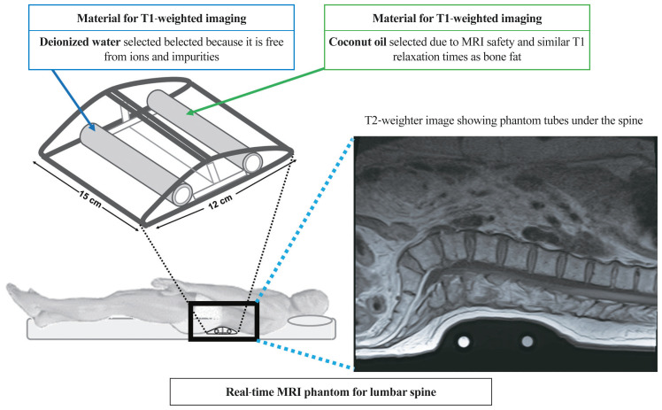
Methods: Forty-two female volunteers (aged 62.3±6.3 years) underwent spine examination with both MRI (including a phantom) and dual-energy X-ray absorptiometry (DXA) following ethical approval. MRI phantom-based F- and W-scoreS1 were defined by normalizing S1 vertebral signal intensities (SIs) by coconut oil and water SIs of the phantom on T1- and T2-weighted imaging, respectively. Using receiver operating characteristic analysis, the diagnostic performances of the new scores for evaluating osteoporosis and vertebral fractures were investigated against standard areal bone mineral density measured with DXA (DXA-aBMD).
Results: The F-scoreS1 and W-scoreS1 were greater (4.11 and 2.43, respectively) in patients with osteoporosis than those without osteoporosis (3.25 and 1.92, respectively) and achieved areas under the curve (AUCs) of 0.82 and 0.76 (p<0.05), respectively, for osteoporosis detection. Similarly, the mean F-scoreS1 and W-scoreS1 were higher (4.11 and 2.63, respectively) in patients with vertebral fractures than in those without fractures (3.30 and 1.82, respectively) and had greater AUCs (0.90 for W-scoreS1 and 0.74 for F-scoreS1) than DXA-aBMD (AUC, 0.26; p<0.03). In addition, the F- and W-scoreS1 demonstrated a strong correlation (r=0.65, p<0.001).
Conclusions: The new S1 vertebral-based MRI scores were developed to detect osteoporotic changes and demonstrated improvements over DXA-aBMD in differentiating patients with vertebral fractures.
Keywords: Magnetic resonance imaging; Osteoporosis; Osteoporotic fracture; Phantom; Sacral region.
Conflict of interest statement
No potential conflict of interest relevant to this article was reported.
Figures


References
-
- Sabnis AB, Chamoli U, Diwan AD. Is L5–S1 motion segment different from the rest?: a radiographic kinematic assessment of 72 patients with chronic low back pain. Eur Spine J. 2018;27:1127–35. - PubMed
-
- Huang W, Gong Z, Wang H, et al. Use of MRI-based vertebral bone quality score (VBQ) of S1 body in bone mineral density assessment for patients with lumbar degenerative diseases. Eur Spine J. 2023;32:1553–60. - PubMed
-
- Zhuang XM, Yu BS, Zheng ZM, Zhang JF, Lu WW. Effect of the degree of osteoporosis on the biomechanical anchoring strength of the sacral pedicle screws: an in vitro comparison between unaugmented bicortical screws and polymethylmethacrylate augmented unicortical screws. Spine (Phila Pa 1976) 2010;35:E925–31. - PubMed
-
- Compston JE, McClung MR, Leslie WD. Osteoporosis. Lancet. 2019;393:364–76. - PubMed
-
- de Villiers TJ, Goldstein SR. Bone health 2022: an update. Climacteric. 2022;25:1–3. - PubMed
Grants and funding
LinkOut - more resources
Full Text Sources
Research Materials
