

Atypical Aggressive Hemangioma of Thoracic Vertebrae Associated With Thoracic Myelopathy-A Case Report and Review of the Literature
- PMID: 39165484
- PMCID: PMC11335424
- DOI: 10.1155/2024/2307950
Atypical Aggressive Hemangioma of Thoracic Vertebrae Associated With Thoracic Myelopathy-A Case Report and Review of the Literature
Abstract
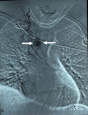
Aggressive thoracic hemangiomas are rare, benign tumors that extend into the spinal canal and cause neurological symptoms. Delayed diagnosis and treatment, due to a paucity of literature on optimal treatment strategies, can increase morbidity. This case report describes a 19-year-old male patient with aggressive thoracic hemangioma who presented with upper back pain and progressive weakness of the lower extremities. The patient underwent preoperative embolization and sclerotherapy, followed by decompression, posterior instrumentation, and stabilization. The final diagnosis was confirmed by biopsy, and there was a significant improvement in neurology after the surgical intervention. The diagnosis of rare lesions, such as aggressive hemangiomas, requires a high level of clinical suspicion and the assistance of imaging modalities in patients with features of compressive myelopathy. A combination of endovascular and surgical approaches can lead to optimal outcomes.
Keywords: aggressive hemangioma; embolization; spinal cord compression; spinal neoplasms; thoracic vertebrae.
Copyright © 2024 Krishna Timilsina et al.
Conflict of interest statement
The authors declare no conflicts of interest.
Figures


References
-
- Chu E. C.-P., Trager R. J., Chen A. T. C., Shum J. S. F. A 68-year-old woman with a remote history of breast cancer presenting with low back pain to a chiropractic clinic in Hong Kong with imaging findings consistent with a vertebral hemangioma and vertebral metastatic lesions. The American Journal of Case Reports . 2022;23:e938034-1–e938034-7. doi: 10.12659/AJCR.938034. - DOI - PMC - PubMed
-
- Kumar V., Kumar A., Dhatt S., Bahadur R. Should angiography be done in suspected tuberculosis of spine in an endemic country? - Hemangioma masquerading as tuberculosis of spine. Clinical Trials in Orthopedic Disorders . 2017;2(2):p. 52. doi: 10.4103/2542-4157.207011. - DOI
Publication types
LinkOut - more resources
Full Text Sources
