. 2024 Aug 15;15(37):15109-15124.
doi: 10.1039/d4sc04339a. Online ahead of print.

Targeting lysosomes by design: novel N-acridine thiosemicarbazones that enable direct detection of intracellular drug localization and overcome P-glycoprotein (Pgp)-mediated resistance

Affiliations

Targeting lysosomes by design: novel N-acridine thiosemicarbazones that enable direct detection of intracellular drug localization and overcome P-glycoprotein (Pgp)-mediated resistance

Busra Kaya et al. Chem Sci. .

Abstract

Innovative N-acridine thiosemicarbazones (NATs) were designed along with their iron(iii), copper(ii), and zinc(ii) complexes. Lysosomal targeting was promoted by specifically incorporating the lysosomotropic Pgp substrate, acridine, into the thiosemicarbazone scaffold to maintain the tridentate N, N, S-donor system. The acridine moiety enables a significant advance in thiosemicarbazone design, since: (1) it enables tracking of the drugs by confocal microscopy using its inherent fluorescence; (2) it is lysosomotropic enabling lysosomal targeting; and (3) as acridine is a P-glycoprotein (Pgp) substrate, it facilitates lysosomal targeting, resulting in the drug overcoming Pgp-mediated resistance. These new N-acridine analogues are novel, and this is the first time that acridine has been specifically added to the thiosemicarbazone framework to achieve the three important properties above. These new agents displayed markedly greater anti-proliferative activity against resistant Pgp-expressing cells than very low Pgp-expressing cells. The anti-proliferative activity of NATs against multiple Pgp-positive cancer cell-types (colon, lung, and cervical carcinoma) was abrogated by the third generation Pgp inhibitor, Elacridar, and also Pgp siRNA that down-regulated Pgp. Confocal microscopy demonstrated that low Pgp in KB31 (-Pgp) cells resulted in acridine's proclivity for DNA intercalation promoting NAT nuclear-targeting. In contrast, high Pgp in KBV1 (+Pgp) cells led to NAT lysosomal sequestration, preventing its nuclear localisation. High Pgp expression in KBV1 (+Pgp) cells resulted in co-localization of NATs with the lysosomal marker, LysoTracker™, that was significantly (p < 0.001) greater than the positive control, the di-2-pyridylketone-4-cyclohexyl-4-methyl-3-thiosemicarbazone (DpC) Zn(ii) complex, [Zn(DpC)2]. Incorporation of acridine into the thiosemicarbazone scaffold led to Pgp-mediated transport into lysosomes to overcome Pgp-resistance.


Conflict of interest statement

The authors declare no competing conflict of interest.

Figures

Fig. 1
Fig. 1. (A) Line drawings of the structures of: (I) Dp44mT; (II) DpC; (III) BpT; and (IV) ApT; (B) line drawings of the structures of the N-acridine thiosemicarbazones (NATs): (I) AOBP; (II) AODP; (III) AOAP; and (IV) AOAPZ. (C) Scheme for the synthesis of NATs and their metal ion complexes.
Fig. 2
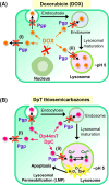
Fig. 2. (A) Schematic demonstrating that P-glycoprotein (Pgp) is found in cells at the plasma membrane and also intracellularly in endosomes and lysosomes after endocytosis. Schematic demonstrating that Pgp induces resistance to Doxorubicin (DOX) through two mechanisms: (i) as an efflux pump at the plasma membrane that induces the release of DOX from tumor cells; and (ii) through the ability of lysosomal Pgp to sequester DOX in the lysosomal lumen, preventing its access to a key pharmacological target, the nucleus. The sequestration of DOX in lysosomes leads to resistance as it does not induce lysosomal membrane permeabilization. (B) Schematic showing that: (i) Pgp at the plasma membrane effluxes DpT thiosemicarbazones such as Dp44mT and DpC from cells; and (ii) lysosomal Pgp results in thiosemicarbazone sequestration in lysosomes that results in lysosomal membrane permeabilization due to the potent redox activity of their Cu(ii) complexes. Lysosomal membrane permeabilization is a catastrophic process leading to apoptosis.
Fig. 3
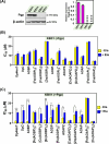
Fig. 3. (A) Pgp is expressed at high levels in KBV1 (+Pgp) cells, while KB31 (−Pgp) cells do not express significant Pgp levels. Results are relative to the protein loading control, β-actin. The western blot shown is typical of 3 experiments, while the densitometry represents the mean ± SD (3 experiments). ****p < 0.0001 vs. KBV1 (+Pgp) cells. (B) Incubation of KB31 (−Pgp) cells with the Pgp inhibitor, Elacridar (Ela), has no significant (p > 0.05) effect on the anti-proliferative activity (IC50) of Dp44mT, DpC, and the NATs and their Fe(iii), Cu(ii), and Zn(ii) complexes. (C) Incubation of KBV1 (+Pgp) cells with the Pgp inhibitor, Ela, significantly inhibits the anti-proliferative activity (IC50) of the positive controls, Dp44mT, DpC, and also AOBP, [Cu(AOBP)2], [Zn(AOBP)2], AODP, AOAPZ, [Zn(AOAPZ)2], and AOAP. In (B) and (C), KB31 (−Pgp) and KBV1 (+Pgp) cells were pre-incubated in the presence and absence of Ela (0.2 μM) for 1 h/37 °C. The cells were then incubated for 24 h/37 °C with the ligands and their complexes in the presence and absence of Ela (0.2 μM). Cellular proliferation was then examined to calculate the IC50. Results are mean ± SD (3 experiments). *p < 0.05; **p < 0.01; ***p < 0.001 as shown on the graph.
Fig. 4
Fig. 4. Confocal microscopy demonstrates AOBP and AOAP co-localize with the nuclear probe, HCS NuclearMask™ Deep Red, in KB31 (−Pgp) cells, while only cytosolic localization of AOBP and AOAP is observed in KBV1 (+Pgp) cells. (A) The KB31(−Pgp) and (B) KBV1 (+Pgp) cell-types were incubated with [Zn(DpC)2], AOBP, and AOAP (25 μM) for 2 h/37 °C and then examined using confocal microscopy. The images are typical from 3 experiments. (C) Quantification of the pixel intensity of [Zn(DpC)2], AOBP, and AOAP at 405 nm was performed with ImageJ. (D) Quantitation of the co-localization of HCS NuclearMask™ Deep Red (638 nm) with [Zn(DpC)2], AOBP, and AOAP (405 nm) was performed with ImageJ. Studies were performed using a 40× objective using a constant acquisition setting with Olympus Fluoview FV3000 software. Images were digitally magnified (5×) for demonstration of the co-localization. Results are mean ± SD (3 experiments). *p < 0.05; **p < 0.01; ***p < 0.001; ****p < 0.0001 as shown on the graph. Scale bar: 10 μm.
Fig. 5
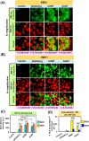
Fig. 5. (A and B) Confocal microscopy demonstrates that AOBP and AOAP co-localize to a significantly greater extent with LysoTracker™ than [Zn(DpC)2] in (A) KB31 (−Pgp) cells and especially (B) KBV1 (+Pgp) cells. Both cell-types were incubated with inherently fluorescent, [Zn(DpC)2], AOBP, and AOAP (25 μM) for 2 h/37 °C resulting in a green (405 nm), punctate pattern. Incubation with LysoTracker™ (red) resulted in a typical, punctated pattern of red (647 nm) fluorescence. (C) Quantification of the pixel intensity of [Zn(DpC)2], AOBP, and AOAP at 405 nm was performed with ImageJ. (D) Quantification of the co-localization of [Zn(DpC)2], AOBP, and AOAP (405 nm) with LysoTracker™ (647 nm) was estimated with ImageJ. Co-localization of AOBP and LysoTracker™ was confirmed by the Pearson's correlation coefficients. The images are typical from three experiments. Studies were performed using a 40× objective using a constant acquisition setting with Olympus Fluoview FV3000 software. Images were digitally magnified (5×) for demonstration of the co-localization. Results are mean ± SD (3 experiments). *p < 0.05; **p < 0.01; ***p < 0.001 as shown on the graph. Scale bar = 10 μm.
Fig. 6
Fig. 6. Schematic illustrating the mechanism of action of AOBP in overcoming Pgp-mediated drug resistance by targeting lysosomes. (A) In Pgp-expressing cells, Pgp on the plasma membrane becomes endocytosed into endosomes, with a proportion of endosomes maturing into lysosomes. The acridine moiety is lysosomotropic, and acts as a Pgp substrate, conferring these properties on AOBP. The AOBP can be transported by Pgp into the lysosomal lumen, where it becomes positively charged and trapped at lysosomal pH. This lysosomal sequestration prevents AOBP from entering the nucleus where it can target DNA. Once in the lysosome, AOBP then binds Cu liberated after lysosomal degradation of proteins to form potent, redox-active Cu complexes that generate ROS and causes lysosomal membrane permeabilization and apoptosis. In addition to entrance of AOBP into the lysosome via lysosomal Pgp, AOBP can theoretically also be effluxed out of the cell by Pgp at the plasma membrane. (B) In cells with very low Pgp expression, the acridine moiety of AOBP can result in strong nuclear association. This latter effect is presumably due to the known ability of acridine to intercalate into DNA, but Pgp overexpression prevents this effect due to lysosomal sequestration of the drug (see (A)).

Similar articles

Cited by

References

    1. Jansson P. J. Yamagishi T. Arvind A. Seebacher N. Gutierrez E. Stacy A. Maleki S. Sharp D. Sahni S. Richardson D. R. J. Biol. Chem. 2015;290:9588–9603. doi: 10.1074/jbc.M114.631283. - DOI - PMC - PubMed
    1. Seebacher N. A. Lane D. J. Jansson P. J. Richardson D. R. J. Biol. Chem. 2016;291:3796–3820. doi: 10.1074/jbc.M115.682450. - DOI - PMC - PubMed
    1. Al-Akra L. Bae D.-H. Sahni S. Huang M. L. Park K. C. Lane D. J. Jansson P. J. Richardson D. R. J. Biol. Chem. 2018;293:3562–3587. doi: 10.1074/jbc.M116.772699. - DOI - PMC - PubMed
    1. Stacy A. E. Palanimuthu D. Bernhardt P. V. Kalinowski D. S. Jansson P. J. Richardson D. R. J. Med. Chem. 2016;59:4965–4984. doi: 10.1021/acs.jmedchem.6b00238. - DOI - PubMed
    1. Seebacher N. A. Richardson D. R. Jansson P. J. Cell Death Dis. 2016;7:e2510. doi: 10.1038/cddis.2016.381. - DOI - PMC - PubMed

LinkOut - more resources