. 2024 Aug;14(8):e1744.
doi: 10.1002/ctm2.1744.

Cyclic-di-GMP induces inflammation and acute lung injury through direct binding to MD2

Affiliations

Cyclic-di-GMP induces inflammation and acute lung injury through direct binding to MD2

Chenchen Qian et al. Clin Transl Med. 2024 Aug.

Abstract

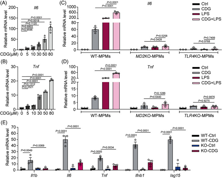
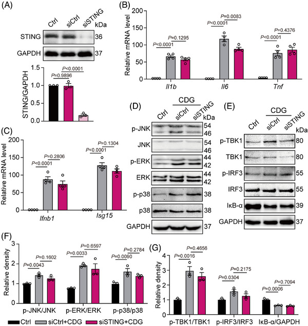
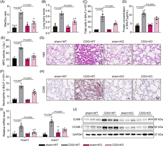
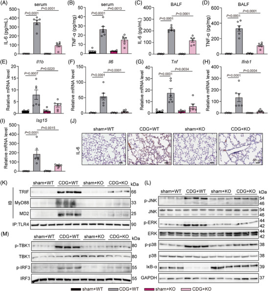
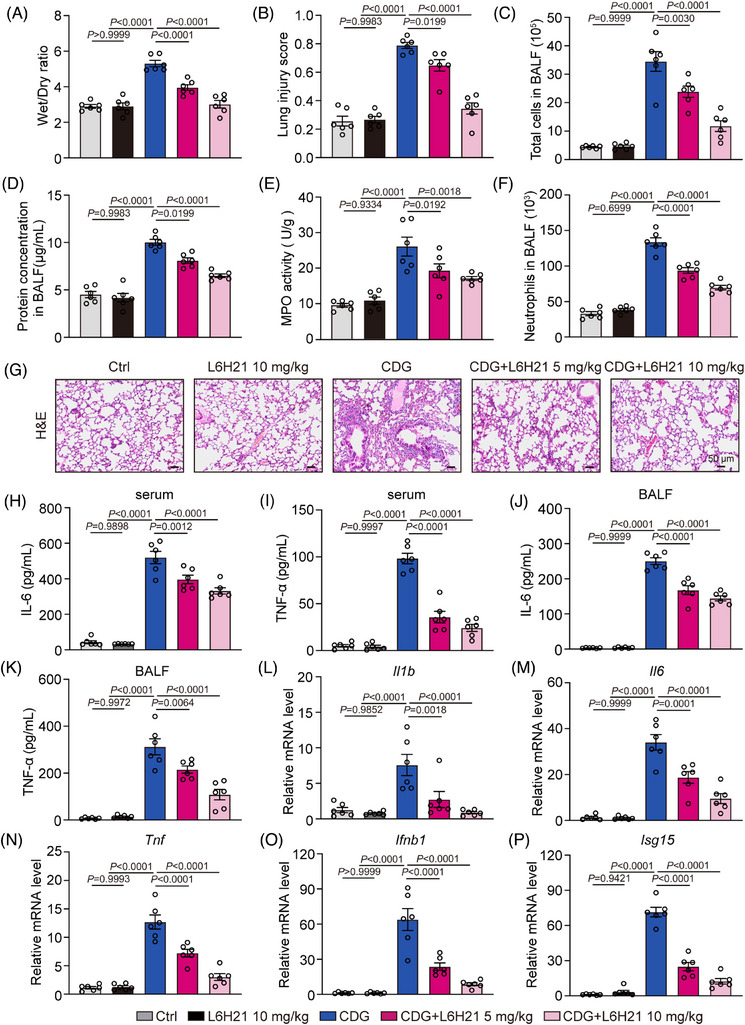
Background: Severe bacterial infections can trigger acute lung injury (ALI) and acute respiratory distress syndrome, with bacterial pathogen-associated molecular patterns (PAMPs) exacerbating the inflammatory response, particularly in COVID-19 patients. Cyclic-di-GMP (CDG), one of the PAMPs, is synthesized by various Gram-positve and Gram-negative bacteria. Previous studies mainly focused on the inflammatory responses triggered by intracellular bacteria-released CDG. However, how extracellular CDG, which is released by bacterial autolysis or rupture, activates the inflammatory response remains unclear.

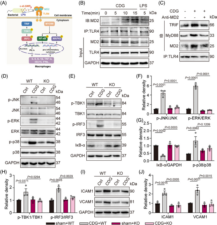
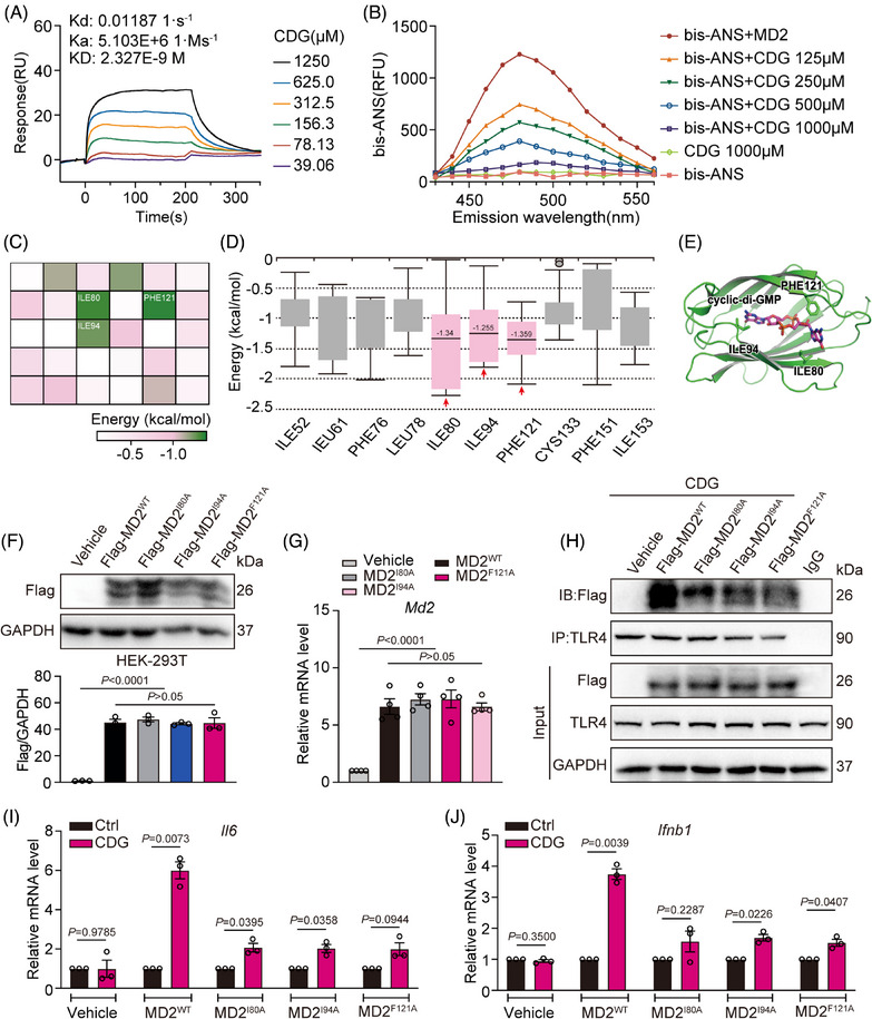
Methods: The interaction between extracellular CDG and myeloid differentiation protein 2 (MD2) was investigated using in vivo and in vitro models. MD2 blockade was achieved using specific inhibitor and genetic knockout mice. Site-directed mutagenesis, co-immunoprecipitation, SPR and Bis-ANS displacement assays were used to identify the potential binding sites of MD2 on CDG.

Results: Our data show that extracellular CDG directly interacts with MD2, leading to activation of the TLR4 signalling pathway and lung injury. Specific inhibitors or genetic knockout of MD2 in mice significantly alleviated CDG-induced lung injury. Moreover, isoleucine residues at positions 80 and 94, along with phenylalanine at position 121, are essential for the binding of MD2 to CDG.

Conclusion: These results reveal that extracellular CDG induces lung injury through direct interaction with MD2 and activation of the TLR4 signalling pathway, providing valuable insights into bacteria-induced ALI mechanisms and new therapeutic approaches for the treatment of bacterial co-infection in COVID-19 patients.

Keywords: COVID‐19; MD2; acute lung injury; cyclic‐di‐GMP.


Conflict of interest statement

The authors declare no conflict of interest.

Figures

FIGURE 1
FIGURE 1
CDG‐induced inflammatory response in macrophages via MD2. (A, B) Mouse primary macrophages (MPMs) derived from C57BL/6J (WT) mice were treated with different concentrations of CDG for 12 h. The mRNA levels of Il6 (A) and Tnf (B) were measured via RT‐qPCR assay. Data were normalized to the levels of Actb (n = 3 in each group, biological replicates). (C, D) MPMs were derived from WT, global Md2 −/− (MD2KO), or global Tlr4 −/− (TLR4KO) mice, respectively. MPMs were treated with CDG (80 µM), LPS (0.5 µg/mL), or CDG (80 µM) combined with LPS (0.5 µg/mL), and then the mRNA levels of Il6 (C) and Tnf (D) were measured via RT‐qPCR assay. Data were normalized to the levels of Actb (n = 3 in each group, biological replicates). E MPMs derived from WT and MD2KO mice were treated with CDG (80 µM) for 12 h. The mRNA levels of Il1b, Il6, Tnf, Ifnb1, and Isg15 were measured via RT‐qPCR assay. Data were normalized to the levels of Actb (n = 3 in each group, biological replicates). Data information: Data are presented as mean ± SEM. One‐way ANOVA followed by Dunnett's multiple comparisons test.
FIGURE 2
FIGURE 2
CDG mediated both the MyD88‐ and TRIF‐dependent pathways in MPMs. (A) Schematic diagram showing the MD2‐TLR4 signalling pathway and downstream MyD88‐ and TRIF‐dependent branches. (B) MPMs from WT mice were stimulated with CDG (80 µM) for 5, 10, or 15 min. Representative immunoblots of co‐immunoprecipitation of MD2 and TLR4 in MPMs were shown. LPS (0.5 µg/mL, 5 min) was used as the positive control. (C) MPMs derived from WT mice were stimulated with CDG (80 µM) for 15 min. MD2 blockade was achieved using MD2‐neutralizing antibody (anti‐MD2, 100 ng/mL). Representative co‐immunoprecipitation images of TRIF/TLR4/MD2 and MyD88/TLR4/MD2 were shown. (D–H) MPMs from WT or MD2KO mice were treated with CDG (80 µM) for 1 h. Activation of MyD88‐ and TRIF‐dependent pathways was measured. Unphosphorylated proteins and/or GAPDH were used as the loading controls (n = 3 in each group, biological replicates). Representative blots (D, E) and densitometric quantification (F–H) are shown. (I, J) MPMs from WT mice were stimulated with CDG (80 µM) for 24 h. The protein levels of ICAM1 and VCAM1 were examined via Western blot analysis. GAPDH was used as the loading control (n = 3 in each group, biological replicates). Representative blots (I) and densitometric quantification are shown (J). Data information: Data are presented as mean ± SEM. One‐way ANOVA followed by Dunnett's multiple comparisons test.
FIGURE 3
FIGURE 3
CDG interacts directly with MD2. (A) Surface plasmon resonance (SPR) analysis between CDG and rMD2. (B) The effects of CDG on the binding of fluorescent Bis‐ANS (5 µM) to rMD2. (C) Heatmap of average binding free energies for the top 30 residues in CDG‐MD2 molecular docking results. (D) Box plot of the per‐residue decomposition energy. € Molecular docking of CDG with MD2 protein was carried out with the programme Tripos molecular modelling packages Sybyl‐x.v1.1.083. (F, G) The overexpression efficiency of MD2WT and MD2Mut (I80A/I94A/F121A) plasmids in HEK‐293T cells and MD2KO‐derived MPMs was determined using Western blot assay (F) and RT‐qPCR assay (G). GAPDH was used as the loading control (n = 3 in each group, biological replicates). Data were normalized to the levels of Actb (n = 4 in each group, biological replicates). (H) HEK‐293T cells were stimulated with CDG (80 µM) for 15 min, and the effects of empty vehicle (Vehicle), MD2WT (MD2WT), or MD2Mut (MD2I80A, MD2I94A, MD2F121A) plasmids on MD2/TLR4 complex formation were assessed using co‐immunoprecipitation. (I, J) MD2KO‐derived MPMs were transfected with empty vehicle (Vehicle), MD2WT (MD2WT), or MD2Mut (MD2I80A, MD2I94A, MD2F121A) plasmids, respectively. Then the cells were exposed to CDG (80 µM) for 12 h. The mRNA levels of Il6 (I) and Ifnb1 (J) were measured via RT‐qPCR assay. Data were normalized to the levels of Actb (n = 3 in each group, biological replicates). Data information: In (F, G), data are presented as mean ± SEM, one‐way ANOVA followed by Dunnett's multiple comparisons test. In (I, J), data are presented as mean ± SEM, Student's t‐test.
FIGURE 4
FIGURE 4
MD2 plays a vital role in CDG‐induced inflammation via a STING‐independent pathway. (A) MPMs were derived from WT mice. Immunoblot showing STING levels (n = 3 in each group, biological replicates) in MPMs following siRNA transfection [siSTING = STING siRNA, siCtrl = Ctrl siRNA]. (B, C) MPMs were derived from WT mice. STING‐silencing MPMs were stimulated with CDG (80 µM) for 12 h. mRNA levels of Il1b, Il6, Tnf, Ifnb1, and Isg15 were measured via RT‐qPCR assay. Data were normalized to the levels of Actb (n = 4 in each group, biological replicates). (D–G) MPMs were derived from WT mice. STING‐silencing MPMs were stimulated with CDG (80 µM) for 1 h. Protein levels of IκB‐α and MAPK (ERK1/2, JNK, and p38), TBK1, and IRF3 were determined by Western blot assay. Unphosphorylated proteins and/or GAPDH were used as the loading control (n = 3 in each group, biological replicates). Representative blots (D, E) and densitometric quantification are shown (F, G). Data information: Data are presented as mean ± SEM. One‐way ANOVA followed by Dunnett's multiple comparisons test.
FIGURE 5
FIGURE 5
MD2 deficiency prevents CDG‐induced lung injury in vivo. (A) Lung wet/dry weight ratio (n = 6 in each group, biological replicates). (B) Quantification of the lung injury scores (n = 6 in each group, biological replicates). (C) Total cell counts in BALF samples were measured using a hemocytometer (n = 6 in each group, biological replicates). (D) Total protein concentration in BALF samples was measured (n = 6 in each group, biological replicates). (E) MPO activity levels in lung lysates (n = 6 in each group, biological replicates). (F) Neutrophils in BALF samples were assessed using Wright‐Giemsa staining (n = 6 in each group, biological replicates). (G) Representative H&E‐staining of lung tissues. Scale bar: 50 µm. (H) Immunohistochemical staining of lung tissues for CD68 macrophage markers. Scale bar: 50 µm. (I) mRNA levels of adhesion factors Icam1 and Vcam1 in lung tissues were measured via RT‐qPCR assay. Data were normalized to the levels of Actb (n = 6 in each group, biological replicates). (J) The protein levels of ICAM1 and VCAM1 were examined in lung tissues. GAPDH was used as the loading control. Data information: Data are presented as mean ± SEM. One‐way ANOVA followed by Dunnett's multiple comparisons test.
FIGURE 6
FIGURE 6
MD2 deficiency protects against CDG‐induced inflammation in vivo. (A–D) Levels of IL‐6 and TNF‐α in serum (A, B) and BALF (C, D) samples from mice challenged with CDG (n = 6 in each group, biological replicates). (E–I) mRNA levels of Il1b, Il6, Tnf, Ifnb1, and Isg15 in lung tissues were measured via RT‐qPCR assay. Data were normalized to the levels of Actb (n = 6 in each group, biological replicates). (J) Representative immunostaining of mouse lung tissues for IL‐6. Scale bar: 50 µm. (K) MD2‐TLR4 pathway activation was assessed in lung lysates from mice. TLR4 was immunoprecipitated (IP), and the associated TRIF, MD2, and MyD88 levels were detected using immunoblotting (IB). (L–M) MD2‐TLR4 pathway activation was assessed in lung lysates from mice. Activation of the MyD88‐dependent (L) and TRIF‐dependent (M) pathways was assessed using Western blot assays. Unphosphorylated proteins and GAPDH were used as the loading controls. Data information: Data are presented as mean ± SEM. One‐way ANOVA followed by Dunnett's multiple comparisons test.
FIGURE 7
FIGURE 7
MD2 inhibitor suppresses CDG‐induced acute lung injury in vivo. (A) WT mice were intraperitoneally administered with L6H21 (5 or 10 mg/kg/12 h) for 36 h prior to the CDG challenge (3 mg/kg) for 6 h. Lung wet/dry weight ratio (n = 6 in each group, biological replicates). (B) Quantification of the lung injury scores (n = 6 in each group, biological replicates). (C) Total cell counts in BALF samples were measured using a hemocytometer (n = 6 in each group, biological replicates). (D) Total protein concentration in BALF samples was measured (n = 6 in each group, biological replicates). (E) MPO activity levels in lung lysates (n = 6 in each group, biological replicates). (F) Neutrophils in BALF samples were assessed using Wright‐Giemsa staining (n = 6 in each group, biological replicates). (G) Representative H&E staining of lung tissues. Scale bar: 50 µm. (H–K) Levels of IL‐6 and TNF‐α in serum (H, I) and BALF (J, K) samples from mice challenged with CDG (n = 6 in each group, biological replicates). (L–P) mRNA levels of Il1b, Il6, Tnf, Ifnb1, and Isg15 in lung tissues were measured via RT‐qPCR assay. Data were normalized to the levels of Actb (n = 6 in each group, biological replicates). Data information: Data are presented as mean ± SEM. One‐way ANOVA followed by Dunnett's multiple comparisons test.

Similar articles

Cited by

References

    1. He Y‐Q, Zhou C‐C, Yu L‐Y, et al. Natural product derived phytochemicals in managing acute lung injury by multiple mechanisms. Pharmacol Res. 2021;163:105224. - PMC - PubMed
    1. Liu C, Xiao K, Xie L. Advances in the use of exosomes for the treatment of ALI/ARDS. Front Immunol. 2022;13:971189. - PMC - PubMed
    1. Fan EKY, Fan J. Regulation of alveolar macrophage death in acute lung inflammation. Respir Res. 2018;19(1):50. - PMC - PubMed
    1. Zhu W, Wang M, Jin L, et al. Licochalcone A protects against LPS‐induced inflammation and acute lung injury by directly binding with myeloid differentiation factor 2 (MD2). Br J Pharmacol. 2023;180(8):1114‐1131. - PubMed
    1. Collaborators G. Global mortality associated with 33 bacterial pathogens in 2019: a systematic analysis for the Global Burden of Disease Study 2019. Lancet. 2022;400(10369):2221‐2248. - PMC - PubMed

LinkOut - more resources