

Editorial
doi: 10.7554/eLife.102430.
Switching on lysosomes
Affiliations
- PMID: 39166980
- PMCID: PMC11338593
- DOI: 10.7554/eLife.102430
Item in Clipboard
Editorial
Switching on lysosomes
Elife.
.
Abstract
The formation of large endolysosomal structures in unfertilized eggs ensures that lysosomes remain dormant before fertilization, and then shift into clean-up mode after the egg-to-embryo transition.
Keywords: ELYSA; cell biology; developmental biology; embryo; endosome; fertilization; lysosome; mouse; oocyte.
© 2024, Adhikari and Carroll.
Conflict of interest statement
DA, JC No competing interests declared
Figures
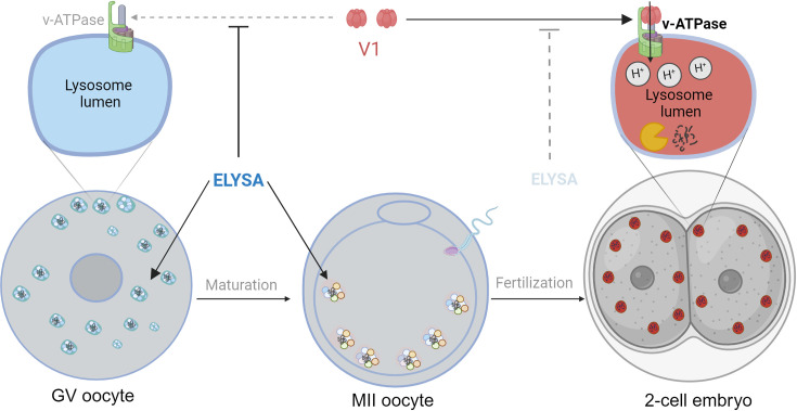
In immature oocytes at the germinal vesicle (GV) stage (grey, left), endosomes (white) and lysosomes (blue) assemble in large spherical structures called ELYSAs. The formation of an ELYSA prevents the assembly of v-ATPase (green; top left inset) on the surface of the lysosomes it contains by blocking the recruitment of the V1 subunit (red), and this in turn prevents acidification of the lysosomes. As the GV oocyte matures into an MII oocyte (middle), ELYSAs fuse with one another to form even bigger structures that can measure 7–8 microns across, and these move towards the periphery of the cell. After the MII oocyte has been fertilized, ELYSAs begin to disassemble in the 2 cell stage embryo (right). This allows V1 to bind to v-ATPase (top right inset), which leads to an influx of protons into the lysosomes (red), creating an acidic environment that increases degradation within the lysosomes. ELYSA: endosomal-lysosomal organellar assembly.
Comment on
- doi: 10.7554/eLife.99358
References
-
- Bomba-Warczak EK, Velez KM, Zhou LT, Guillermier C, Edassery S, Steinhauser ML, Savas JN, Elizabeth Duncan F. Exceptional longevity of mammalian ovarian and oocyte macromolecules throughout the reproductive lifespan. eLife. 2024;13:RP93172. doi: 10.7554/eLife.93172. - DOI
-
- Jentoft IMA, Bäuerlein FJB, Welp LM, Cooper BH, Petrovic A, So C, Penir SM, Politi AZ, Horokhovskyi Y, Takala I, Eckel H, Moltrecht R, Lénárt P, Cavazza T, Liepe J, Brose N, Urlaub H, Fernández-Busnadiego R, Schuh M. Mammalian oocytes store proteins for the early embryo on cytoplasmic lattices. Cell. 2023;186:5308–5327. doi: 10.1016/j.cell.2023.10.003. - DOI - PubMed
Publication types
MeSH terms
LinkOut - more resources
Full Text Sources
