. 2024 Aug 21;15(1):7176.
doi: 10.1038/s41467-024-51445-z.

Tumor-derived RHOA mutants interact with effectors in the GDP-bound state

Affiliations

Tumor-derived RHOA mutants interact with effectors in the GDP-bound state

Yuan Lin et al. Nat Commun. .

Abstract

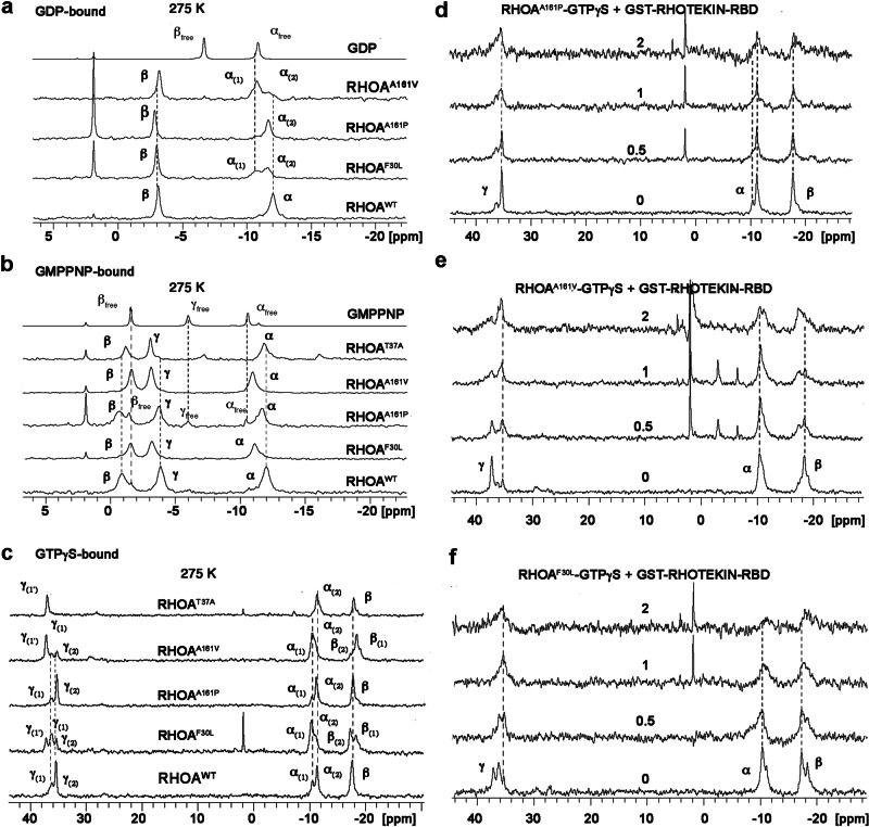
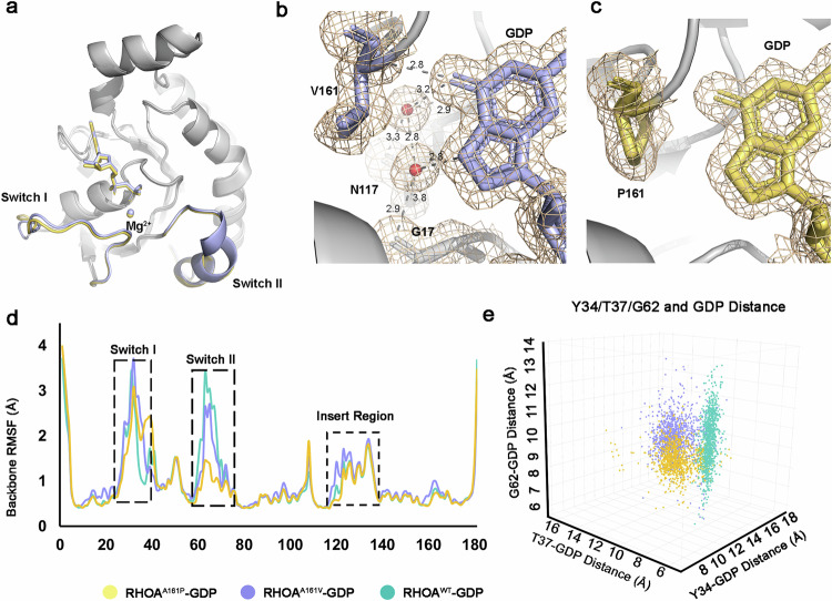
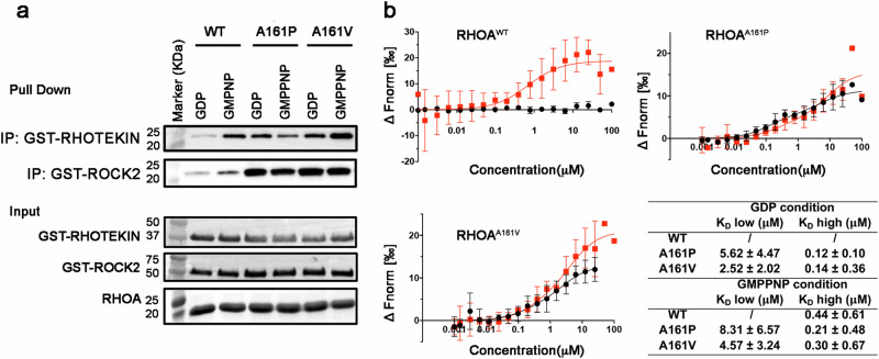
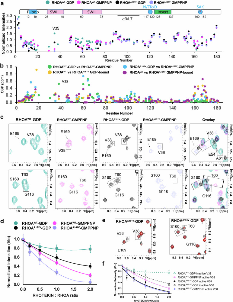
RHOA mutations are found at diverse residues in various cancer types, implying mutation- and cell-specific mechanisms of tumorigenesis. Here, we focus on the underlying mechanisms of two gain-of-function RHOA mutations, A161P and A161V, identified in adult T-cell leukemia/lymphoma. We find that RHOAA161P and RHOAA161V are both fast-cycling mutants with increased guanine nucleotide dissociation/association rates compared with RHOAWT and show reduced GTP-hydrolysis activity. Crystal structures reveal an altered nucleotide association in RHOAA161P and an open nucleotide pocket in RHOAA161V. Both mutations perturb the dynamic properties of RHOA switch regions and shift the conformational landscape important for RHOA activity, as shown by 31P NMR and molecular dynamics simulations. Interestingly, RHOAA161P and RHOAA161V can interact with effectors in the GDP-bound state. 1H-15N HSQC NMR spectra support the existence of an active population in RHOAA161V-GDP. The distinct interaction mechanisms resulting from the mutations likely favor an RHOAWT-like "ON" conformation, endowing GDP-bound state effector binding activity.


Conflict of interest statement

The authors declare no competing interests.

Figures

Fig. 1
Fig. 1. Analysis of wild-type and mutant RHOA biochemical properties.
a Nucleotide dissociation assays for RHOAWT (teal), RHOAF30L (cyan), RHOAA161P (orange), RHOAA161V (purple) with RHOA-BODIPY-FL-GDP: GTPγS ratio of 1:10 in the presence of Mg2+. Data represents mean ± SD with n = 4 biological repeats. b Statistical analysis (unpaired t-test) of dissociation rates obtained from fitting data in a. Data represents mean ± SD. c Pull-down assays with MBP-LARG-DH/PH to detect binding of purified wild-type and mutant RHOA proteins preloaded with GDP or GMPPNP or in the presence of EDTA. The pull-down has been done with or without supplemented nucleotides. The pull-down has been repeated three times with similar results. d Measurement of RHOA intrinsic or GST-p190-GAP stimulated GTPase activity. GTPase activity of wild-type and mutant RHOA at different concentrations was measured with or without 2 μM GST-P190-GAP. Data represents mean ± SD with n = 4–6 biological repeats. e Statistical analysis (unpaired t-test) of EC50s from fitting data in d. Data represents mean ± SD. RHOAT37A is significantly different from the others and the P value is not shown (P < 0.00001). Source data are provided as a Source Data file.
Fig. 2
Fig. 2. 31P NMR spectra of GDP-, GMPPNP-, and GTPγS-bound RHOAWT and mutants.
a Comparison of 31P NMR spectra of GDP-bound RHOAWT (0.84 mM), RHOAF30L (0.71 mM), RHOAA161P (0.40 mM), and RHOAA161V (0.63 mM) at 275 K. Both RHOAA161V and RHOAF30L showed an additional state at the α-phosphate that possibly has an open conformation around the α-phosphate. This state was barely detectable in RHOAA161P. b Comparison of 31P NMR spectra of GMPPNP-bound RHOAWT (0.67 mM), RHOAF30L (0.62 mM), RHOAA161P (0.45 mM), RHOAA161V (0.44 mM), RHOAT37A (0.51 mM) at 275 K. c Comparison of 31P NMR spectra of GTPγS-bound RHOAWT (0.60 mM), RHOAF30L (0.63 mM), RHOAA161P (0.56 mM), RHOAA161V (0.67 mM), and RHOAT37A (0.7 mM) at 275 K. At the γ-phosphate, RHOAA161P was very similar to RHOAWT. In contrast, RHOAA161V and RHOAF30L displayed two inactive states, state 1 as in RHOAWT and state 1’ as in RHOAT37A. To better understand the nature of the states, 31P NMR spectra of RHOAA161P (d), RHOAA161V (e), and RHOAF30L (f) in complex with GST-RHOTEKIN-RBD were collected at 275 K. RHOAWT titrations with GST-RHOTEKIN-RBD and GST control have been published previously. The molar ratio of GST-RHOTEKIN-RBD/RHOA increases from 0, 0.5, 1, to 2, starting from ~0.5 mM RHOA proteins. All spectra were aligned with the inorganic phosphate Pi peak.
Fig. 3
Fig. 3. Structural analysis and MD simulation of RHOAA161P-GDP and RHOAA161V-GDP.
a Overall fold of GDP-bound RHOAA161P and RHOAA161V. The switch regions are colored orange (RHOAA161P) or purple (RHOAA161V), encompassing residues 28–40 and 60–78, respectively. b Two conserved water molecules mediate a hydrogen-bond network between the nucleotide guanine base and RHOA, as exemplified in RHOAA161V-GDP. Electron densities around the GDP, the two water molecules, residue 161, and other surrounding residues are contoured at 1σ (wheat) from the 2Fo-Fc map. c These two water molecules are missing in RHOAA161P-GDP. d Backbone RMSF plot for RHOA-GDP systems. e The distances of important residues and GDP. Y34, T37, and G62 distances were calculated using Cα of the residues and β-phosphate. Source data are provided as a Source Data file.
Fig. 4
Fig. 4. GDP-bound mutant RHOAA161V and RHOAA161P interact with effectors.
a Pull-down assay with GST-RHOTEKIN-RBD and GST-ROCK2-RBD to detect the binding of purified wild-type and mutant RHOA proteins preloaded with GDP or GMPPNP. The pull-down has been repeated five times with similar results. b Microscale thermophoresis analysis of the direct binding between GDP- (black) or GMPPNP-bound (red) RHOA proteins and RHOTEKIN-RBD. ΔFnorm [‰] = 10*(Fnorm(bound) − Fnorm(unbound)). Data represents mean ± SD with n = 8 biological repeats for RHOAWT-GMPPNP and n = 3 for all other samples. KD values obtained from the binding curves are listed and shown in μM. Source data are provided as a Source Data file.
Fig. 5
Fig. 5. RHOAA161V-GDP showed a population of active conformation in the 1H-15N HSQC spectra.
a Plot of peak intensities against residue number for non-overlapping assigned peaks. Intensities were normalized to the average peak intensity from assigned, non-overlapping peaks in the RHOAWT-GDP spectrum. The domain structure of truncated RHOA (residues 1–181) is aligned at the top of the plot and the red asterisk (*) is the mutational site. b Plot of CSPs for assigned peaks against residue number. For V38 and S160, the “active” peak (most similar to RHOAWT-GMPPNP) in the RHOAA161V-GDP spectrum was used to calculate the CSPs. c 1H-15N HSQC spectra of GDP- and GMPPNP-bound RHOAWT and RHOAA161V indicated that RHOAA161V-GDP had two populations, as exemplified by residue V38 (top panel) and S160 (bottom panel). d Average intensities of 1H-15N HSQC spectra, normalized to the corresponding RHOA samples prior to RHOTEKIN addition, were plotted against ratios of titrated RHOTEKIN-RBD to RHOA. The error bars are the standard deviation of the averaged intensities. e Titration of 2X RHOTEKIN-RBD to RHOAA161V-GDP indicated that for the doubled peaks for V38 (left panel) and S160 (right panel). Black: apoprotein; Red: with 2X RHOTEKIN-RBD. f Titration curve of peak intensities for V38. The error bars are the same as in d. The “active” peak (solid black curve) in RHOAA161V-GDP had a greater decrease in intensity upon binding than the other (dotted black curve). In d and f, the spectrum of each sample at each condition was collected once. Source data are provided as a Source Data file.

Similar articles

Cited by

References

    1. Etienne-Manneville, S. & Hall, A. Rho GTPases in cell biology. Nature420, 629–635 (2002). 10.1038/nature01148 - DOI - PubMed
    1. Hodge, R. G. & Ridley, A. J. Regulating Rho GTPases and their regulators. Nat. Rev. Mol. cell Biol.17, 496–510 (2016). 10.1038/nrm.2016.67 - DOI - PubMed
    1. Haga, R. B. & Ridley, A. J. Rho GTPases: Regulation and roles in cancer cell biology. Small GTPases7, 207–221 (2016). 10.1080/21541248.2016.1232583 - DOI - PMC - PubMed
    1. Vetter, I. R. & Wittinghofer, A. The guanine nucleotide-binding switch in three dimensions. Science294, 1299–1304 (2001). 10.1126/science.1062023 - DOI - PubMed
    1. Cherfils, J. & Zeghouf, M. Regulation of small GTPases by GEFs, GAPs, and GDIs. Physiol. Rev.93, 269–309 (2013). 10.1152/physrev.00003.2012 - DOI - PubMed

LinkOut - more resources