. 2024 Aug 21;15(1):7185.
doi: 10.1038/s41467-024-51582-5.

Replay-triggered brain-wide activation in humans

Affiliations

Replay-triggered brain-wide activation in humans

Qi Huang et al. Nat Commun. .

Abstract

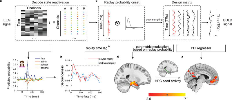
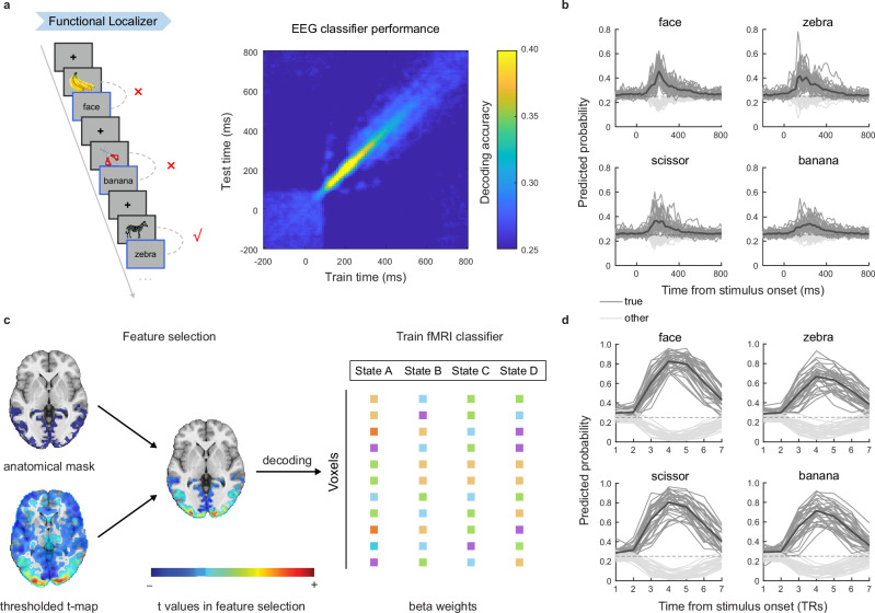
The consolidation of discrete experiences into a coherent narrative shapes the cognitive map, providing structured mental representations of our experiences. In this process, past memories are reactivated and replayed in sequence, fostering hippocampal-cortical dialogue. However, brain-wide engagement coinciding with sequential reactivation (or replay) of memories remains largely unexplored. In this study, employing simultaneous EEG-fMRI, we capture both the spatial and temporal dynamics of memory replay. We find that during mental simulation, past memories are replayed in fast sequences as detected via EEG. These transient replay events are associated with heightened fMRI activity in the hippocampus and medial prefrontal cortex. Replay occurrence strengthens functional connectivity between the hippocampus and the default mode network, a set of brain regions key to representing the cognitive map. On the other hand, when subjects are at rest following learning, memory reactivation of task-related items is stronger than that of pre-learning rest, and is also associated with heightened hippocampal activation and augmented hippocampal connectivity to the entorhinal cortex. Together, our findings highlight a distributed, brain-wide engagement associated with transient memory reactivation and its sequential replay.


Conflict of interest statement

The authors declare no competing interests.

Figures

Fig. 1
Fig. 1. Experimental design of a cued sequential mental simulation task with simultaneous EEG-fMRI.
Subjects, undergoing simultaneous EEG-fMRI recordings, were required to construct a sequence by learning pairwise associations of four discrete visual stimuli. They were then cued to mentally simulate the learned sequence in either a forward or reverse order. As in previous replay studies,,,,,, stimuli were first presented in a random order during functional localizer phase, prior to learning. We included a resting state both before (PRE Rest) and after learning (POST Rest) and this allowed us to measure changes in spontaneous neural activity induced by learning.
Fig. 2
Fig. 2. Simultaneous EEG-fMRI analysis framework for studying sequential replay.
a EEG-based stimuli classifiers were trained using whole-brain channel features during the functional localizer and later used to decode stimuli reactivations during specific task phases, such as rest or during mental simulation. b Temporal Delayed Linear Modelling (TDLM) was applied to the decoded time series to measure the sequential reactivation of states (e.g., visual stimuli) separately for forward and reverse order. c After identifying a time lag of interest (e.g., the peak of sequenceness), we derived an EEG-based replay probability time course. This was then convolved with the hemodynamic response function (HRF) and down-sampled to match the fMRI time resolution, serving as an additional regressor in an fMRI-based GLM analysis. d Based on the new GLM, we determined when (via EEG) and where (via fMRI) replay occurs. e Using an fMRI-derived ROI (green trace, hippocampus), this EEG-based replay probability can be used (by multiplying with ROI neural activity) to detect changes in functional connectivity with other brain regions as a function of replay probability (i.e., psychophysiological interaction, PPI). Data shown here (decoding, EEG replay and coupled fMRI pattern) are from representative subjects. Results are presented with Punc. < 0.01 for illustrative purpose and reported using the MNI coordinate system.
Fig. 3
Fig. 3. EEG-based and fMRI-based decoding during functional localizer.
a The mean cross validated decoding accuracy of EEG-based classifiers. As in previous studies,,,,,,, classifiers were trained independently at each time point and tested on all time points, starting from 200 ms before stimulus onset to 800 ms post onset (10 ms time bin) during the functional localizer task (left panel). Decoding accuracy peaked at 210 ms post-stimulus onset. n = 33. b The time course (−200 – 800 ms) of mean EEG-based decoding probability trained and tested at the same post-stimulus onset (black line), separately for each stimulus. The dark grey lines represent the decoding probability of a particular classifier for a given image (black line represent the mean probability across subjects), while the light grey lines represent the mean decoding probability of the same classifier for other images. n = 33. c Feature selection procedure in fMRI-based decoding. Following Wittkuhn and Schuck, we selected the subject-specific anatomical masks combined with thresholding t-maps (t > 3) to identify voxels that selectively response to functional localizer. Note that the result presented here was selected from a representative subject for illustrative purpose only. d The time course (in TR, starting from stimulus onset) of mean fMRI-based decoding probability trained and tested at the same post-stimulus time, separately for each stimulus. The dark grey lines represent the decoding probability of a particular classifier for a given image (black line represent the mean probability across subjects), while light grey lines represent the mean decoding probability of the same classifier for other images. n = 33. Source data are provided as a Source Data file.
Fig. 4
Fig. 4. EEG-based and fMRI-based replay during cued mental simulation.
a The illustration of two analysis methods for detecting replays. TDLM is used primarily with MEG,,,,,, and more recently also with EEG. The other is a regression method, as per Wittkuhn and Schuck and primarily used with fMRI. Please note that this panel is solely for illustrative purposes. For results based on actual data, refer to Supplementary Fig. 5 and Panel c. b EEG-based replay with TDLM, separately for forward (cued “1”, top row) and backward (cued “4”, bottom row) mental simulation conditions. There were significant forward (but not reverse) replays during both forward and backward mental simulation. Sequence strength on the peak time lag (30 ms) is shown on the right, separately for forward and backward mental simulation conditions (two-sided paired t-test, forward condition: t(32) = 2.80, P = 0.009; backward condition: t(32) = 3.09, P = 0.004). The grey dash line represents the permutation threshold, defined as the 95th percentile of the permutated transitions of interest controlling for multiple comparisons. n = 33. c fMRI-based neural sequence with regression method, separately for forward and backward mental simulation conditions. There was no significant evidence for sequential activation in the correct order (all Pcorr. ≥ 0.06, two-sided one-sample t-test against zero, FDR corrected). The bar plot in the upper right corner shows mean slope coefficients for each period (two-sided paired t-test, forward condition: t(32) = 1.14, P = 0.260; backward condition: t(32) = −0.175, P = 0.862). None of these coefficients were significantly different compared to zero. See Supplementary Fig. 5 for assessing fMRI replay using TDLM, as well as results from single subject for illustration purpose. n = 33. d The parametric modulation of EEG-based replay probability in the whole-brain fMRI during mental simulation showed significant activations in hippocampus and mPFC. We use whole-brain FWE correction at the cluster level (P < 0.05) with a cluster-inducing voxel threshold of Punc. < 0.001. e The psychophysiological interaction (PPI) between hippocampal activity (anatomically defined) and EEG-based replay probability revealed significant functional connectivity change in mPFC, PCC and visual cortex. See Supplementary Fig. 7c-d for mPFC-based PPI results. We use whole-brain FWE correction at the cluster level (P < 0.05) with a cluster-inducing voxel threshold of Punc. < 0.01. Each dot is one subject. The grey lines connect results from the same subject. Shaded areas in b and c show SEM across subjects. Error bars in b and c show SEM across subjects. * P < 0.05, ** P < 0.01, ns., not significant. Abbreviation: HPC - hippocampus. Source data are provided as a Source Data file.
Fig. 5
Fig. 5. EEG-based reactivation during PRE and POST Rest.
a In EEG-based task reactivations, there was a significant higher reactivation strength during POST than PRE Rest (two-sided paired t-test: t(31) = 2.75, P = 0.010). n = 32, excluding an outlier (beyond three deviation of the mean). b Parametric modulation of EEG-based reactivation probability in whole-brain fMRI during POST Rest showed significant activations in bilateral anterior hippocampus (whole-brain FWE correction at the cluster level (P < 0.05) with a cluster-forming voxel threshold of Punc. < 0.001). c ROI analysis. The EEG-based reactivation explained hippocampal activation (anatomically defined) during POST Rest, and it was stronger from PRE to POST Rest in hippocampus (two-sided one-sample t-test: PRE: t(32) = 1.08, Pcorr. = 0.287; POST: t(32) = 3.83, Pcorr. < 0.001; two-sided paired t-test: POST vs. PRE: t(32) = 2.44, Pcorr. = 0.030; FDR corrected). n = 33. d, The task reactivation-aligned BOLD signal in hippocampus during POST Rest. Upon alignment to the onsets of task-related reactivation, we observed a significant increase in hippocampal BOLD activity, peaking at the 2nd TR post-EEG-based reactivation (two-sided one-sample t-test: TR = 1: t(32) = 2.57, P = 0.015; TR = 2: t(32) = 3.02, P = 0.005), and also found at onset of fMRI-based reactivation (TR = 0: two-sided one-sample t-test: t(32) = 2.45, P = 0.020). n = 33. e The PPI between hippocampal activity (anatomically defined) and EEG-based reactivation probability showed increased functional connectivity with EC (anatomical mask depicted in blue) during POST Rest. We thresholded at Punc. < 0.01, K > 10 for visualization. f ROI analysis. The PPI revealed a significant increase in hippocampal-seed connectivity with the EC (anatomically defined) during POST Rest when EEG-based reactivation increased. (two-sided one-sample t-test: PRE: t(32) = 1.48, P = 0.148; POST: t(32) = 2.75, P = 0.010). n = 33. g There was a positive correlation between EEG-based and fMRI-based reactivation in explaining hippocampal activity during POST Rest, but not PRE Rest (robust correlation, PRE: r = 0.09, P = 0.602; POST: r = 0.38, P = 0.029). n = 33. The solid lines reflect the robust linear fit. Each dot is one subject. The grey lines connect results from the same subject. Shaded areas in d show SEM across subjects. Error bars in a, c and f show SEM across subjects. * P < 0.05, ** P < 0.01, *** P < 0.001, ns., not significant. Abbreviation: HPC - hippocampus. Source data are provided as a Source Data file.

References

    1. Wilson, M. A. & McNaughton, B. L. Reactivation of hippocampal ensemble memories during sleep. Science265, 676–679 (1994). - PubMed
    1. Nádasdy, Z., Hirase, H., Czurkó, A., Csicsvari, J. & Buzsáki, G. Replay and time compression of recurring spike sequences in the hippocampus. J. Neurosci.19, 9497–9507 (1999). - PMC - PubMed
    1. Liu, Y., Mattar, M. G., Behrens, T. E. J., Daw, N. D. & Dolan, R. J. Experience replay is associated with efficient nonlocal learning. Science372, eabf1357 (2021). - PMC - PubMed
    1. Widloski, J. & Foster, D. J. Flexible rerouting of hippocampal replay sequences around changing barriers in the absence of global place field remapping. Neuron110, 1547–1558 (2022). - PMC - PubMed
    1. Schwartenbeck, P. et al. Generative replay for compositional visual understanding in the prefrontal-hippocampal circuit. Cell186, 4885–4897.e4814 (2023). - PMC - PubMed