. 2024 Aug 21;29(1):112.
doi: 10.1186/s11658-024-00629-y.

CLDN6 inhibits breast cancer growth and metastasis through SREBP1-mediated RAS palmitoylation

Affiliations

CLDN6 inhibits breast cancer growth and metastasis through SREBP1-mediated RAS palmitoylation

Qiu Jin et al. Cell Mol Biol Lett. .

Abstract

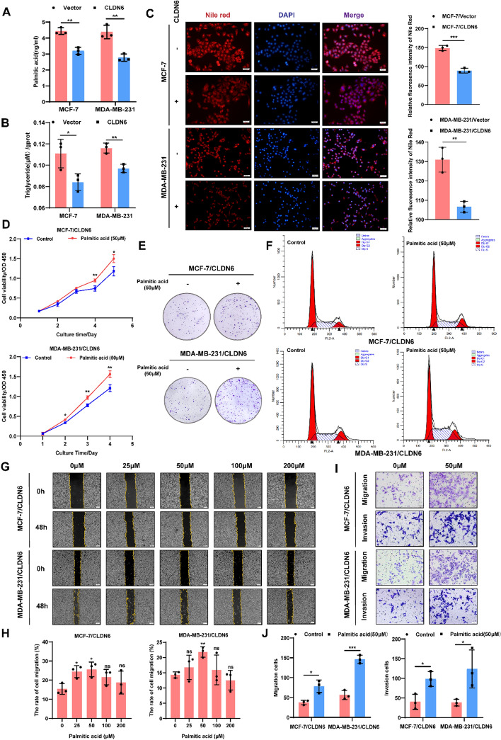
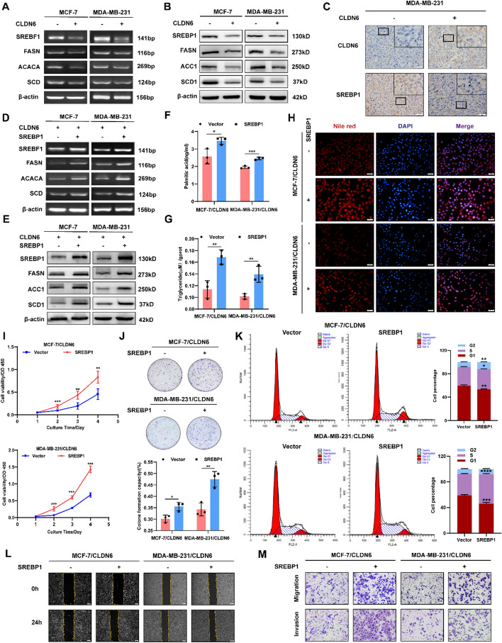
Background: Breast cancer (BC) ranks as the third most fatal malignant tumor worldwide, with a strong reliance on fatty acid metabolism. CLDN6, a candidate BC suppressor gene, was previously identified as a regulator of fatty acid biosynthesis; however, the underlying mechanism remains elusive. In this research, we aim to clarify the specific mechanism through which CLDN6 modulates fatty acid anabolism and its impact on BC growth and metastasis.

Methods: Cell function assays, tumor xenograft mouse models, and lung metastasis mouse models were conducted to evaluate BC growth and metastasis. Human palmitic acid assay, triglyceride assay, Nile red staining, and oil red O staining were employed to investigate fatty acid anabolism. Reverse transcription polymerase chain reaction (RT-PCR), western blot, immunohistochemistry (IHC) assay, nuclear fractionation, immunofluorescence (IF), immunoprecipitation and acyl-biotin exchange (IP-ABE), chromatin immunoprecipitation (ChIP), dual luciferase reporter assay, and co-immunoprecipitation (Co-IP) were applied to elucidate the underlying molecular mechanism. Moreover, tissue microarrays of BC were analyzed to explore the clinical implications.

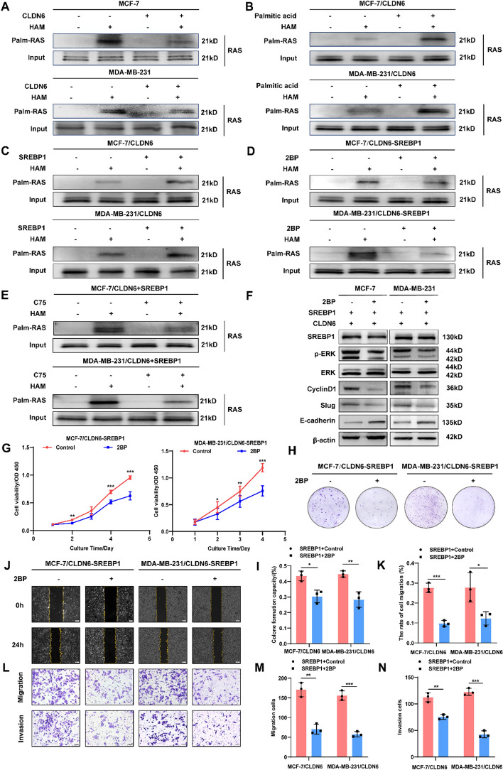
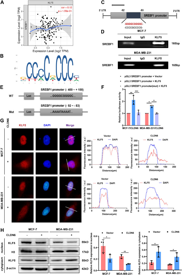
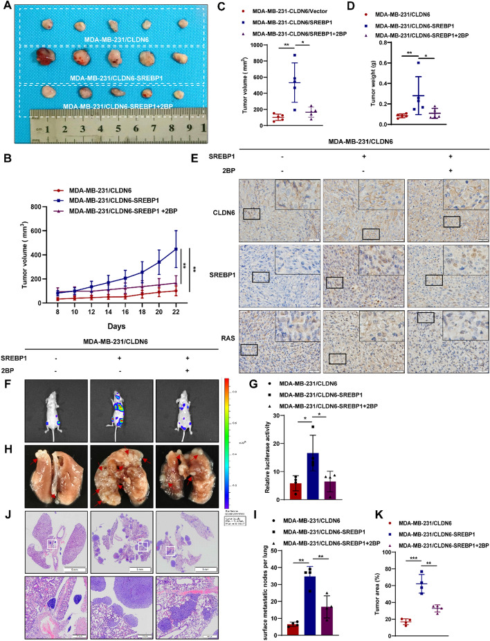
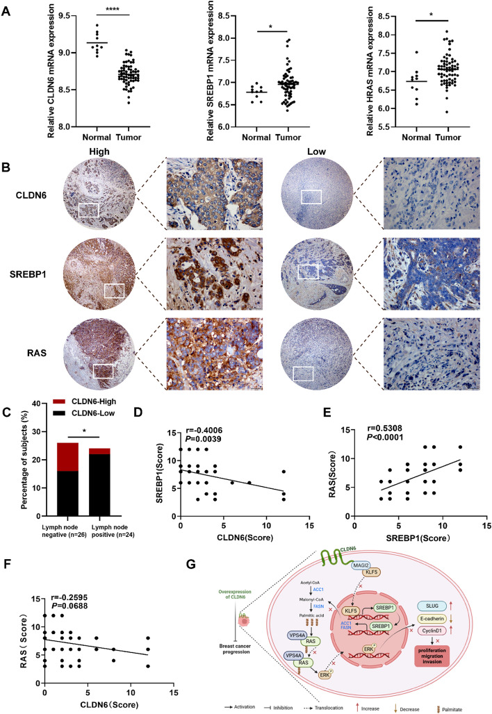
Results: We identified that CLDN6 inhibited BC growth and metastasis by impeding RAS palmitoylation both in vitro and in vivo. We proposed a unique theory suggesting that CLDN6 suppressed RAS palmitoylation through SREBP1-modulated de novo palmitic acid synthesis. Mechanistically, CLDN6 interacted with MAGI2 to prevent KLF5 from entering the nucleus, thereby restraining SREBF1 transcription. The downregulation of SREBP1 reduced de novo palmitic acid synthesis, hindering RAS palmitoylation and subsequent endosomal sorting complex required for transport (ESCRT)-mediated plasma membrane localization required for RAS oncogenic activation. Besides, targeting inhibition of RAS palmitoylation synergized with CLDN6 to repress BC progression.

Conclusions: Our findings provide compelling evidence that CLDN6 suppresses the palmitic acid-induced RAS palmitoylation through the MAGI2/KLF5/SREBP1 axis, thereby impeding BC malignant progression. These results propose a new insight that monitoring CLDN6 expression alongside targeting inhibition of palmitic acid-mediated palmitoylation could be a viable strategy for treating oncogenic RAS-driven BC.

Keywords: Breast cancer; CLDN6; Fatty acid; Palmitoylation; RAS; SREBP1.


Conflict of interest statement

The authors declare that they have no competing interests.

Figures

Fig. 1
Fig. 1
Inhibitory effects of CLDN6 on fatty acid anabolism-induced BC progression. A, B Palmitic acid and triglyceride content in BC cells with CLDN6 overexpression (n = 3). C Nile red staining detected the effect of CLDN6 on neutral lipid content. Scale bar, 50 μm. D-F The impact of palmitic acid on cell viability (D), the clonogenicity (E) and cell cycle progression (F) in BC cells with CLDN6 overexpression. GJ The palmitic acid-induced alterations in cellular migratory and invasive capacity of BC cells with CLDN6 overexpression (n = 3). Scale bar, 200 μm (G) and 50 μm (I). *P < 0.05, **P < 0.01, ***P < 0.001 denoted the presence of statistically significant disparities
Fig. 2
Fig. 2
CLDN6 exerts an inhibitory effect on de novo fatty acid synthesis by downregulating SREBP1 expression. A, B The effect of CLDN6 overexpression on SREBP1, FASN, ACC1, and SCD1 expression at mRNA and protein levels in BC cells (n = 3). C Visualization of the expression levels of CLDN6 and SREBP1 in xenograft tumor tissues. Scale bar, 20 μm. D-E The effect of SREBP1 overexpression on key lipogenic enzymes expression at mRNA and protein levels in CLDN6-overexpressing BC cells (n = 3). FH The impact of SREBP1 overexpression on palmitic acid, triglyceride and neutral lipid in BC cells with CLDN6 overexpression (n = 3). Scale bar, 50 μm. IK The impact of SREBP1 overexpression on cell viability (I), clonogenicity (J), and cell cycle progression (K) of BC cells with CLDN6 overexpression. L, M The SREBP1 overexpression-induced alterations in cellular migratory (L) and invasive (M) capacity of BC cells with CLDN6 overexpression (n = 3). Scale bar, 200 μm (L) and 50 μm (M). *P < 0.05, **P < 0.01, ***P < 0.001 denoted the presence of statistically significant disparities
Fig. 3
Fig. 3
CLDN6 governs de novo palmitic acid synthesis to suppress RAS palmitoylation. AE The alterations of RAS palmitoylation in BC cells with CLDN6 overexpression (A), palmitic acid treatment (B), SREBP1 overexpression (C), 2BP treatment (D), and C75 treatment (E). F Western blot of the protein expression in RAS/ERK signaling pathway (n = 3). G-N The promotion of SREBP1 on cell viability (G), clonogenicity (H, I), and migratory and invasive (JN) capacity of BC cells was reversed by 2BP (n = 3). Scale bar, 200 μm (J) and 50 μm (L). *P < 0.05, **P < 0.01, ***P < 0.001 denoted the presence of statistically significant disparities
Fig. 4
Fig. 4
CLDN6 modulates the localization of palmitoylated RAS via VPS4A. A-D The localization of RAS (red) in BC cells with CLDN6 overexpression(A), palmitic acid treatment (B), SREBP1 overexpression (C), and 2BP treatment (D-E). Scale bar, 20 μm. F The interaction between VPS4A and HRAS. G, H The impact of CLDN6 overexpression on palmitoylation (G) and localization (H) of HRAS (green) in BC cells. Scale bar, 20 μm. I, J The regulatory impact of CLDN6 overexpression on the binding between HRAS and VPS4A (I) and the colocalization of HRAS (red) and VPS4A (green) (J). Scale bar, 20 μm. K-L The alteration of the binding between HRAS and VPS4A (K) and the colocalization of HRAS (red) with VPS4A (green) (L) in SREBP1-overexpressing BC cells induced by 2BP treatment. Scale bar, 20 μm
Fig. 5
Fig. 5
CLDN6 restrains SREBF1 transcription via modulating KLF5 nuclear translocation. A The SREBF1 expression in the Timer database was positively associated with the KLF5 expression in stromal BC. B, C The most likely binding site of KLF5 to the SREBF1 promoter predicted by the JASPAR database. D ChIP was used to detect the binding of KLF5 to SREBF1 promoter in BC cells. E, F Dual-luciferase reporter assay of BC cells transfected with WT or Mut SREBF1 promoter reporter plasmid and KLF5 plasmid (n = 3). G The impact of CLDN6 overexpression on KLF5 (red) positioning in BC cells. Scale bar, 20 μm. H The modulation of CLDN6 on the KLF5 nuclear and cytoplasmic distribution (n = 3). *P < 0.05, **P < 0.01, ***P < 0.001 denoted the presence of statistically significant disparities
Fig. 6
Fig. 6
CLDN6 sequesters KLF5 in the cytoplasm through its interaction with MAGI2. AC The interaction among CLDN6, MAGI2, and KLF5 in MDA-MB-231/CLDN6 cells. D The interaction between CLDN6 and MAGI2 or KLF5 in MDA-MB-231/CLDN6δPBM cells. E The alteration of SREBP1 expression in MAGI2-knockdown BC cells (n = 3). F-G The interaction between CLDN6 and KLF5 in BC cells with MAGI2 knockdown. H, I The effect of MAGI2-knockdown on localization (red) (H) and distribution of KLF5 (I) (n = 3) in BC cells with CLDN6 overexpression. Scale bar, 20 μm. *P < 0.05, **P < 0.01, ***P < 0.001 denoted statistically significant differences
Fig. 7
Fig. 7
In vivo, CLDN6 inhibits BC progression through RAS palmitoylation. A Images of subcutaneous xenograft tumors (n = 5). BD Tumor volume (B, C) and weight (D) of subcutaneous xenograft tumors. E Visualization of the expression levels and localization of CLDN6, SREBP1, and RAS in transplanted tumor tissue. Scale bar, 20 μm. F, G Analysis of lung metastasis using bioluminescent techniques (n = 4). H, I Lung metastatic nodes were indicated with red arrowheads. J, K The lung metastasis of BC was observed through H&E staining. Scale bar, 5 mm and 500 μm. *P < 0.05, **P < 0.01, ***P < 0.001 denoted statistically significant differences
Fig. 8
Fig. 8
Clinical association among the expression of CLDN6, SREBP1, and RAS in patients with BC. A The levels of CLDN6, SREBP1, and HRAS mRNA expression were compared between normal tissues and BC tissues using GSE103512 (normal, 10 and tumor, 65). B Visualization of the low and high expression of CLDN6, SREBP1 and RAS in human BC tissues (n = 50). Scale bar, 200 μm (left) and 20 μm (right). C The distribution of CLDN6 expression levels in patients with BC with and without lymph node involvement. DF Correlation analysis was conducted on a BC tissue microarray, examining the relationship between CLDN6 and SREBP1 (D), SREBP1 and RAS (E), as well as CLDN6 and RAS (F) (n = 50). G A suggested framework elucidating the regulatory process by which CLDN6 hinders the progression of BC via SREBP1-facilitated RAS palmitoylation. *P < 0.05, **P < 0.01, ***P < 0.001 denoted statistically significant differences

References

    1. Sung H, Ferlay J, Siegel RL, Laversanne M, Soerjomataram I, Jemal A, et al. Global cancer statistics 2020: GLOBOCAN estimates of incidence and mortality worldwide for 36 cancers in 185 countries. CA A Cancer J Clin. 2021;71(3):209–49. - PubMed
    1. Loke SY, Lee ASG. The future of blood-based biomarkers for the early detection of breast cancer. Eur J Cancer. 2018;92:54–68. - PubMed
    1. Rajput S, Sharma PK, Malviya R. Biomarkers and treatment strategies for breast cancer recurrence. Curr Drug Targets. 2023;24(15):1209–20. - PubMed
    1. Park M, Kim D, Ko S, Kim A, Mo K, Yoon H. Breast cancer metastasis: mechanisms and therapeutic implications. Int J Mol Sci. 2022;23(12):6806. - PMC - PubMed
    1. Wang T, Fahrmann JF, Lee H, Li YJ, Tripathi SC, Yue C, et al. JAK/STAT3-regulated fatty acid β-oxidation is critical for breast cancer stem cell self-renewal and chemoresistance. Cell Metab. 2018;27(1):136-150.e5. - PMC - PubMed

MeSH terms

LinkOut - more resources