. 2024 Nov 6;32(11):3915-3931.
doi: 10.1016/j.ymthe.2024.08.018. Epub 2024 Aug 21.

TALEN-edited allogeneic inducible dual CAR T cells enable effective targeting of solid tumors while mitigating off-tumor toxicity

Affiliations

TALEN-edited allogeneic inducible dual CAR T cells enable effective targeting of solid tumors while mitigating off-tumor toxicity

Sonal Dharani et al. Mol Ther. .

Abstract

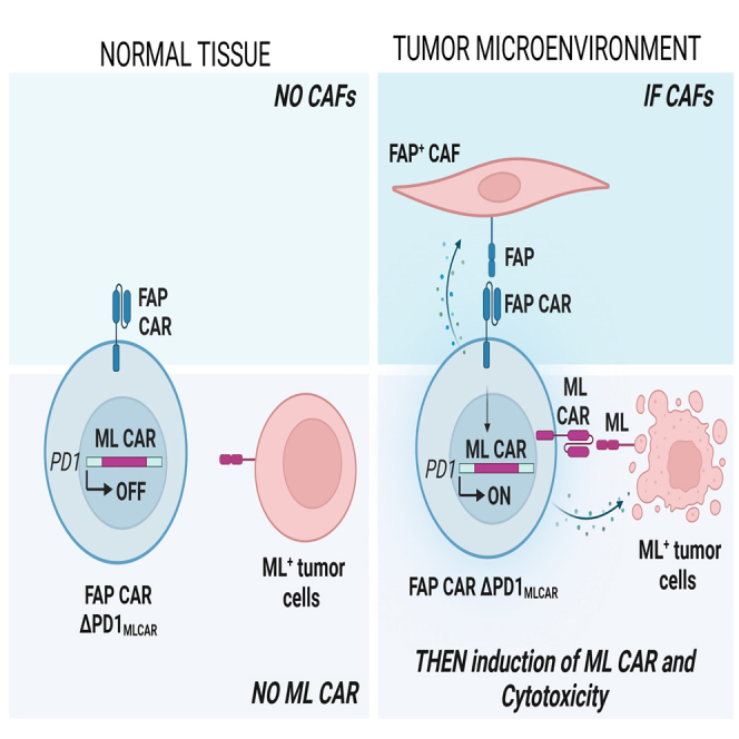
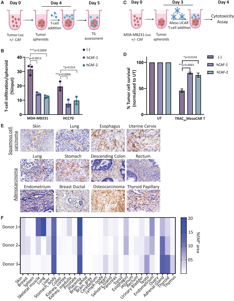
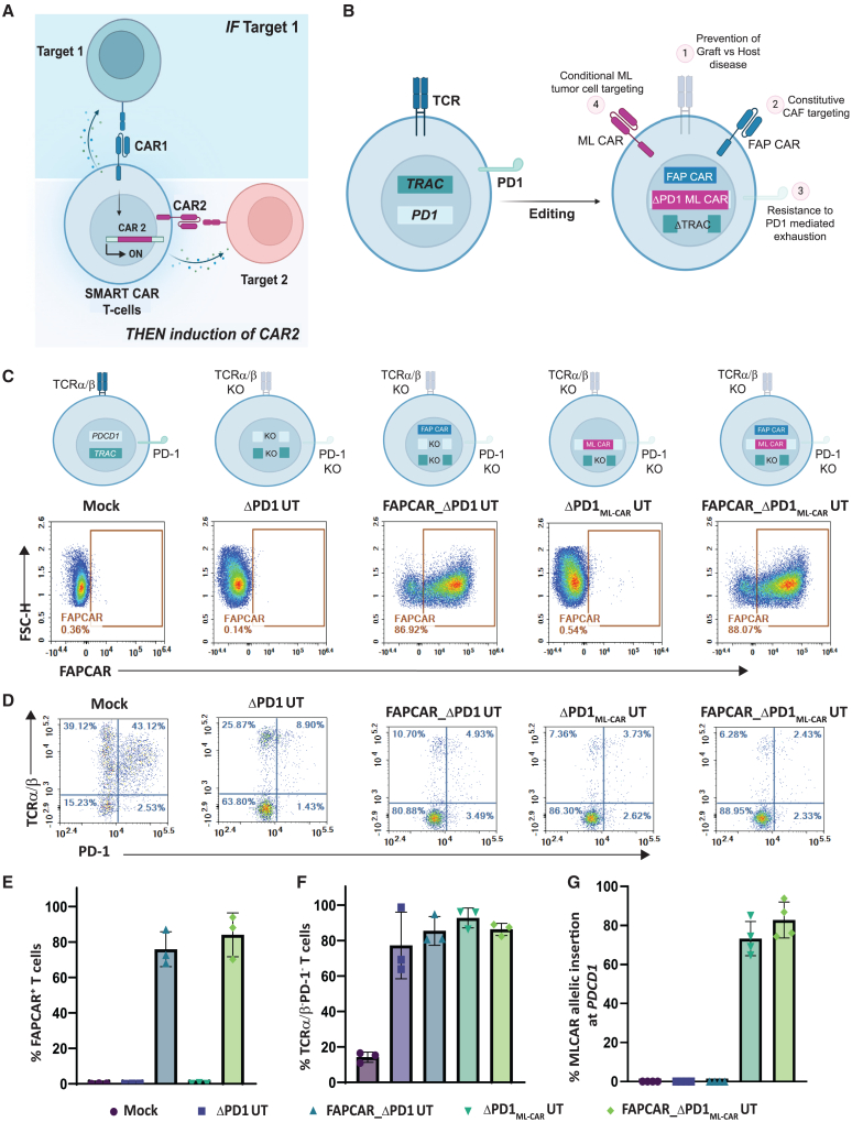
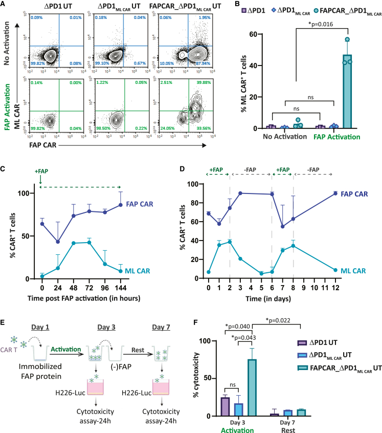
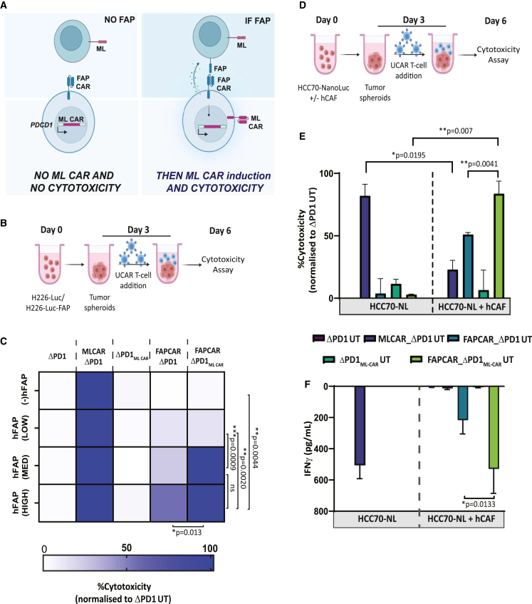
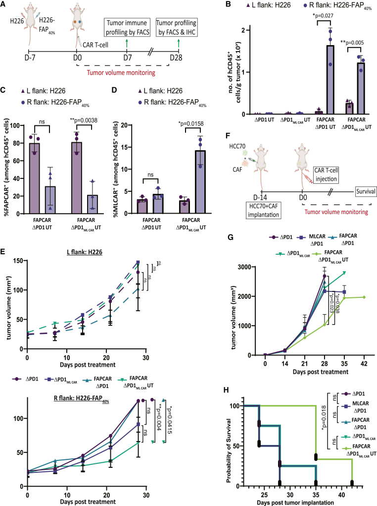
Adoptive cell therapy using chimeric antigen receptor (CAR) T cells has proven to be lifesaving for many cancer patients. However, its therapeutic efficacy has been limited in solid tumors. One key factor for this is cancer-associated fibroblasts (CAFs) that modulate the tumor microenvironment (TME) to inhibit T cell infiltration and induce "T cell dysfunction." Additionally, the sparsity of tumor-specific antigens (TSA) and expression of CAR-directed tumor-associated antigens (TAA) on normal tissues often results in "on-target off-tumor" cytotoxicity, raising safety concerns. Using TALEN-mediated gene editing, we present here an innovative CAR T cell engineering strategy to overcome these challenges. Our allogeneic "Smart CAR T cells" are designed to express a constitutive CAR, targeting FAP+ CAFs in solid tumors. Additionally, a second CAR targeting a TAA such as mesothelin is specifically integrated at a TCR signaling-inducible locus like PDCD1. FAPCAR-mediated CAF targeting induces expression of the mesothelin CAR, establishing an IF/THEN-gated circuit sensitive to dual antigen sensing. Using this approach, we observe enhanced anti-tumor cytotoxicity, while limiting "on-target off-tumor" toxicity. Our study thus demonstrates TALEN-mediated gene editing capabilities for design of allogeneic IF/THEN-gated dual CAR T cells that efficiently target immunotherapy-recalcitrant solid tumors while mitigating potential safety risks, encouraging clinical development of this strategy.

Keywords: CAR T cell; CAR logic gate; TALEN; allogeneic; cell therapy; gene editing; immunotherapy; on-target off-tumor toxicity; solid tumors.


Conflict of interest statement

Declaration of interests S.Dharani, H.C., A.J., J.V., P.D., L.P., and S.Das are current employees and equity holders at Cellectis. TALEN is a Cellectis patented technology. J.P.F. is a former Cellectis employee and is a current employee of Bristol Myers Squibb.

Figures

None
Graphical abstract
Figure 1
Figure 1
Tumor localized FAP+ CAFs inhibit CAR T cell intra-tumoral infiltration and anti-tumor cytotoxicity (A) Schematic illustrating T cell infiltration assay in tumor spheroids plated with or without CAFs. (B) Graph depicting flow cytometry quantitation of T cells infiltrated per tumor spheroids with or without CAFs, represented as percentage of T cell input. Bars show means ± SD, n = 3; p values determined by Student t test (two-tailed, unpaired). ns, not significant, ∗p ≤ 0.05, ∗∗p ≤ 0.01, ∗∗∗p ≤ 0.001. (C) Schematic of TRACKO ML CAR T cell cytotoxicity assay against tumor spheroids of TNBC cell line MDA-MB-231-Luc alone or co-cultured with TNBC-derived CAFs. (D) Bar graph representing percentage MDA-MB-231-Luc tumor cell survival post cytotoxicity assay outlined in (C), at Effector:Target ratio = 5:1. Bars show means ± SD, n = 3; p values determined by Student t test (two-tailed, unpaired). ns, not significant, ∗p ≤ 0.05, ∗∗p ≤ 0.01. (E) Immunohistochemistry for detection of human FAP protein in tissue microarray of patient samples from different cancers. (F) Heatmap depicting percentage positive area stained for human FAP protein in immunohistochemical analysis of healthy human tissue microarray.
Figure 2
Figure 2
TALEN and AAV-mediated multiplex genome editing generates universal, PD-1-resistant dual inducible CAR T cells against solid tumor targets (A) Schematic depicting IF/THEN-gate logic strategy wherein activation of constitutive expression of FAP CAR specifically in TME induces activation of ML CAR integrated through TALEN-mediated targeted disruption at the PDCD1 locus, resulting in enhanced tumor localized dual FAP CAR and ML CAR anti-tumor activity. (B) Pictorial representation of universal dual inducible CAR T cell developed by TALEN-mediated multiplex editing of TRAC and PDCD1 gene loci to downregulate surface TCRα/β and PD-1 expression, lentiviral FAP CAR random integration for stable surface expression AAV6 DNA repair matrix-mediated disruptive integration of ML CAR at PDCD1 gene locus, for inducible expression downstream of FAP CAR activation. (C) Flow cytometry plots showing frequency of CAR expression among viable engineered T cells. (D) Flow cytometry plots showing frequency of TCRα/β (−)/PD-1 (−) viable engineered T cells post activation with PMA/Ionomycin. (E) Graph representing quantitation of percentage of FAP CAR positive viable T cells, as determined in (C). Bars show the means ± SD, n = 3 donors. (F) Graph representing quantitation of percentage of TCRα/β (−)/PD-1 (−) viable T cells, as determined in (D). Bars show the means ± SD, n = 3 donors. (G) Graph representing quantitation of percentage of ML CAR allelic insertion at PDCD1 locus in positive viable T cells post ΔLNGFR enrichment, as determined by droplet digital PCR (ddPCR). Bars show the means ± SD, n = 3 donors.
Figure 3
Figure 3
FAP CAR activation-dependent stringent regulation of ML CAR expression and activity in multiplex engineered FAPCAR_ΔPD1ML CAR UT cells (A) Flow cytometry plots depicting FAP CAR and ML CAR expression in indicated UT cell groups, with or without human FAP (FAP) protein-mediated FAP CAR activation for 48 h. (B) Graph representing quantitation of percentage of ML CAR-positive viable T cells, as determined in (A). Bars show the means ± SD, n = 3 donors; p values determined by Student t test (two-tailed, unpaired). ns, not significant, ∗p ≤ 0.05. (C) Graph depicting kinetics of FAP CAR and ML CAR expression following FAP protein-mediated activation of FAP CAR on FAPCAR_ΔPD1ML CAR UT cells at 0 h. Each data point represents mean ± SD, n = 3 donors. (D) Graph representing time course of FAP CAR and MLCAR expression upon FAP CAR stimulation and withdrawal of stimulus (FAP protein) from FAPCAR_ΔPD1ML CAR UT cells. Each data point represents mean ± SD, n = 3 donors. (E) Schematic for assessing ML CAR cytotoxicity of indicated UT cells against ML+FAP NCI-H226-LUC tumor cells upon FAP CAR stimulation with FAP for 3 days, and subsequent withdrawal of stimulus (FAP protein) for 4 days. (F) Bar graph representing percentage ML+FAP NCI-H226-LUC tumor cell killing at different time points defined in (E). Cytotoxicity was measured 24 h post incubation of UT cells taken from indicated time points in (E) with target cells at Effector:Target ratio = 1:1. Bars show the means ± SD, n = 2 independent experiments; p values determined by Student t test (two-tailed, unpaired). ns, not significant, ∗p ≤ 0.05.
Figure 4
Figure 4
FAPCAR_ΔPD1ML CAR UT cells display efficient FAP CAR activation-dependent dual CAR tumor killing in vitro (A) Pictorial representation of strategy designed to test activity and sensitivity of the IF/THEN logic gate in FAPCAR_ΔPD1ML CAR UT cells. As depicted, tumor cells with no FAP expression should not induce FAP CAR-mediated ML CAR expression and should therefore survive, despite being ML positive. On the other hand, ML+FAP+ double-positive cells should induce FAP CAR-mediated ML CAR expression, resulting in dual CAR activity and enhanced killing of target cells. (B) Schematic of engineered UT cell cytotoxicity assay against tumor spheroids NCI-H226-Luc cells with or without varying proportions of FAP+ cells, co-incubated for 72 h at E:T = 2.5:1. (C) Heatmap representing percentage target tumor cell cytotoxicity when treated with indicated UT groups, as outlined in (B). Brackets indicate statistical comparison of cytotoxic activity between different target cell groups, n = 2 independent experiments, two donors per experiment, three technical replicates per donor per experiment; p values determined by Student t test (two-tailed, unpaired). ns, not significant, ∗p ≤ 0.05, ∗∗p ≤ 0.01, ∗∗∗p ≤ 0.001. (D) Schematic of engineered UT cell cytotoxicity assay against 3D spheroids of TNBC cell line HCC70-NanoLuc alone or co-cultured with TNBC-derived CAFs at 1:2 ratio. Effector and target spheroids co-incubated for 72 h at 5:1 ratio. (E) Bar graph representing percentage of HCC70-GFP tumor cell cytotoxicity post cytotoxicity assay outlined in (D). Data representative of n = 2 independent experiments, two donors per experiment, three technical replicates per donor per experiment. Bars show the means ± SD; p values determined by Student t test (two-tailed, unpaired). ns, not significant, ∗p ≤ 0.05, ∗∗p ≤ 0.01. (F) Bar graph representing IFNγ release in cytotoxicity assay outlined in (D), as measured by ELISA in co-culture supernatant on day 6. Data representative of n = 2 independent experiments, two donors per experiment, three technical replicates per donor per experiment. Bars show the means ± SD; p values determined by Student t test (two-tailed, unpaired). ns, not significant, ∗p ≤ 0.05.
Figure 5
Figure 5
FAPCAR_ΔPD1ML CAR UT cells efficiently target FAP+ML+ tumors and impede CAF+ tumor growth with no “off-target” toxicity in vivo (A) Schematic of UCAR T cell treatment and analysis of bilateral subcutaneous tumors implanted in NSG mice. Data representative of two independently conducted studies, each with different T cell donors. (B) Bar graph representing quantitation of total number of hCD45+ cells per gram of tumors from mice treated with indicated UT cells 7 days post administration, as determined by flow cytometry. p values determined by Student t test (two-tailed, paired), n = 3 mice per cohort. ns, not significant, ∗p ≤ 0.05, ∗∗p ≤ 0.01. (C) Bar graph representing percentage of FAP CAR+ cells among total hCD45+ cells in tumors from mice treated with indicated UT cells 7 days post administration, as determined by flow cytometry. p values determined by Student t test (two-tailed, paired), n = 3 mice per cohort. ns, not significant, ∗p ≤ 0.05, ∗∗p ≤ 0.01. (D) Bar graph representing percentage of ML CAR+ cells among total hCD45+ cells in tumors from mice treated with indicated UT cells 7 days post administration, as determined by flow cytometry. p values determined by Student t test (two-tailed, paired), n = 3 mice per cohort. ns, not significant, ∗p ≤ 0.05. (E) Top panel: Graph representing growth kinetics of subcutaneous left flank NCI-H226 tumors of remaining mice treated as indicated over time until study endpoint. p values determined by Student t test (two-tailed, unpaired), n = 5 mice per cohort. ns, not significant. Bottom panel: Graph representing growth kinetics of subcutaneous right flank NCI-H226-FAP40% tumors of remaining mice treated as indicated over time until study endpoint. p values determined by Student t test (two-tailed, unpaired), n = 5 mice per cohort. ns, not significant, ∗∗p ≤ 0.01. (F) Schematic of UT cell treatment and analysis of orthotopic TNBC tumors co-implanted with patient-derived CAFs in NSG mouse mammary fat pad. Data representative of two independently conducted studies, each with different T cell donors. (G) Graph representing growth kinetics of orthotopic TNBC tumors in mice treated as indicated over time. p values determined by Student t test (two-tailed, unpaired), n = 3–5 mice per cohort. ns, not significant, ∗p ≤ 0.05. (H) Kaplan-Meier curve for survival analysis of orthotopic TNBC tumor-implanted NSG mice treated as indicated (n = 3–5 per cohort). p values determined by log rank (Mantel-Cox) test. ns, not significant, ∗p ≤ 0.05.

Similar articles

Cited by

References

    1. Imai C., Mihara K., Andreansky M., Nicholson I.C., Pui C.H., Geiger T.L., Campana D. Chimeric receptors with 4-1BB signaling capacity provoke potent cytotoxicity against acute lymphoblastic leukemia. Leukemia. 2004;18:676–684. doi: 10.1038/sj.leu.2403302. - DOI - PubMed
    1. Brentjens R.J., Rivière I., Park J.H., Davila M.L., Wang X., Stefanski J., Taylor C., Yeh R., Bartido S., Borquez-Ojeda O., et al. Safety and persistence of adoptively transferred autologous CD19-targeted T cells in patients with relapsed or chemotherapy refractory B-cell leukemias. Blood. 2011;118:4817–4828. doi: 10.1182/blood-2011-04-348540. - DOI - PMC - PubMed
    1. Westin J.R., Kersten M.J., Salles G., Abramson J.S., Schuster S.J., Locke F.L., Andreadis C. Efficacy and safety of CD19-directed CAR-T cell therapies in patients with relapsed/refractory aggressive b-cell lymphomas: Observations from the JULIET, ZUMA-1, and TRANSCEND trials. Am. J. Hematol. 2021;96:1295–1312. doi: 10.1002/ajh.26301. - DOI - PMC - PubMed
    1. Li D., Li X., Zhou W.L., Huang Y., Liang X., Jiang L., Yang X., Sun J., Li Z., Han W.D., Wang W. Genetically engineered T cells for cancer immunotherapy. Signal Transduct. Targeted Ther. 2019;4:35. doi: 10.1038/s41392-019-0070-9. - DOI - PMC - PubMed
    1. Schuster S.J., Svoboda J., Chong E.A., Nasta S.D., Mato A.R., Anak Ö., Brogdon J.L., Pruteanu-Malinici I., Bhoj V., Landsburg D., et al. Chimeric antigen receptor T cells in refractory B-cell lymphomas. N. Engl. J. Med. 2017;377:2545–2554. doi: 10.1056/NEJMoa1708566. - DOI - PMC - PubMed

MeSH terms