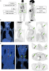
Role and potential of 18F-fluorodeoxyglucose-positron emission tomography-computed tomography in large-vessel vasculitis: a comprehensive review
- PMID: 39170047
- PMCID: PMC11335723
- DOI: 10.3389/fmed.2024.1432865
Role and potential of 18F-fluorodeoxyglucose-positron emission tomography-computed tomography in large-vessel vasculitis: a comprehensive review
Abstract
Large-vessel vasculitis (LVV) is a group of diseases characterized by inflammation of the aorta and its main branches, which includes giant cell arteritis (GCA), polymyalgia rheumatica (PMR), and Takayasu's arteritis (TAK). These conditions pose significant diagnostic and management challenges due to their diverse clinical presentations and potential for serious complications. 18F-fluorodeoxyglucose positron emission tomography-computed tomography (18F-FDG-PET-CT) has emerged as a valuable imaging modality for the diagnosis and monitoring of LVV, offering insights into disease activity, extent, and response to treatment. 18F-FDG-PET-CT plays a crucial role in the diagnosis and management of LVV by allowing to visualize vessel involvement, assess disease activity, and guide treatment decisions. Studies have demonstrated the utility of 18F-FDG-PET-CT in distinguishing between LVV subtypes, evaluating disease distribution, and detecting extracranial involvement in patients with cranial GCA or PMR phenotypes. Additionally, 18F-FDG-PET-CT has shown promising utility in predicting clinical outcomes and assessing treatment response, based on the correlation between reductions in FDG uptake and improved disease control. Future research should focus on further refining PET-CT techniques, exploring their utility in monitoring treatment response, and investigating novel imaging modalities such as PET-MRI for enhanced diagnostic accuracy in LVV. Overall, 18F-FDG-PET-CT represents a valuable tool in the multidisciplinary management of LVV, facilitating timely diagnosis and personalized treatment strategies to improve patient outcomes.
Keywords: PET-CT; Takayasu’s arteritis; giant cell arteritis; large-vessel vasculitis; polymyalgia rheumatica.
Copyright © 2024 Collada-Carrasco, Gómez-León, Castillo-Morales, Lumbreras-Fernández, Castañeda and Rodríguez-Laval.
Conflict of interest statement
The authors declare that the research was conducted in the absence of any commercial or financial relationships that could be construed as a potential conflict of interest.
Figures

References
-
- González-Gay MA, García-Porrúa C. Epidemiology of the vasculitides. Rheum Dis Clin N Am. (2001) 27:729–49. - PubMed
Publication types
LinkOut - more resources
Full Text Sources
Miscellaneous

