. 2024 Aug 2;10(15):e34968.
doi: 10.1016/j.heliyon.2024.e34968. eCollection 2024 Aug 15.

JDF promotes the apoptosis of M2 macrophages and reduces epithelial-mesenchymal transition and migration of liver cancer cells by inhibiting CSF-1/PI3K/AKT signaling pathway

Affiliations

JDF promotes the apoptosis of M2 macrophages and reduces epithelial-mesenchymal transition and migration of liver cancer cells by inhibiting CSF-1/PI3K/AKT signaling pathway

Xiaolin Liu et al. Heliyon. .

Abstract

Background: The interaction between cancer cells and the tumor microenvironment is of critical importance in liver cancer. Jiedu Granule formula (JDF) has been shown to minimize the risk of recurrence and metastasis following liver cancer resection. Investigating the mechanism underlying the therapeutic effects of JDF can extend its field of application and develop novel treatment approaches.

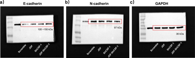
Methods: We established a rat liver orthotopic transplantation tumor model, and recorded the prognostic effects of JDF adjuvant therapy on the recurrence and metastasis of liver cancer. Liver and lung tissues were collected for immunofluorescence staining and H&E staining, respectively. In addition, THP-1 cells were incubated with PMA and IL-4 to induce them to differentiate into M2 macrophages. CSF-1 expression was knocked down using lentivirus to determine the function of CSF-1. Liver cancer cells were cultured with a conditioned medium (CM) or co-cultured with macrophages. Cell viability was determined using the MTT assay. The levels of CSF-1, CSF-1R, E-cadherin, N-cadherin, PI3K, AKT, and cleaved caspase-3 were detected using ELISA, Western blotting and qPCR. The ability of cells to migrate was assessed using cell scratch and transwell assays. Apoptosis was evaluated using flow cytometry.

Results: The JDF treatment decreased the risk of liver cancer metastasis after surgery and the infiltration of CD206/CD68 cells in liver cancer tissue. In cell experiments, JDF showed effects in suppressing M2 macrophages activity and downregulating the expression of CSF-1 and CSF-1R. The concentration of CSF-1 in the supernatant was also lower in the JDF-treated group. Futhermore, M2-CM was found to promote cancer cell migration and epithelial-mesenchymal transition (EMT); however, these effects were weakened after administering JDF. Knocking down endogenous CSF-1 in M2 macrophages resulted in a comparable suppression of cancer cell migration and EMT. Additionally, JDF treatment inhibited activation of the PI3K/AKT pathway, thus promoting the apoptosis of M2 macrophages.

Conclusions: Treatment with JDF reduced the EMT and migratory capacity of liver cancer cells, which might be attributed to the inhibition of M2 macrophage infiltration and interruption of the CSF-1/PI3K/AKT signaling pathway. This mechanism may hold significant implications for mitigating the risk of metastatic spread in the aftermath of hepatic surgery.

Keywords: CSF-1/CSF-1R/PI3K/AKT; Epithelial-mesenchymal transition; JDF; Liver cancer; M2 macrophages; Migration and invasion.


Conflict of interest statement

The authors declare that they have no known competing financial interests or personal relationships that could have appeared to influence the work reported in this paper.

Figures

Image 1
Graphical abstract
Fig. 1
Fig. 1
Jiedu Granule formula (JDF) reduced macrophage infiltration in liver cancer, and inhibited lung metastasis. The rats were treated with JDF 3 days after resection for 2 weeks. JDF-L: 12.74 g/kg/day, JDF-M: 38.22 g/kg/day, and JDF-H: 63.7 g/kg/day. A. The results of H&E staining of lung tissue; scale bar: 200 μm; 400 μm. B. Immunofluorescence double-labeling of CD206 and CD68 in liver cancer tissue, scale bar: 130 μm; **P < 0.01 vs. Saline.
Fig. 2
Fig. 2
JDF (3 mg/mL) decreased the level of expression of CSF-1/CSF-1R in M2 macrophages. THP-1 cells were incubated with PMA (phorbol 12-myristate 13-acetate) and IL-4 to induce differentiation into M2 macrophages. A. The M2 macrophage marker CD206 was detected by flow cytometry. M2 macrophages were treated with JDF for 24 h. B. Cell activity was assessed using the MTT assay. C-D. The protein levels of CSF-1 and CSF-1R in M2 macrophages were determined using western blotting. E. The extracellular concentration of CSF-1 produced by M2 macrophages was detected by enzyme-linked immunosorbent assay (ELISA). The original blots for the protein immunoblotting were provided in Supplementary Fig. S3. *P < 0.05 and **P < 0.01 vs. Saline.
Fig. 3
Fig. 3
The conditioned medium (CM) of JDF-M2 inhibited MHCC97H cell migration. A. A cell scratch assay was used to measure the migration ability of MHCC97H cancer cells. B. Transwell assay was used to measure the migration ability of MHCC97H cancer cells. **P < 0.01 vs. Control, ##P < 0.01 vs. M2.
Fig. 4
Fig. 4
Treatment with JDF (3 mg/mL) and knocking down of shCSF-1 decreased the level of expression of CSF-1/CSF-1R in M2 macrophages. The shCSF-1 lentivirus was transfected. A. qPCR was used to determine the transcription level of CSF-1. B. Western blotting was used to determine the level of CSF-1 protein. C. The M2 macrophage marker CD206 was detected by flow cytometry. D. The extracellular CSF-1 concentration was determined by ELISA. E. Western blotting was used to determine the expression of CSF-1 and CSF-1R proteins after treatment with JDF. The original blots for the protein immunoblotting were provided in Supplementary Fig. S4. **P < 0.01 vs. Scramble, ##P < 0.01 vs. shCSF-1.
Fig. 5
Fig. 5
JDF-CM and shCSF-1-CM inhibited the migration of MHCC97H cells. A. Cell scratch assay was used to measure the migration ability of MHCC97H cancer cells. B. Transwell assay was used to measure the migration ability of MHCC97H cancer cells. **P < 0.01 vs. Scramble, #P < 0.05 vs. shCSF-1.
Fig. 6
Fig. 6
Treatment with JDF (3 mg/mL) reduced the migration of MHCC97H cells when co-cultured with M2 macrophages by knocking down CSF-1. A. Transwell assay was used to measure the migration ability of MHCC97H cancer cells. B. Western blotting was used to determine the expression of CSF-1 protein in MHCC-97H cells. C. qPCR detected the expression of CSF-1 mRNA in M2 macrophages. D. The concentration of CSF-1 in the lower chamber culture system was determined by ELISA. E. Immunofluorescence detection of CSF-1R+ M2 macrophages. The original blots for the protein immunoblotting were provided in Supplementary Fig. S5. **P < 0.01 vs. Scramble; #P < 0.05 and ##P < 0.01 vs. shCSF-1; ^ P < 0.05 and ^^ P < 0.01 vs. JDF.
Fig. 7
Fig. 7
Treatment with JDF (3 mg/mL) inhibited the CSF-1/PI3K/AKT signaling pathway and promoted apoptosis of M2 macrophages. A. Western blotting was used to determine the activity of PI3K/AKT pathway. B–C. The apoptosis of M2 macrophages was measured by flow cytometry. D. Western blotting was used to determine the expression of cleaved caspase-3. The original blots for the protein immunoblotting were provided in Supplementary Fig. S6. **P < 0.01 vs. Scramble; #P < 0.05 and ##P < 0.01 vs. shCSF-1; ^ P < 0.05 vs. JDF; $ P < 0.05 and $$ P < 0.01 vs. JDF-shCSF-1.
figs1
figs1
figs2
figs2
figs3
figs3
figs4
figs4
figs5
figs5
figs6
figs6

References

    1. Bray F., Laversanne M., Sung H., Ferlay J., Siegel R.L., Soerjomataram I., et al. Global cancer statistics 2022: GLOBOCAN estimates of incidence and mortality worldwide for 36 cancers in 185 countries. CA A Cancer J. Clin. 2024;74(3):229–263. doi: 10.3322/caac.21834. - DOI - PubMed
    1. Siegel R.L., Miller K.D., Jemal A. Cancer statistics, 2020. CA A Cancer J. Clin. 2020;70(1):7–30. doi: 10.3322/caac.21590. - DOI - PubMed
    1. Feng K., Yan J., Li X., Xia F., Ma K., Wang S., et al. A randomized controlled trial of radiofrequency ablation and surgical resection in the treatment of small hepatocellular carcinoma. J. Hepatol. 2012;57(4):794–802. doi: 10.1016/j.jhep.2012.05.007. - DOI - PubMed
    1. Xu X.L., Liu X.D., Liang M., Luo B.M. Radiofrequency ablation versus hepatic resection for small hepatocellular carcinoma: systematic review of randomized controlled trials with meta-analysis and trial sequential analysis. Radiology. 2018;287(2):461–472. doi: 10.1148/radiol.2017162756. - DOI - PubMed
    1. Peng Y., Liu F., Wei Y., Li B. Outcomes of laparoscopic repeat liver resection for recurrent liver cancer: a system review and meta-analysis. Medicine (Baltim.) 2019;98(41) doi: 10.1097/MD.0000000000017533. - DOI - PMC - PubMed

LinkOut - more resources