. 2024 Jul 1;10(15):e33663.
doi: 10.1016/j.heliyon.2024.e33663. eCollection 2024 Aug 15.

FYN-mediated phosphorylation of BCKDK at Y151 promotes GBM proliferation by increasing the oncogenic metabolite N-acetyl-L-alanine

Affiliations

FYN-mediated phosphorylation of BCKDK at Y151 promotes GBM proliferation by increasing the oncogenic metabolite N-acetyl-L-alanine

Ling Zou et al. Heliyon. .

Abstract

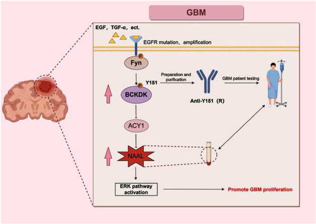
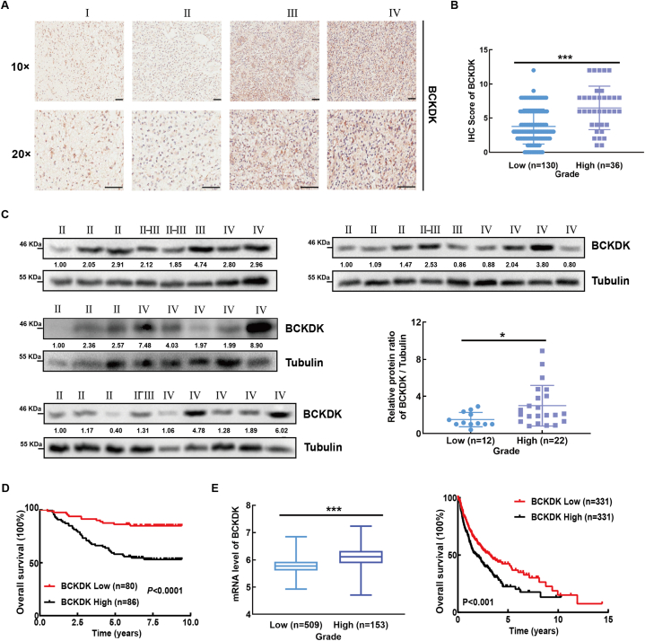
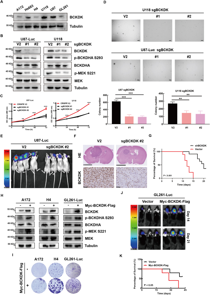
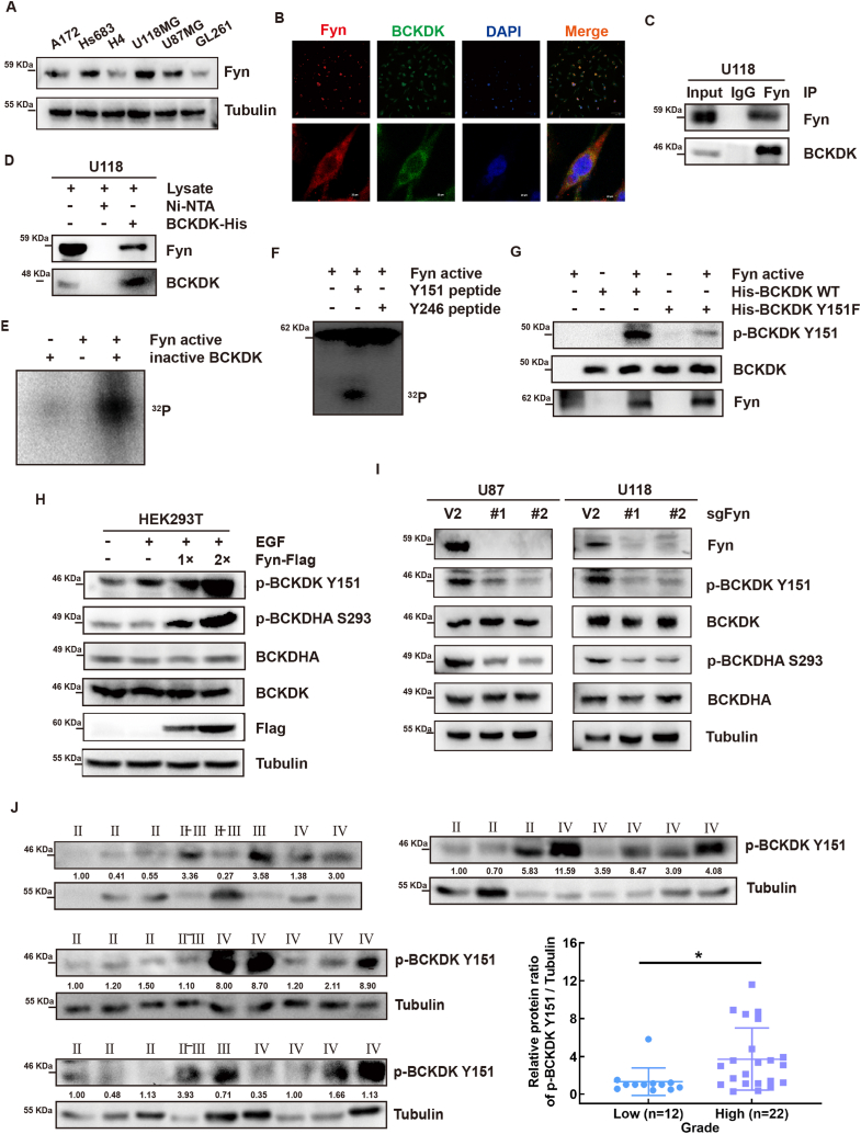
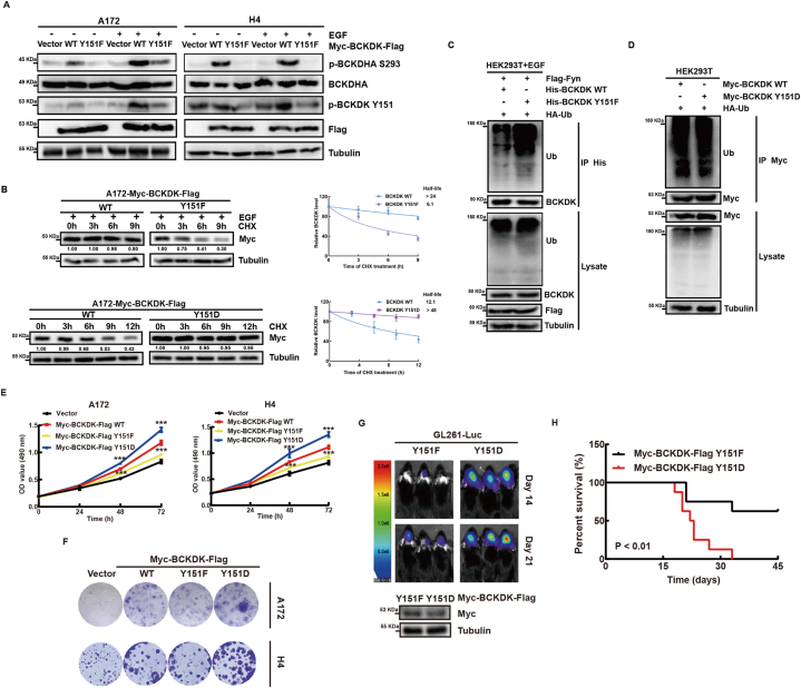
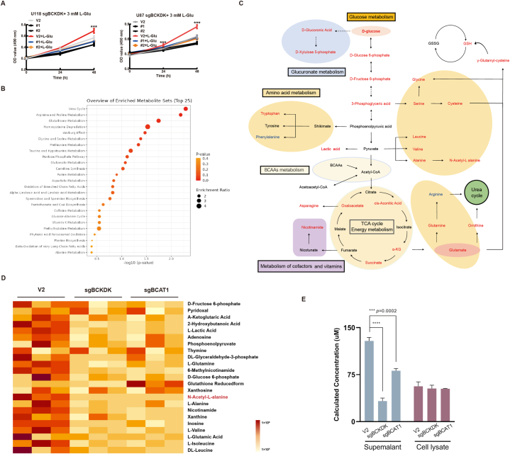
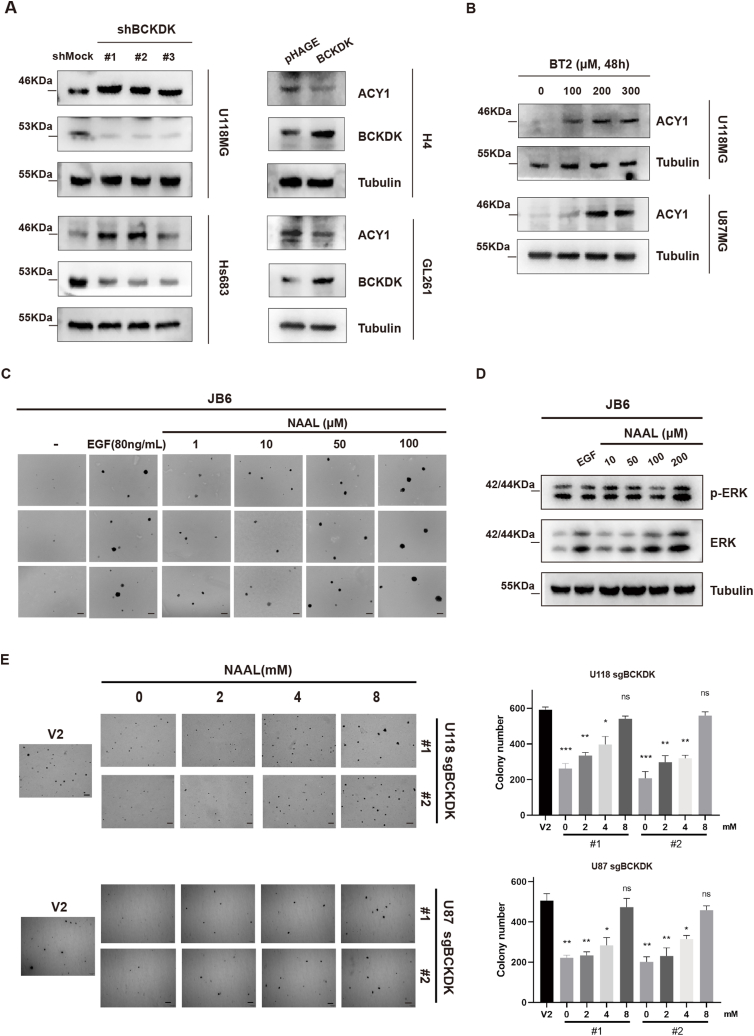
Branched chain α-keto acid dehydrogenase kinase (BCKDK) is a key enzyme involved in the metabolism of branched-chain amino acids (BCAAs). Its potential as a therapeutic target and prognostic factor for a variety of cancers has been widely reported. In this study, we investigated the expression of BCKDK in clinical glioma samples and found that BCKDK was significantly overexpressed in glioblastoma (GBM) and was associated with its poor prognosis. We further found that BCKDK is phosphorylated by tyrosine protein kinase Fyn at Y151, which increases its catalytic activity and stability, and demonstrate through in vivo and in vitro experiments that BCKDK phosphorylation promotes GBM cell proliferation. In addition, we found that the levels of the metabolite N-acetyl-L-alanine (NAAL) in GBM cells with high BCKDK were higher than those in the silencing group, and silencing or inhibition of BCKDK promotes the expression of ACY1, an enzyme that catalyzes the hydrolysis of NAAL into acetic acid and alanine. Exogenous addition of NAAL can activate the ERK signaling pathway and promote the proliferation of GBM cells. Taken together, we identified a novel mechanism of BCKDK activation and found NAAL is a novel oncogenic metabolite. Our study confirms the importance of the Fyn-BCKDK-ACY1-NAAL signalling axis in the development of GBM and suggests that p-BCKDK (Y151) and NAAL can serve as potential predictors of GBM progression and prognosis.

Keywords: ACY1; BCKDK; Fyn; Glioblastoma; N-acetyl-L-alanine.


Conflict of interest statement

The authors declare the following financial interests/personal relationships which may be considered as potential competing interests:Feng Zhu reports financial support was provided by National Natural Science Foundation of China. If there are other authors, they declare that they have no known competing financial interests or personal relationships that could have appeared to influence the work reported in this paper.

Figures

Image 1
Graphical abstract
Fig. 1
Fig. 1
BCKDK is highly expressed in GBM tissue and is positively correlated with poor prognosis of patients. A, Immunohistochemical (IHC) staining of BCKDK from 166 cases of glioma patients in the TMA. Representatives of IHC staining of BCKDK from different grades of glioma (upper panel) (10 × , 20 × , scale bar = 100 μm). B, The correlation between the protein level of BCKDK and the grades of glioma (down left panel). Kaplan-Meier analysis of OS. C, BCKDK protein level of 34 cases glioma patients were analyzed by WB. Statistical analysis on the correlation between protein level of BCKDK and grade (right down panel). The ratio values represent the levels normalized to BCKDK ratio Tubulin in each sample. D, The mRNA levels of BCKDK in different grades of glioma. E, Kaplan-Meier analysis of OS in glioma patients. The Data was obtained from TCGA database and analyzed by GraphPad Prism 7.0 software. (*p < 0.05, ***p < 0.001).
Fig. 2
Fig. 2
BCKDK promotes GBM proliferation ex vivo and in vivo. A, the protein levels of BCKDK in 6 glioma cell lines were analyzed by WB. B, the effect of BCKDK knockdown on the expression of its downstream signaling molecules was examined by WB. The data are representative pictures of three replicates. C, Growth curve of BCKDK knockdown cell lines were determined by MTT assay. The data are presented as the mean ± SD of three replications. D, Soft agar of BCKDK knockdown cell lines. Scale bar: 200 μm. The data are presented as the mean ± SD of three replications. E, Tumor size by in vivo bioluminescence in xenograft tumor-bearing BALB/c-nu mice with silenced BCKDK cell lines. F, The intracranial tumor sections of control group and experimental group of nude mice were stained with HE. G, the OS curves of BALB/c-nu mice with GBM xenografts are built according to the Kaplan-Meier methods. n = 8 (***p < 0.001). H, the effect of stable overexpression of BCKDK on the expression of its downstream signaling molecules was examined by WB. The data are representative pictures of three replicates. I, Colony formation assay of control and BCKDK stable cells. J, Tumor size of BCKDK stable overexpression cell lines in xenograft mice was detected using bioluminescence on day 14 and day 21. K, the OS curves of C57 mice with GBM xenografts are built according to the Kaplan-Meier methods. n = 8 (*p < 0.05).
Fig. 3
Fig. 3
Fyn binds with and phosphorylates BCKDK at Y151 site in vitro and enhances the activity of BCKDK ex vivo. A, the protein expression of Fyn in 6 glioma cell lines were analyzed by WB. B, Representative immunofluorescence for BCKDK (green) and Fyn (red) in U118 cells. Scale bar, 100 μm and 20 μm. C, In vitro pulldown assay was performed to detect Ni-NTA-His-BCKDK bound with endogenous Fyn in U118 cells. D, Endogenous BCKDK was co-immunoprecipitated with Fyn in U118 cells. E, Active Fyn phosphorylated inactive BCKDK in vitro in the presence of γ-32P-ATP as visualized by autoradiography. F, Active Fyn phosphorylated BCKDK Y151 peptides in vitro in the presence of γ-32P-ATP. G, BCKDK-WT protein and BCKDK-Y151F proteins as substrates for active Fyn in vitro were detected by WB using p-BCKDK (Y151) antibody. H, the phosphorylation and the activity of BCKDK was detected by WB with the p-BCKDK Y151 and p-BCKDHA S293 antibody in HEK293T cells transfected with Fyn-Flag plasmids under EGF (80 ng/ mL, 15 min) stimulation. I, the phosphorylation and the activity of BCKDK was detected by WB in cell silencing Fyn. The data are representative pictures of three replicates. J, p-BCKDK Y151 protein level of glioma tissues were analyzed by WB. Statistical analysis on the correlation between protein level of p-BCKDK Y151 and grade (*p < 0.05). . (For interpretation of the references to color in this figure legend, the reader is referred to the Web version of this article.)
Fig. 4
Fig. 4
Fyn phosphorylates BCKDK at the Y151 site to increase its activity and stability to promote glioma proliferation. A, WB of BCKDK-WT and BCKDK-Y151F stable A172 or H4 cells with or without EGF (80 ng/ mL) stimulation for 30 min. B, the levels of BCKDK were detected by WB in BCKDK-WT and BCKDK-Y151F stable A172 cells treated with CHX and EGF stimulation or in BCKDK-WT and BCKDK-Y151D stable A172 cells only treated with CHX. The data are presented as the mean ± SD of three replications. C and D, the ubiquitination of the BCKDK were analyzed by WB in HEK293T cells transfected with indicated plasmids. E, Growth curves of control and BCKDK stable cell lines by MTT assay. The data are presented as the mean ± SD of three replications. (**p < 0.01, ***p < 0.001). F, Colony formation assay of control and BCKDK stable cells. G, Live images of C57 mice with luciferase-labeled GL261 cells expressing BCKDK-Y151F or BCKDK-Y151D xenografts on day 14 and day 21 are shown by bioluminescence. H, Kaplan-Meier survival curve of C57 mice. n = 8 (**p < 0.01).
Fig. 5
Fig. 5
BCKDK promotes metabolic reprogramming in gliomas and metabolite NAAL production A, Growth curves of control cells and BCKDK-silenced cells treated with Glu by MTT assay. Data are expressed as the mean ± SD of three replicates. B, Classification and enrichment map of the differential metabolite KEGG after silencing BCKDK and BCAT1. C, The metabolite signaling network of sgBCKDK and sgBCAT1 cells co-varying is presented, with red font indicating a decrease in the level of the metabolite, blue indicating an increase, and black font indicating no metabolite detected. D, Heatmap showing control versus sgBCKDK and sgBCAT1 differential metabolite content. E, Statistical plots of NAAL levels in NAAL standards, U118 sgBCKDK, sgBCAT1 cells and culture supernatants determined by UPLC/ESI-Q TRAP-MS-MS. . (For interpretation of the references to color in this figure legend, the reader is referred to the Web version of this article.)
Fig. 6
Fig. 6
BCKDK inhibit ACY1 expression and NAAL is an oncogenic metabolite A, WB detection of ACY1 expression in silenced (right) or overexpressed (left) BCKDK cell lines; B, WB detection of ACY1 expression in U118 and U87 cells after treatment with different concentrations of BT2 for 48 h. C, Soft agar assay of JB6 cells treated EGF (80 ng/ mL) or different concentrations NAAL. The data are presented as the mean ± SD of three replications. Scale bar: 400 μm. D, WB assay for p-ERK levels in EGF (80 ng/ mL) and NAAL-stimulated JB6 Cl41 cells for 48 h; E, Soft agar observation of JB6 cells in response to NAAL stimulation, EGF (80 ng/mL) stimulation was used as a positive control, and image acquisition was performed on day 21 post-stimulation (scale bar: 400 μm). The data are presented as the mean ± SD of three replications. (*p < 0.05, **p < 0.01, ***p < 0.001).

Similar articles

Cited by

References

    1. Ohgaki H., Kleihues P. Population-based studies on incidence, survival rates, and genetic alterations in astrocytic and oligodendroglial gliomas. J. Neuropathol. Exp. Neurol. 2005;64(6):479–489. - PubMed
    1. Louis D.N., et al. The 2016 World Health Organization classification of tumors of the central nervous system: a summary. Acta Neuropathol. 2016;131(6):803–820. - PubMed
    1. Wen P.Y., Reardon D.A. Neuro-oncology in 2015: progress in glioma diagnosis, classification and treatment. Nat. Rev. Neurol. 2016;12(2):69–70. - PubMed
    1. Bush N.A., Chang S.M., Berger M.S. Current and future strategies for treatment of glioma. Neurosurg. Rev. 2017;40(1):1–14. - PubMed
    1. Ostrom Q.T., et al. The epidemiology of glioma in adults: a "state of the science" review. Neuro Oncol. 2014;16(7):896–913. - PMC - PubMed

LinkOut - more resources