

Radiologic Evaluation of the Distal End Radius Indices in Indian Paediatric Population
- PMID: 39170654
- PMCID: PMC11333421
- DOI: 10.1007/s43465-024-01209-0
Radiologic Evaluation of the Distal End Radius Indices in Indian Paediatric Population
Abstract
Background: The distal end radius's bony anatomy in relation to three variables-Radial Inclination, Volar tilt, and radial height-has been discussed commonly in the adult population and is not very well defined in the growing skeleton. In children aged 8-16 years old, we measured the osseous distal end radius according to radiography standards. The research comprised 130 patients, 65 males and 65 females aged 8-16. In each child, the norms for radial inclination, volar tilt, and radial epiphyseal height were established. This research defines these radiographic parameters for the paediatric population in India for the first time.
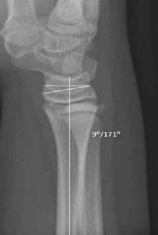
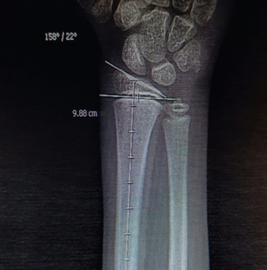
Methods: This research is an Unicentric Cross-sectional observational analytical study. We studied 130 normal wrist posteroanterior and lateral radiographs of the Indian paediatric population aged 8-16 years who reported to our OPD and calculated the three parameters-(1) Radial height, (2) Volar Tilt, and (3) Radial Inclination. Mean measurement values were analysed statistically.
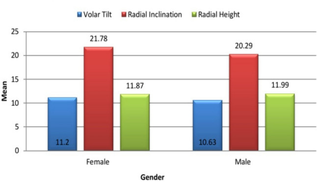
Results: The Mean distal end radius volar tilt is 10.92° ± 1.76° SD with a range from 4° to 15°. The Mean distal end radial inclination is 21.04° ± 2.10° SD with a range from 15° to 25°. The Mean distal end radial height is 11.93 ± 1.44 mm SD with a range from 9.0 to 14.50 mm.
Conclusion: In true Postero-anterior and lateral wrist radiographs of the Indian paediatric population, we have established normal values that may be utilized as a guide for the evaluation and treatment of a variety of traumatic and non-traumatic problems in Indian children.
Keywords: Distal radius; Epiphysis; Radial inclination; Volar tilt.
© Indian Orthopaedics Association 2024. Springer Nature or its licensor (e.g. a society or other partner) holds exclusive rights to this article under a publishing agreement with the author(s) or other rightsholder(s); author self-archiving of the accepted manuscript version of this article is solely governed by the terms of such publishing agreement and applicable law.
Conflict of interest statement
Conflict of InterestOn behalf of all authors, the corresponding author states that there is no conflict of interest.
Figures


References
-
- Luk, K. D., Saw, L. B., Grozman, S., Cheung, K. M., & Samartzis, D. (2014). Assessment of skeletal maturity in scoliosis patients to determine clinical management: A new classification scheme using distal radius and ulna radiographs. The Spine Journal,14(2), 315–325. 10.1016/j.spinee.2013.10.045 - DOI - PubMed
-
- Kirmani, S., Christen, D., Van Lenthe, G. H., Fischer, P. R., Bouxsein, M. L., McCready, L. K., Melton, L. J., III., Riggs, B. L., Amin, S., Müller, R., & Khosla, S. (2009). Bone structure at the distal radius during adolescent growth. Journal of Bone and Mineral Research,24(6), 1033–1042. 10.1359/jbmr.081255 - DOI - PMC - PubMed
-
- Caffey, J. (1956). Pediatric X-ray Diagnosis (3rd ed., p. 691). Chicago: The Year Book Publishers.
LinkOut - more resources
Full Text Sources