

Morphologic and Molecular Identification of Human Ocular Infection Caused by Pelecitus Nematodes, Thailand
- PMID: 39173658
- PMCID: PMC11346972
- DOI: 10.3201/eid3009.231692
Morphologic and Molecular Identification of Human Ocular Infection Caused by Pelecitus Nematodes, Thailand
Abstract
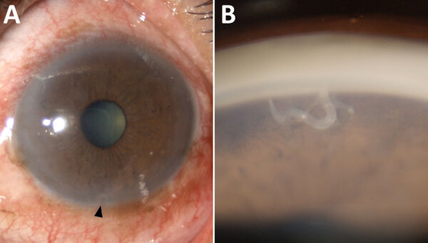
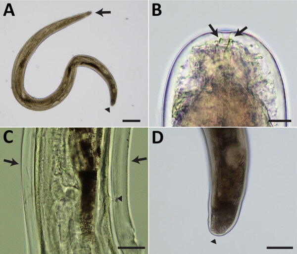
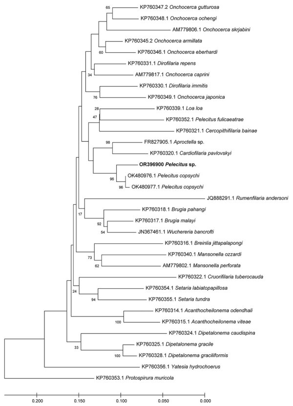
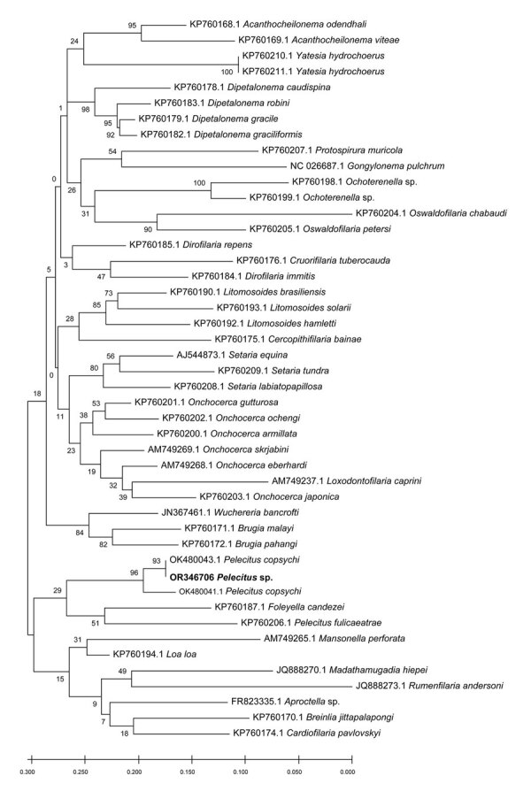
Nematodes of the Onchocercidae family, such as Pelecitus spp., are filarial parasites of medical and veterinary importance. Although infections are widely distributed among avian species, only 2 cases of human Pelecitus ocular infection, both in South America, have been reported. We describe a 61-year-old man in northeast Thailand diagnosed with an ocular infection. Morphologic characteristics suggested the causative agent was a female Pelecitus nematode: coiled body, rounded anterior and posterior extremities, a distinct preesophageal cuticular ring, lateral alae, a postdeirid, and a protuberant vulva. Sequences of the 12S rDNA gene indicated 95%-96% identity and cox1 gene 92%-96% identity with published P. copsychi sequences. P-distance for cox1 sequences between the causative agent and P. copsychi was 6.71%. Phylogenetic trees of 12S rDNA and cox1 genes indicated the species differed from but is closely associated with P. copsychi. Healthcare providers should be aware of the threat of ocular infection from Pelecitus spp. nematodes.
Keywords: Pelecitus; Thailand; arboviruses; filariae; helminths; nematodes; ocular parasitosis; parasites; postdeirid; preesophageal cuticular ring; zoonoses.
Figures
Similar articles
-
Description and molecular characterisation of Pelecitus copsychi Uni, Mat Udin & Martin n. sp. (Nematoda: Onchocercidae) from the white-rumped shama Copsychus malabaricus (Scopoli) (Passeriformes: Muscicapidae) of Pahang, Malaysia.Curr Res Parasitol Vector Borne Dis. 2022 Jan 30;2:100078. doi: 10.1016/j.crpvbd.2022.100078. eCollection 2022. Curr Res Parasitol Vector Borne Dis. 2022. PMID: 36589876 Free PMC article.
-
Preliminary study on buffy coat smear and molecular detection of microfilaria in domestic chickens (Gallus gallus domesticus) raised in Southern Thailand.Vet World. 2024 Apr;17(4):888-894. doi: 10.14202/vetworld.2024.888-894. Epub 2024 Apr 20. Vet World. 2024. PMID: 38798302 Free PMC article.
-
A new species of Pelecitus (Filarioidea: Onchocercidae) from the endangered Tehuantepec jackrabbit Lepus flavigularis.J Parasitol. 2004 Aug;90(4):803-7. doi: 10.1645/GE-213R1. J Parasitol. 2004. PMID: 15357073
-
Detection of DNA of filariae closely related to Mansonella perstans in faecal samples from wild non-human primates from Cameroon and Gabon.Parasit Vectors. 2020 Jun 16;13(1):313. doi: 10.1186/s13071-020-04184-1. Parasit Vectors. 2020. PMID: 32546281 Free PMC article.
-
Molecular epidemiology, phylogeny and evolution of the filarial nematode Wuchereria bancrofti.Infect Genet Evol. 2014 Dec;28:33-43. doi: 10.1016/j.meegid.2014.08.018. Epub 2014 Aug 29. Infect Genet Evol. 2014. PMID: 25176600 Free PMC article. Review.
Cited by
-
Insights into the genetic diversity and species distribution of Oswaldocruzia nematodes (Trichostrongylida: Molineidae) in Europe: apparent absence of geographic and population structuring in amphibians.Parasite. 2025;32:27. doi: 10.1051/parasite/2025020. Epub 2025 Apr 23. Parasite. 2025. PMID: 40273322 Free PMC article.
References
-
- Bartlett CM, Greiner EC. A revision of Pelecitus Railliet & Henry, 1910 (Filarioidea, Dirofilariinae) and evidence for the “capture” by mammals of filarioids from birds [in French]. Bull Mus Natl Hist Nat Sect A: Zool Biol Ecol Anim. 1986;8:47–99. 10.5962/p.287619 - DOI
-
- Uni S, Mat Udin AS, Tan PE, Rodrigues J, Martin C, Junker K, et al. Description and molecular characterisation of Pelecitus copsychi Uni, Mat Udin & Martin n. sp. (Nematoda: Onchocercidae) from the white-rumped shama Copsychus malabaricus (Scopoli) (Passeriformes: Muscicapidae) of Pahang, Malaysia. Curr Res Parasitol Vector Borne Dis. 2022;2:100078. 10.1016/j.crpvbd.2022.100078 - DOI - PMC - PubMed
Publication types
MeSH terms
Substances
LinkOut - more resources
Full Text Sources
Miscellaneous
