. 2024 Sep;300(9):107701.
doi: 10.1016/j.jbc.2024.107701. Epub 2024 Aug 22.

A heterocyclic compound inhibits viral release by inducing cell surface BST2/Tetherin/CD317/HM1.24

Affiliations

A heterocyclic compound inhibits viral release by inducing cell surface BST2/Tetherin/CD317/HM1.24

Perpetual Nyame et al. J Biol Chem. 2024 Sep.

Abstract

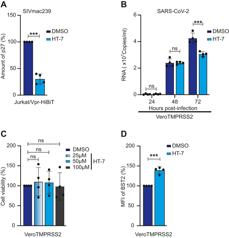
The introduction of combined antiretroviral therapy (cART) has greatly improved the quality of life of human immunodeficiency virus type 1 (HIV-1)-infected individuals. Nonetheless, the ever-present desire to seek out a full remedy for HIV-1 infections makes the discovery of novel antiviral medication compelling. Owing to this, a new late-stage inhibitor, Lenacapavir/Sunlenca, an HIV multi-phase suppressor, was clinically authorized in 2022. Besides unveiling cutting-edge antivirals inhibiting late-stage proteins or processes, newer therapeutics targeting host restriction factors hold promise for the curative care of HIV-1 infections. Notwithstanding, bone marrow stromal antigen 2 (BST2)/Tetherin/CD317/HM1.24, which entraps progeny virions is an appealing HIV-1 therapeutic candidate. In this study, a novel drug screening system was established, using the Jurkat/Vpr-HiBiT T cells, to identify drugs that could obstruct HIV-1 release; the candidate compounds were selected from the Ono Pharmaceutical compound library. Jurkat T cells expressing Vpr-HiBiT were infected with NL4-3, and the amount of virus release was quantified indirectly by the amount of Vpr-HiBiT incorporated into the progeny virions. Subsequently, the candidate compounds that suppressed viral release were used to synthesize the heterocyclic compound, HT-7, which reduces HIV-1 release with less cellular toxicity. Notably, HT-7 increased cell surface BST2 coupled with HIV-1 release reduction in Jurkat cells but not Jurkat/KO-BST2 cells. Seemingly, HT-7 impeded simian immunodeficiency virus (SIV) and severe acute respiratory syndrome coronavirus 2 (SARS-CoV-2) release. Concisely, these results suggest that the reduction in viral release, following HT-7 treatment, resulted from the modulation of cell surface expression of BST2 by HT-7.

Keywords: BST2; HIV-1; SARS-CoV-2; SIVmac239; Vpu; drug screening; tetherin.


Conflict of interest statement

Conflict of interest The authors declare that they have no conflict of interest with the contents of this article.

Figures

Figure 1
Figure 1
Vpr-HiBiT co-assembled with Gag into the virions.A, Jurkat cells expressing the indicated nanoluciferase (NLuc), Vpr-fused NLuc, Vpr-fused repetitive NLuc (x2, x3, x4), and Vpr-fused HiBiT were infected with VSV-G-pseudotyped-NL4-3/KFS. The NLuc activity in the cell and the cell supernatant was measured 3 days post-infection. The virus and the virus-free cells were fractionated from the cell supernatant. B, Jurkat cells were infected with VSV-G-pseudotyped-NL4-3/KFS. The amount of p24 was measured 3 days post-infection. C, 293T cells encoding the LTR-driven Vpr-fused HiBiT were transfected with pNL4-3/KFS. The NLuc activity in the cell and the cell supernatant was measured 2 days post-transfection. D, 293T cells were transfected with pNL4-3/KFS, and the amounts of p24 were measured 2 days post-transfection. E, Jurkat cells encoding the LTR-driven indicated luciferase (firefly: FLuc, renilla: RLuc) were infected with VSV-G-pseudotyped-NL4-3/KFS. The luciferase activities were measured 3 days post-infection. F, Jurkat/Vpr-HiBiT cells were infected with VSV-G-pseudotyped-NL4-3/KFS. The infected cells were treated with each 9 nM and 840 nM of the drugs (efavirenz, EFV; nevirapine, NVP) at 0 h and 15 h post-infection. The NLuc activities were measured 3 days post-infection. G, structure of PF-3450074. H, Jurkat/Vpr-HiBiT cells were infected with VSV-G-pseudotyped-NL4-3/KFS. About 15 h post-infection, the infected cells were treated with PF-3450074 at concentrations (100 μM, 50 μM, 25 μM, 12.5 μM, 6.3 μM, 3.2 μM, 1.6 μM, and 0.8 μM). The NLuc activities were measured 3 days post-infection. The error bars denote the means ± standard deviation of three independent experiments (n = 3). The One-way ANOVA by Sidak's multiple comparison test (B, C, and D) and the Two-way ANOVA using Tukey's multiple comparison test (A, E, and F) were applied to compare the data. The IC50 and CC50 values were analyzed by GraphPad Prism (Nonlinear regression) (G)∗, p < 0.01; ∗∗, p < 0.001; ∗∗∗, p < 0.0001; n.s., not significant.
Figure 2
Figure 2
Ten small compounds are candidates for virus release inhibition.AD, Jurkat/Vpr-HiBiT cells were infected with VSV-G-pseudotyped-NL4-3/KFS. The infected cells were treated with 5 μM of the compounds selected from the Ono Pharmaceutical compound library (drug discovery initiative of Tokyo University) at 15 h post-infection. The NLuc activity and the amount of p24 in the cell supernatants (AD) and the cells (D) were measured 3 days post-infection. B and C, The uninfected cells were treated with the compounds. Two days after compound treatment, the cell viability was examined by MTT assay.
Figure 3
Figure 3
Derivatives inhibit the virus release.AC, structures of the three candidates and nine derivative compounds. A BTD group is shown in the purple dot box. D and E, Jurkat/Vpr-HiBiT cells were infected with VSV-G-pseudotyped-NL4-3/KFS. The infected cells were treated with the indicated compounds at 15 h post-infection. The NLuc activity was measured 3 days post-infection. The uninfected cells were treated with the indicated compounds. Two days after compound treatment, the cell viability was examined by MTT assay. F, the Prism software determined EC50 and CC50.
Figure 4
Figure 4
Gag localization is affected by HT-7. Jurkat/Vpr-HiBiT cells were infected with VSV-G-pseudotyped-NL4-3/GagVenus. The infected cells were treated with 50 μM HT-7 at 15 h post-infection. A and B, the cells were analyzed by Flow cytometry. C, the cells were observed using confocal microscopy. Images were acquired at the mid-section and the top-section of the cells. Gray arrows indicate Gag accumulation in the polarized area. The scale bar indicates 10 μm. D, the cells with Gag localized predominantly to the PM (green), to the intracellular compartments (gray), or in the cytosol (black) were counted. About 100 cells that showed Gag signals were examined. E, the polarized (dark green) or nonpolarized (light green) cells with Gag localized to the PM were counted. Each cell was imaged from top to bottom and the number of cells with Gag accumulated on the PM was counted.
Figure 5
Figure 5
Cellular surface expression of PSGL-1 nor its interaction with HIV-1 Gag is not affected by HT-7.A, Jurkat cells were infected with VSV-G-pseudotyped-NL4-3/GagVenus for 15 h, after which infected cells were washed and then treated with 50 μM HT-7. The infected cells were incubated for 48 h, then cells were analyzed by flow cytometry. The error bars denote the means ± standard deviation of three independent experiments (n = 3). The One-way ANOVA by Sidak's multiple comparison test (B, C, and D) and the Two-way ANOVA using Tukey's multiple comparison test (A, E, and F) were applied to compare the data. ∗, p < 0.01; ∗∗, p < 0.001; ∗∗∗, p < 0.0001; n.s., not significant. B, representative diagram of HIV-1 Gag co-clustering with PSGL-1 in the presence or absence of 50 μM HT-7. Jurkat/Vpr-HiBiT cells were infected with or without VSV-G-pseudotyped-NL4-3/GagVenus, VSV-G-pseudotyped-Fyn (10)/fullMA/GagVenus and VSV-G-pseudotyped-Fyn (10)/6A2T/GagVenus for 15 h, and the cells were observed by confocal laser microscopy. The green color indicates Gag-YFP signals and the red color indicates PSGL-1 signals. The scale bar indicates 10 μm. C, each dot denotes single cells, n = 28. Error bars indicate the mean ± standard deviation. The correlation (r) between the MFI of PSGL-1 and HIV-1 Gag in control and treated conditions was calculated by Pearson’s correlation test. p values were determined by Two-way ANOVA using Tukey’s multiple comparison test. ∗, p < 0.01; ∗∗, p < 0.001; ∗∗∗, p < 0.0001; n.s., not significant. D, by employing Image J software, the MFI of HIV-1 GagVenus and PSGL-1 was determined. Each dot denotes an individual cell, n = 36. Error bars show the means ± standard deviation of repeated tests. The correlation (r) between the MFI of PSGL-1 and HIV-1 Gag in control and treated conditions was calculated by Pearson’s correlation test. ∗, p < 0.01; ∗∗, p < 0.001; ∗∗∗, p < 0.0001; n.s., not significant.
Figure 6
Figure 6
Knockout of BST2 canceled HT-7’s release inhibitory effect. The MFI of BST2 on Jurkat/Vpr-HiBiT cells (A) and infected Jurkat/Vpr-HiBiT cells (B) treated with 50 μM HT-7 was determined following analysis by flow cytometry. The NLuc activity (C) and the amount of p24 (D) in the supernatant from the infected Jurkat/Vpr-HiBiT cells were analyzed 3 days post-infection. Error bars show the mean ± standard deviation of repeated independent experiments. E, the BST2-positive cell population was analyzed by flow cytometry. (F) The sequences of BST2 in Jurkat/Vpr-HiBiT and Jurkat/Vpr-HiBiT/KO-BST2 cells were analyzed by Sanger sequencing. A guide RNA for BST2 knockout is designed as shown by the arrow. The inserted nucleotides are shown as red color. G, the MFI of BST2 on Jurkat/Vpr-HiBiT or Jurkat/Vpr-HiBiT/BST2-KO cells was determined following analysis by flow cytometry. H, the NLuc activity in the supernatant from the infected Jurkat/Vpr-HiBiT or Jurkat/Vpr-HiBiT/KO-BST2 cells was analyzed 3 days post-infection. (C, D, G, and H) The error bar illustrates the mean ± standard deviation of the repeated independent experiment. Direct comparisons between control and treated conditions were done by Two-way ANOVA using Tukey’s multiple comparison test. ∗, p < 0.01; ∗∗, p < 0.001; ∗∗∗, p < 0.0001; n.s., not significant.
Figure 7
Figure 7
HT-7 alleviates SIVmac239 and SARS-CoV-2 release.A, Jurkat Vpr-HiBiT cells were infected with VSV-G-pseudotyped SIVmac239ΔEnv for 15 h. Infected cells were treated with 50 μM HT-7. After 2 days of incubation, p27 amounts in the cell supernatant were analyzed. B, VeroE6/TMPRSS2 cells were infected with SARS-CoV-2 B.1.1 and were treated with 50 μM of HT-7. At 0, 24, 48, and 72 h post-infection, RNA copy in the cell supernatant was quantified by RT-qPCR. C, the uninfected VeroE6 TMPRSS2 cells were treated with HT-7 (25 μM, 50 μM, and 100 μM). Three days after HT-7 treatment, the cell viability was examined by MTT assay. D, the MFI of VeroE6 TMPRSS2 cells was analyzed by flow cytometry. Error bars show the mean ± standard deviations of four experiments. Comparisons of control and treated conditions were evaluated by the unpaired t test (A and D), Two-way ANOVA using Tukey’s multiple comparison test (B), and one-way ANOVA using Sidak’s multiple comparison test (C). ∗, p < 0.01; ∗∗, p < 0.001; ∗∗∗, p < 0.0001; n.s., not significant.

References

    1. Diseases G.B.D., Injuries C. Global burden of 369 diseases and injuries in 204 countries and territories, 1990-2019: a systematic analysis for the Global Burden of Disease Study 2019. Lancet. 2020;396:1204–1222. - PMC - PubMed
    1. Deeks S.G., Lewin S.R., Havlir D.V. The end of AIDS: HIV infection as a chronic disease. Lancet. 2013;382:1525–1533. - PMC - PubMed
    1. Pei X., Xiao J., Wei G., Zhang Y., Lin F., Xiong Z., et al. Oenothein B inhibits human non-small cell lung cancer A549cell proliferation by ROS-mediated PI3K/Akt/NF-kappaB signaling pathway. Chem. Biol. Interact. 2019;298:112–120. - PubMed
    1. Koh Y., Nakata H., Maeda K., Ogata H., Bilcer G., Devasamudram T., et al. Novel bis-tetrahydrofuranylurethane-containing nonpeptidic protease inhibitor (PI) UIC-94017 (TMC114) with potent activity against multi-PI-resistant human immunodeficiency virus in vitro. Antimicrob. Agents Chemother. 2003;47:3123–3129. - PMC - PubMed
    1. Lv Z., Chu Y., Wang Y. HIV protease inhibitors: a review of molecular selectivity and toxicity HIV. AIDS (Auckl) 2015;7:95–104. - PMC - PubMed

MeSH terms

LinkOut - more resources