Review
. 2024:203:39-58.
doi: 10.1016/B978-0-323-90820-7.00002-1.

Periodic paralysis

Affiliations
Review

Periodic paralysis

Stephen C Cannon. Handb Clin Neurol. 2024.

Abstract

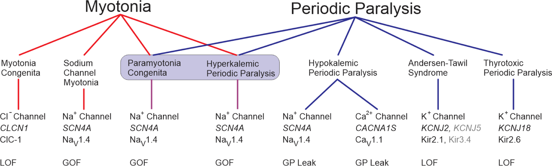
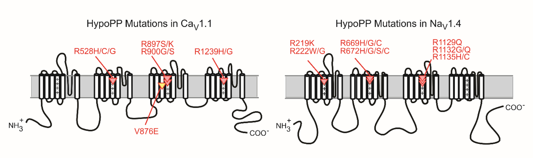
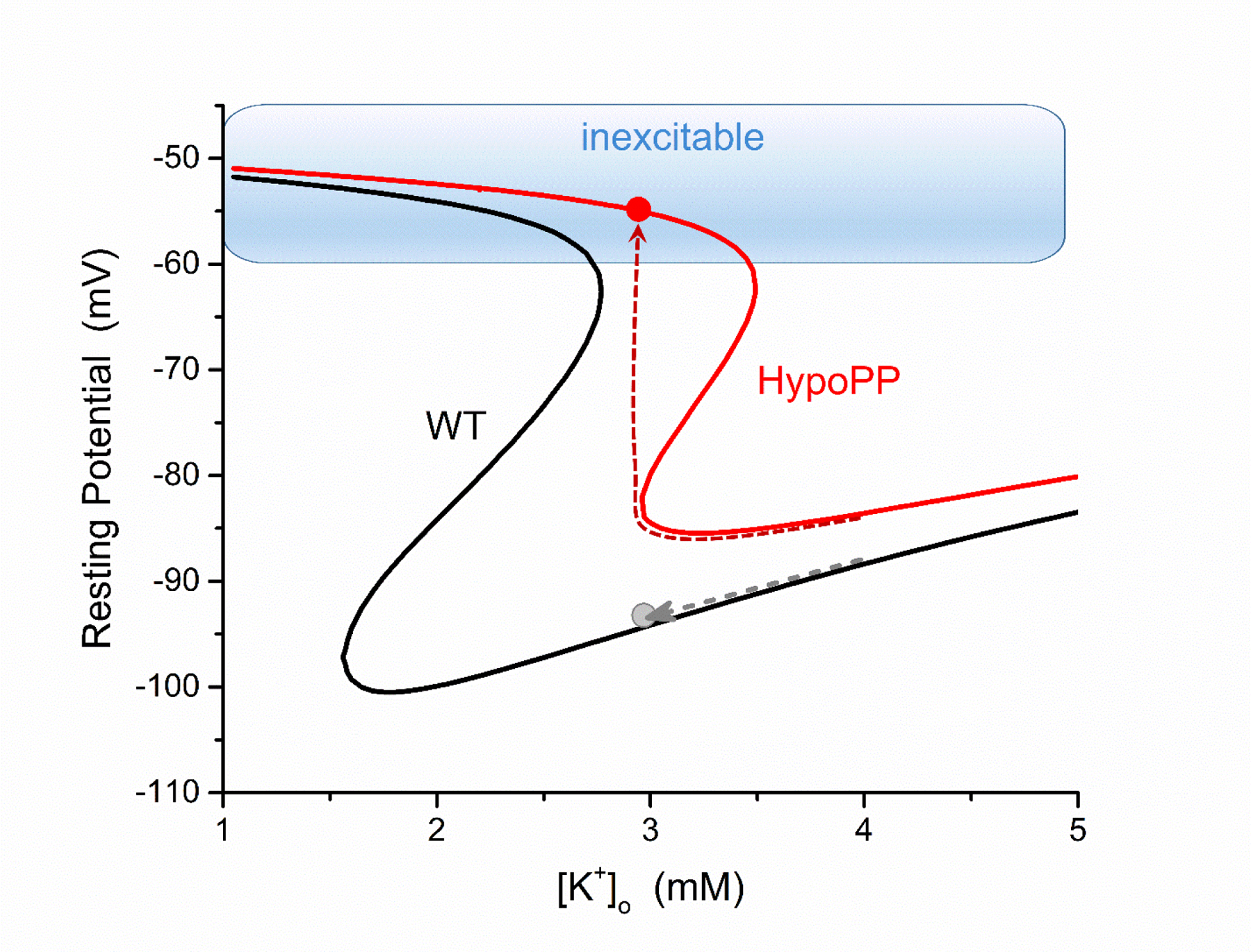
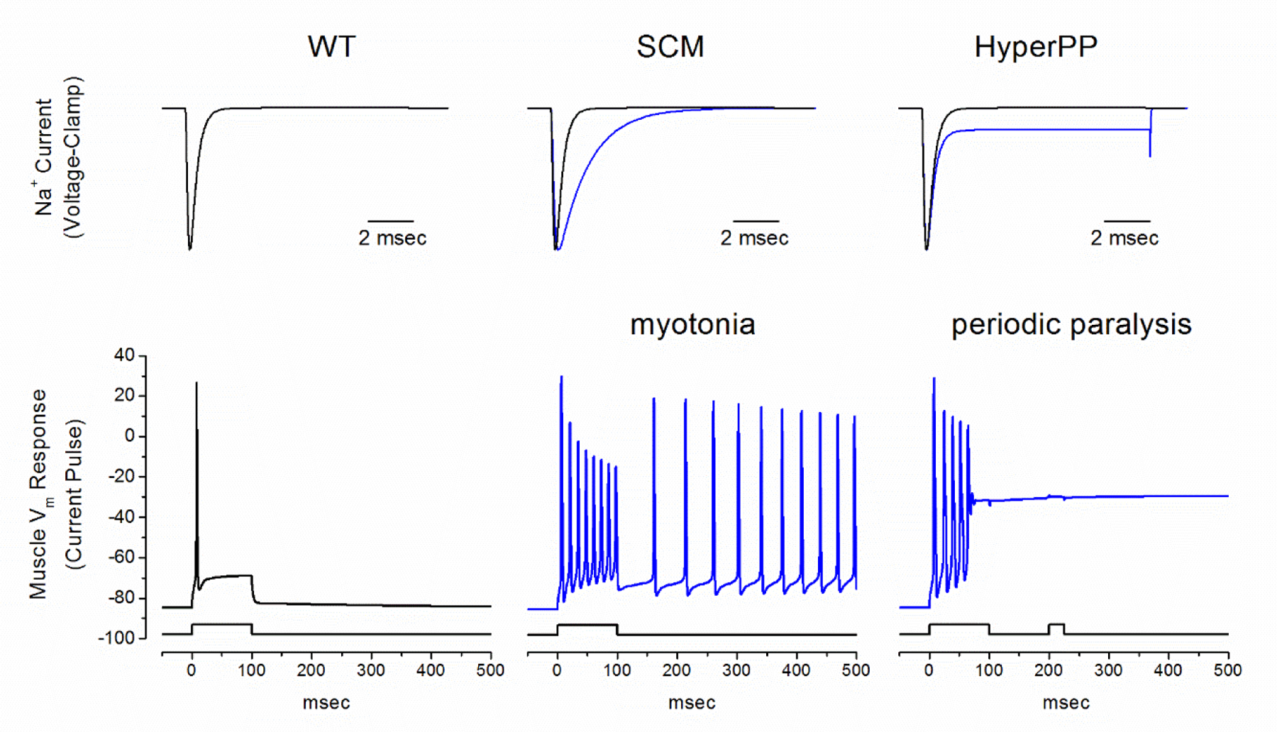
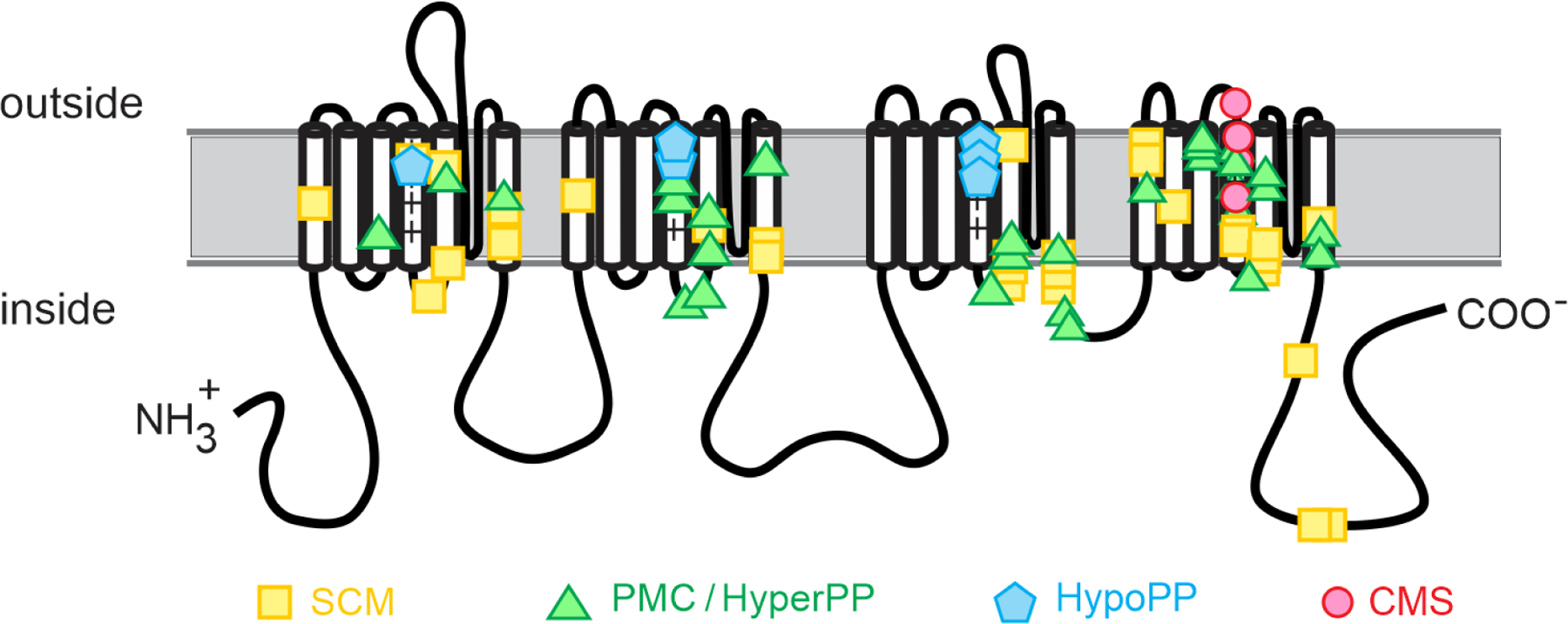
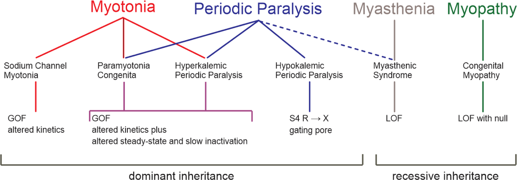
Periodic paralysis is a rare, dominantly inherited disorder of skeletal muscle in which episodic attacks of weakness are caused by a transient impairment of fiber excitability. Attacks of weakness are often elicited by characteristic environmental triggers, which were the basis for clinically delineating subtypes of periodic paralysis and are an important distinction for optimal disease management. All forms of familial periodic paralysis are caused by mutations of ion channels, often selectively expressed in skeletal muscle, that destabilize the resting potential. The missense mutations usually alter channel function through gain-of-function changes rather than producing a complete loss-of-function null. The knowledge of which channel gene harbors a variant, whether that variant is expected to (or known to) alter function, and how altered function impairs fiber excitability aides in the interpretation of patient signs and symptoms, the interpretation of gene test results, and how to optimize therapeutic intervention for symptom management and improve quality of life.

Keywords: CACNA1S; Calcium channel; Channelopathy; KCNJ2; Muscle; Myotonia; Potassium channel; SCN4A; Sodium channel.


Figures

Figure 1.
Figure 1.
Spectrum of diseases and associated gene defects for ion channelopathies of skeletal muscle. The clinical presentation ranges from myotonia (left) to periodic paralysis (right) with diseases in the center for which both myotonia and periodic paralysis may occur in the same patient. Paramyotonia congenita and hyperkalemic periodic paralysis (shaded box) are highly overlapping diseases for which features typical of either condition may occur for different affected members in a single family. The row below each disease indicates the channel name, gene name, and nomenclature for the channel protein subunit. KNCJ5 and Kir3.4 are listed in gray because this is an ultra-rare cause of Andersen-Tawil syndrome. Abbreviations: LOF loss-of-function, GOF gain-of-function, GP gating pore.
Figure 2.
Figure 2.
HypoPP mutations in CaV1.1 and NaV1.4. Schematic representations for the membrane-folding topology of the CaV1.1 (left) and NaV1.4 (right) channel subunits illustrate the clustering of HypoPP missense mutations in S4 segments of voltage sensor domains. The V876E mutation in CaV1.1 (yellow) is the only exception to the pattern of R → X missense substitution for these 25 HypoPP mutations. The “+” symbols emphasize the repeating pattern of positively charged amino acids (usually arginine = R, occasionally lysine = K) in S4 segments.
Figure 3.
Figure 3.
Model simulation of the muscle fiber resting potential as a function of extracellular [K+] for a wild type (WT, black) and a HypoPP (red) fiber. In simulated HypoPP fibers (either a CaV1.1 or NaV1.4 mutation), the gating pore leakage current causes a rightward shift of the VrestK+ relation. Starting from a normal [K+] of 4 mmol/L, both fibers are polarized and excitable. As the extracellular [K+] is reduced (dashed lines), the HypoPP fiber will paradoxically depolarize and from hypokalemia whereas a WT fiber hyperpolarizes. From a depolarized Vrest62 mV, the fiber is refractory from sodium channel inactivation and unable to generate an action potential. (Modified from (Cannon, 2018).
Figure 4.
Figure 4.
Model simulation of myotonia and periodic paralysis resulting from gain-of-function defects in NaV1.4. Top row shows simulated Na+ currents recorded under voltage clamp for WT (black) and mutant NaV1.4 currents (blue) in SCM (middle) and HyperPP/PMC (right). The simulated fiber response to current injection (bottom row) is normally a single action potential with full repolarization after termination of the stimulus (left, for WT). A slower rate of inactivation in SCM mutant channels (middle) enhances excitability and gives rise to a sustained burst of myotonic discharges that persist after termination of the stimulus. On the other hand, the response with a steady-state defect of inactivation in HyperPP mutant channels (right) also begins with a myotonic burst, but then settles to a sustained plateau depolarization. The fiber is refractory at this depolarized state, such that a second current stimulus fails to elicit an action potential (right). Because the mutant NaV1.4 channel has a steady-state gain-of-function defect, the fiber will remain depolarized and inexcitable, resulting in flaccid paralysis. Abbreviations: WT wild type, SCM sodium channel myotonia. (Reproduced from (Cannon, 2018).
Figure 5.
Figure 5.
Structure-function relationship for missense mutations of the skeletal muscle sodium channel, NaV1.4. The HypoPP mutations are all R→X substitutions in S4 transmembrane segments of voltage sensors in domains I – III, which produces the canonical gating pore leakage current. The homologous mutations in domain IV do not produce a gating pore current and instead alter channel inactivation to cause PMC/HyperPP. In general, the PMC/HyperPP mutations are in transmembrane segments of voltage-sensors or of the inner vestibule of the pore (cytoplasmic end of S5 or S6), or in the intracellular III-IV loop that forms the inactivation gate. These regions form the voltage sensors, gates, and receptors for gates that regulate conduction through the pore by activation and inactivation. The mutations associated with CMS are all in the S4 segment of the domain IV voltage sensor, which is tightly coupled to channel inactivation. The CMS mutations all produce loss-of-function changes (enhanced inactivation) for this recessively inherited phenotype. Abbreviations: SCM sodium channel myotonia, PMC paramyotonia congenita, HyperPP hyperkalemic periodic paralysis, HypoPP hypokalemic periodic paralysis, CMS congenital myasthenic syndrome.
Figure 6.
Figure 6.
Summary of the phenotypes, diseases, and class of channel defect for the NaV1.4 channelopathies of skeletal muscle. In general, GOF changes cause dominantly inherited phenotypes whereas the LOF changes are associated with recessive inheritance. Abbreviations: GOF gain-of-function, LOF loss-of-function, S4 fourth transmembrane segment of a voltage sensor domain, R→X missense substitution from arginine (R) to some other amino acid (X). (Modified from (Cannon, 2018).

References

    1. Adrian RH & Bryant SH (1974). On the repetitive discharge in myotonic muscle fibres. Journal of Physiology 240: 505–515. - PMC - PubMed
    1. Aickin CC, Betz WJ & Harris GL (1989). Intracellular chloride and the mechanism for its accumulation in rat lumbrical muscle. J Physiol 411: 437–455. - PMC - PubMed
    1. Arimura K, Arimura Y, Ng AR, et al. (2007). Muscle membrane excitability after exercise in thyrotoxic periodic paralysis and thyrotoxicosis without periodic paralysis. Muscle & nerve 36: 784–788. - PubMed
    1. Arnold WD, Feldman DH, Ramirez S, et al. (2015). Defective fast inactivation recovery of Nav 1.4 in congenital myasthenic syndrome. Ann Neurol 77: 840–850. - PMC - PubMed
    1. Bassi MT, Bresolin N, Tonelli A, et al. (2004). A novel mutation in the ATP1A2 gene causes alternating hemiplegia of childhood. J Med Genet 41: 621–628. - PMC - PubMed

MeSH terms

LinkOut - more resources