. 2024 Aug 22;15(1):6519.
doi: 10.1038/s41467-024-50747-6.

Cathepsin C inhibition reduces neutrophil serine protease activity and improves activated neutrophil-mediated disorders

Affiliations

Cathepsin C inhibition reduces neutrophil serine protease activity and improves activated neutrophil-mediated disorders

Yuka Nishibata et al. Nat Commun. .

Abstract

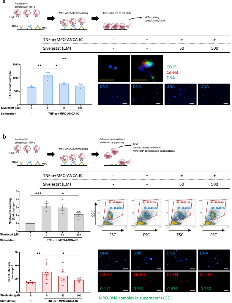
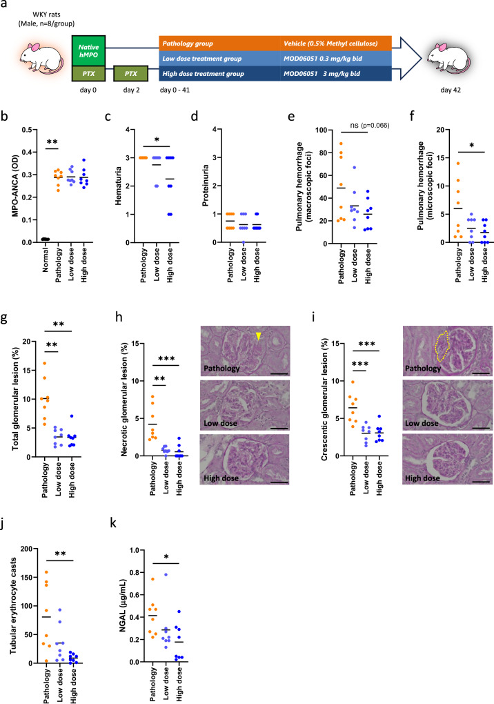
Cathepsin C (CatC) is an enzyme which regulates the maturation of neutrophil serine proteases (NSPs) essential for neutrophil activation. Activated neutrophils are key players in the innate immune system, and are also implicated in the etiology of various inflammatory diseases. This study aims to demonstrate a therapeutic potential for CatC inhibitors against disorders in which activated neutrophil-derived neutrophil extracellular traps (NETs) play a significant role. We demonstrate that a CatC inhibitor, MOD06051, dose-dependently suppresses the cellular activity of NSPs, including neutrophil elastase (NE), in vitro. Neutrophils derived from MOD06051-administered rats exhibit significantly lower NE activity and NET-forming ability than controls. Furthermore, MOD06051 dose-dependently ameliorates vasculitis and significantly decreases NETs when administered to a rat model of myeloperoxidase (MPO)-antineutrophil cytoplasmic antibody-associated vasculitis (AAV). These findings suggest that CatC inhibition is a promising strategy to reduce neutrophil activation and improve activated neutrophil-mediated diseases such as MPO-AAV.


Conflict of interest statement

T.S., W.S., T.N., and Y.T. are employees of Alivexis, Inc. Y.N., S.M., and A.I. were given research funds from Alivexis, Inc. The remaining authors declare no competing interests.

Figures

Fig. 1
Fig. 1. Inhibition of cellular NSP activity by MOD06051.
a Human CD34+ Bone Marrow Stem/Progenitor Cells were differentiated into neutrophils in the presence of 0–10 µM MOD06051. NE activity in in vitro-differentiated neutrophils was determined. NE activity was inhibited by MOD06051 with an IC50 value of 18 nM, which was calculated based on repeated biological experiments (N1 and N2). Each experiment was conducted in duplicate. To estimate all NE activity in samples, an excess dose (200 µM) of sivelestat (sv) was used. b Normal rats were administered with MOD06051 (0, 0.3, 1, 3, and 10 mg/kg) orally bid daily (n = 5/group). Two weeks later, NE activity in PMNs derived from the bone marrow was assessed. Results showed an ED50 dose around 0.3 mg/kg bid and that the administration of 3 mg/kg bid reached the trough level. To estimate all specific NE activity in samples, the maximum dose (10 mg/kg) of AZD7986 was used for reference. Values are displayed as the mean with standard error of the mean (SEM), and p-values were calculated using One-way ANOVA followed by Dunnett’s analysis. c Normal rats were administered with MOD06051 (0, 0.3, and 3 mg/kg) orally (n = 5/group). Two hours later, CatC activity in whole blood was assessed using a membrane-permeable CatC-specific fluorescent substrate. Values are displayed as the mean with SEM and p-values were calculated using One-way ANOVA followed by Dunnett’s analysis. d Human CD34+ Bone Marrow Stem/Progenitor Cells were differentiated into neutrophils in the presence of 0–10 µM MOD06051. PR3 activity in in vitro-differentiated neutrophils was determined. PR3 activity was inhibited by MOD06051 with an IC50 value of 29 nM, which was calculated based on repeated biological experiments (N1 and N2). Each experiment was conducted in duplicate. To estimate all NE activity in samples, an excess dose (200 µM) of sivelestat (sv) was used. Cell surface expression of PR3 on in vitro-differentiated neutrophils treated with and without MOD06051 was assessed by FCM. Representative histograms (e) and dose-dependent shift of mean fluorescence intensity (MFI; f) based on four independent experiments are shown. *p < 0.05, **p < 0.01, ****p < 0.0001. Source data are provided as a Source Data file.
Fig. 2
Fig. 2. Reduction in MPO-ANCA-IC-induced NET formation in neutrophils derived from MOD06051-administered normal rats.
Normal rats were administered orally bid with MOD06051 (3 mg/kg) or vehicle (0.5% methylcellulose) daily (n = 3/group). Two weeks later, neutrophils were separated from the blood using Polymorphprep and treated with 100 ng/ml rat TNF-α or 100 ng/ml mouse C5a for 15 min at 37 °C. Thereafter, these cells were incubated in MPO-ANCA-IC-immobilized 4-well slide chambers for 3 h at 37 °C. Cells were collected by pipetting in chambers and resuspended in EDTA-PBS were allowed to react with Sytox Green, and then submitted for FCM to detect Sytox Green+ NET-forming neutrophils. a The representative FCM profiles that reproduced in each group. b Comparison of NET-forming rate in TNF-α-primed neutrophils and C5a-primed neutrophils between MOD06051-administered rats and vehicle controls. Values are displayed as the mean with SEM, and p-values were calculated using a one-sided Student’s t test. c Neutrophils isolated from a non-treated normal rat were stimulated and harvested similarly and then subjected to fluorescent immunostaining for Cit-H3 and FCM using Sytox Green. Moreover, MPO-DNA complexes in supernatants were quantified by ELISA. *p < 0.05, **p < 0.01. Bar, 50 µm. Source data are provided as a Source Data file.
Fig. 3
Fig. 3. Suppression of NET formation of TNF-α-primed neutrophils by pharmaceutical inhibition of NE.
Neutrophils were separated from the peripheral blood of healthy volunteers (n = 3) using Polymorphprep and treated with sivelestat (0, 50, and 500 µM) for 15 min, followed by 5 ng/ml TNF-α (a and b) for 15 min at 37 °C. Thereafter, these cells were incubated in MPO-ANCA-IC-immobilized 4-well slide chambers for 3–4 h at 37 °C. After the removal of supernatants and chambers, cells were fixed on the slide by paraformaldehyde and then stained for CD15 (neutrophil marker), Cit-H3 (NETs marker), and DNA (using DAPI) (a). Three microphotographs were taken in a randomized manner per well, and the DAPI-positive DNA area per neutrophil was calculated using ImageJ 1.50i. Alternatively, cells were collected by pipetting in chambers and resuspended in EDTA-PBS, and then analyzed by FCM to examine FSC/SSC profiles (b). In this analysis, swollen neutrophils [R1 gate (whole neutrophils) - R5 gate (normal size neutrophils)] were focused on. Furthermore, the cells were subjected to Cit-H3 immunofluorescent staining, and ELISA was used to quantify MPO-DNA complexes in supernatants. Values are displayed as the mean with SEM, and p-values were calculated using One-way ANOVA followed by Dunnett’s analysis for (a)and (b). Yellow bar, 25 µm; White bar, 50 µm. *p < 0.05, **p < 0.01, ***p < 0.001. Source data are provided as a Source Data file.
Fig. 4
Fig. 4. Suppression of NET formation of C5a-primed neutrophils by pharmaceutical inhibition of NE.
Neutrophils were separated from the peripheral blood of healthy volunteers (n = 3) using Polymorphprep and treated with sivelestat (0, 50, and 500 µM) for 15 min, followed by 100 ng/ml C5a (a and b) for 15 min at 37 °C. Thereafter, these cells were incubated in MPO-ANCA-IC-immobilized 4-well slide chambers for 3–4 h at 37 °C. After the removal of supernatants and chambers, cells were fixed on the slide by paraformaldehyde and then stained for CD15 (neutrophil marker), Cit-H3 (NETs marker), and DNA (using DAPI) (a). Three microphotographs were taken in a randomized manner per well, and the DAPI-positive DNA area per neutrophil was calculated using ImageJ 1.50i. Alternatively, cells were collected by pipetting in chambers and resuspended in EDTA-PBS, and then analyzed by FCM to examine FSC/SSC profiles (b). In this analysis, swollen neutrophils [R1 gate (whole neutrophils) - R5 gate (normal size neutrophils)] were focused on. Furthermore, the cells were subjected to Cit-H3 immunofluorescent staining, and ELISA was used to quantify MPO-DNA complexes in supernatants. Values are displayed as the mean with SEM, and p-values were calculated using One-way ANOVA followed by Dunnett’s analysis for (a) and (b). Yellow bar, 25 µm; White bar, 50 µm. *p < 0.05, **p < 0.01, ***p < 0.001, ****p < 0.0001. Source data are provided as a Source Data file.
Fig. 5
Fig. 5. Effects of MOD06051 on MPO-AAV rats.
a MPO-AAV rats were divided into three groups (n = 8/group), and MOD06051 (0.3 or 3 mg/kg) or vehicle (0.5% methylcellulose) was administered orally bid daily for 42 days. b Serum MPO-ANCA titer at day 42 was determined by ELISA. Normal rats (n = 8) were used as controls. Urine was collected on day 40, and hematuria (c) and proteinuria (d) were detected using dipsticks. Macroscopic foci of pulmonary hemorrhage (e) on the surface of the lungs were counted at necropsy. Microscopic foci of pulmonary hemorrhage (f) with clear regional bleeding were counted in the maximal section of the lung under a low-power field of view. Glomeruli exhibiting NCGN were counted, and the glomerular lesion rate was calculated (g): total glomerular lesion, (h): necrotic glomerular lesion, and (i): crescentic glomerular lesion). The arrowhead in (h) (periodic acid-Schiff (PAS) staining) indicates a necrotic lesion, and the dotted line in (i) (PAS staining) indicates a crescent. Tubular erythrocyte casts in the renal cortex (j), which represented glomerular bleeding, were also counted. Urinary NGAL (k) was measured with the ELISA kit on day 40. Values are displayed as the mean with SEM. P-values were calculated using One-way ANOVA followed by Dunnett’s analysis for (e, f, i, and j)and Kruskal-Wallis followed by Dunn’s analysis for (b–d, g, h, and k). Pathology: vehicle control; Low dose: 0.3 mg/kg, bid; High dose: 3 mg/kg, bid. *p < 0.05, **p < 0.01, ***p < 0.001. Bar, 50 µm. Source data are provided as a Source Data file.
Fig. 6
Fig. 6. Suppression of NET formation by MOD06051 in MPO-AAV rats.
a NET-forming neutrophils in the blood at day 42 were detected by FCM using Sytox Green (n = 8/group). Normal rats (n = 8) were used as controls. b NET-forming neutrophils were detected as MPO+ Cit-H3+ DAPI+ substances (arrowhead) in glomeruli of MPO-AAV rats. c The rate of NET-forming neutrophils (arrowhead) in neutrophils infiltrating into glomeruli (%) was calculated. d The number of MPO+ neutrophils (arrowheads) infiltrated into 10 glomeruli were counted, and neutrophils per glomerulus (dotted circle) were calculated. e The number of CD68+ macrophages infiltrated into and around glomeruli were counted. Arrowheads indicate macrophages infiltrated into the glomerulus. Extraglomerular macrophage infiltration was scored as follows; 0: none, 1: 1–10, 2: 11–25, 3: 26–50, 4: > 50 macrophages infiltrated around the glomerulus. Values are displayed as the mean with SEM. P-values were calculated using One-way ANOVA followed by Dunnett’s analysis for (c) and Kruskal-Wallis followed by Dunn’s analysis for (a, d, and e). *p < 0.05, **p < 0.01, ***p < 0.001, ****p < 0.0001. Bar, 50 µm. Source data are provided as a Source Data file.

Similar articles

Cited by

References

    1. Nemeth, T., Sperandio, M. & Mocsai, A. Neutrophils as emerging therapeutic targets. Nat. Rev. Drug Discov.19, 253–275 (2020). 10.1038/s41573-019-0054-z - DOI - PubMed
    1. Pham, C. T. Neutrophil serine proteases fine-tune the inflammatory response. Int. J. Biochem. Cell Biol.40, 1317–1333 (2008). 10.1016/j.biocel.2007.11.008 - DOI - PMC - PubMed
    1. Korkmaz, B. et al. Therapeutic targeting of cathepsin C: from pathophysiology to treatment. Pharm. Ther.190, 202–236 (2018).10.1016/j.pharmthera.2018.05.011 - DOI - PubMed
    1. Mutua, V. & Gershwin, L. J. A review of neutrophil extracellular traps (NETs) in disease: potential anti-NETs therapeutics. Clin. Rev. Allergy Immunol.61, 194–211 (2021). 10.1007/s12016-020-08804-7 - DOI - PMC - PubMed
    1. Brinkmann, V. et al. Neutrophil extracellular traps kill bacteria. Science303, 1532–1535 (2004). 10.1126/science.1092385 - DOI - PubMed

MeSH terms

LinkOut - more resources