. 2024 Aug 22;14(1):19507.
doi: 10.1038/s41598-024-70220-0.

Hedgehog components are overexpressed in a series of liver cancer cases

Affiliations

Hedgehog components are overexpressed in a series of liver cancer cases

Caroline Brandi Schlaepfer Sales et al. Sci Rep. .

Abstract

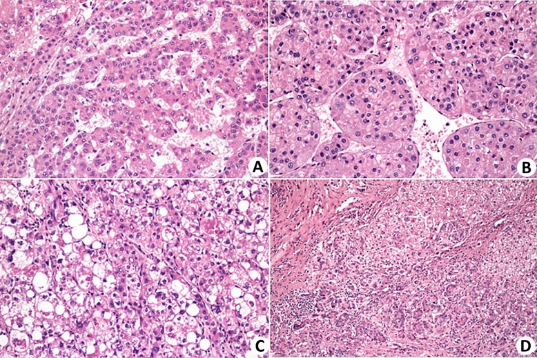
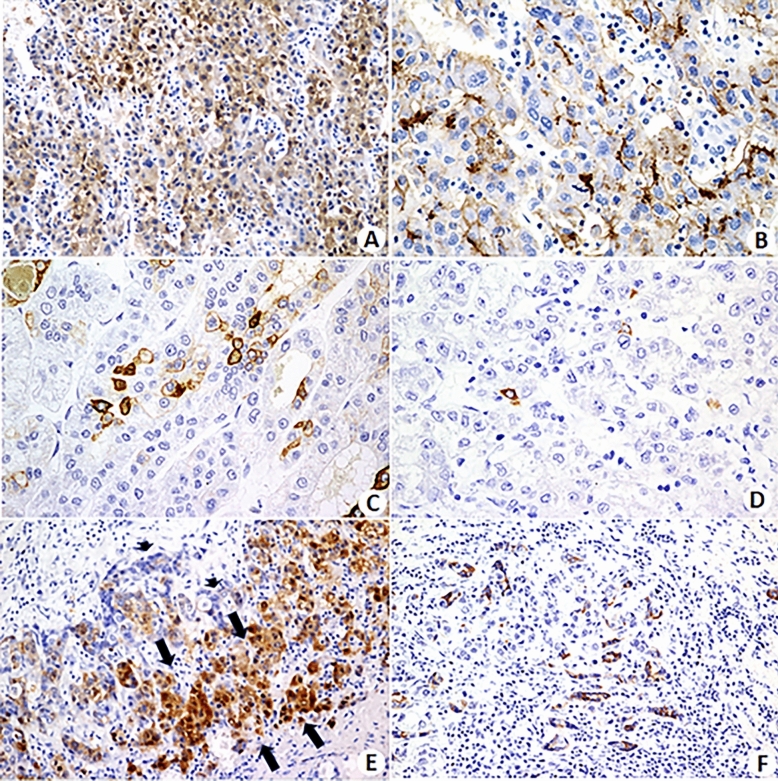
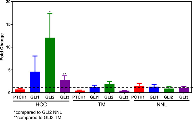
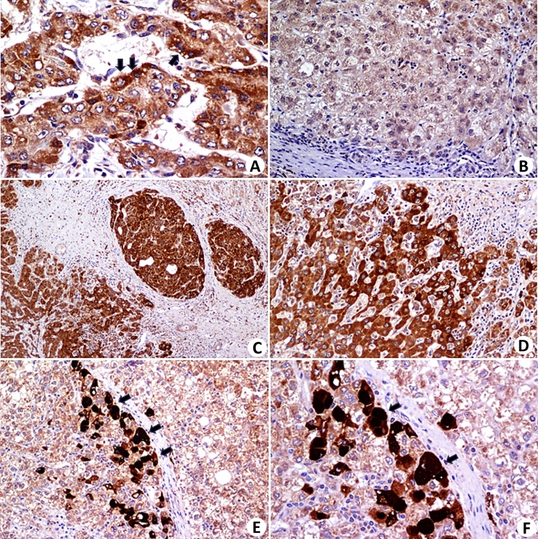
Liver cancers, including hepatocellular carcinoma (HCC), are the sixth most common cancer and the third leading cause of cancer-related death worldwide, representing a global public health problem. This study evaluated nine patients with HCC. Six of the cases involved hepatic explants, and three involved hepatic segmentectomy for tumor resection. Eight out of nine tumors were HCC, with one being a combined hepatocellular-cholangiocarcinoma tumor. Conventional markers of hepatocellular differentiation (Hep Par-1, arginase, pCEA, and glutamine synthetase) were positive in all patients, while markers of hepatic precursor cells (CK19, CK7, EpCAM, and CD56) were negative in most patients, and when positive, they were detected in small, isolated foci. Based on in silico analysis of HCC tumors from The Cancer Genome Atlas database, we found that Hedgehog (HH) pathway components (GLI1, GLI2, GLI3 and GAS1) have high connectivity values (module membership > 0.7) and are strongly correlated with each other and with other genes in biologically relevant modules for HCC. We further validated this finding by analyzing the gene expression of HH components (PTCH1, GLI1, GLI2 and GLI3) in our samples through qPCR, as well as by immunohistochemical analysis. Additionally, we conducted a chemosensitivity analysis using primary HCC cultures treated with a panel of 18 drugs that affect the HH pathway and/or HCC. Most HCC samples were sensitive to sunitinib. Our results offer a comprehensive view of the molecular landscape of HCC, highlighting the significance of the HH pathway and providing insight into focused treatments for HCC.

Keywords: Chemotherapy; Hedgehog proteins; Hepatocarcinoma; Molecular biology.


Conflict of interest statement

The authors declare no competing interests.

Figures

Figure 1
Figure 1
Histopathological aspects of HCC. (A) Classic trabecular pattern of HCC, histological grade II, composed of cells with moderate atypia, arranged in thin trabeculae (H&E ×100). (B) HCC, macrotrabecular subtype. The trabeculae are thick, with more than 10 cells (H&E, ×200). (C) Steatohepatitic subtype of HCC. The neoplastic cells exhibited steatosis, ballooning, and Mallory-Denk bodies (H&E, ×100). (D) Combined HCC-CCA. Biphenotypic neoplasia with areas of hepatocellular differentiation alongside areas of cholangiocellular differentiation (H&E ×100).
Figure 2
Figure 2
Immunohistochemical staining. (A) Immunohistochemistry for arginase shows positive staining in neoplastic hepatocytes. (B) Immunohistochemical reaction is positive for pCEA, depicting bile canaliculi formed by neoplastic hepatocytes. (C) Cytokeratin 7 is densely positive in small cells with morphological characteristics of hepatic precursor cells and "blushed" in intermediate type hepatocytes. (D) A few isolated CK19 positive cells with morphological characteristics of hepatic precursor cells. (E) Zone of transition between a hepatocellular carcinoma (HCC) and intrahepatic cholangiocarcinoma (iCCA) in the case of combined HCC-iCCA. Neoplastic hepatocytes are positive for Hep-Par, a marker of hepatocytes (arrows), whereas the cholangiolar component of the tumor is negative. (F) Immunostaining for CK7 shows positivity in the biliary component of a combined HCC-CCA tumor. (Magnifications—A: ×100; B: ×400; C: ×200; D: ×200, E: ×100, and F: ×40).
Figure 3
Figure 3
Fold changes in PTCH1, GLI1, GLI2 and GLI3 expression in patients with HCC, including tumors, tumor lateral margin (TM) at the interface with the nonneoplastic liver, and distant nonneoplastic liver tissue (NNL) far from the tumor. The data are shown as the means ± S.E.M.s of nine patients.
Figure 4
Figure 4
Immunohistochemistry with an anti-GLI1 monoclonal antibody. (A,C,D,E, and F) show the protein expression in HCC cells from three patients. The intensity of the expression was heterogeneous, with very dense areas in some cells (Figures E and F). The expression appears to be more intense in the interface zone of the neoplasm with the stroma in (A,E, and F) (arrows). Figure (B) shows a nonneoplastic hepatic parenchymal nodule without protein expression or with very weak expression (Magnifications—A: ×400; B: ×100; C: ×40; D: ×100, E: ×40, and F: ×200).

References

    1. Bray, F. et al. Global cancer statistics 2022: GLOBOCAN estimates of incidence and mortality worldwide for 36 cancers in 185 countries. CA Cancer J. Clin.74(3), 229–263. 10.3322/caac.21834 (2024). 10.3322/caac.21834 - DOI - PubMed
    1. Bidkhori, G. et al. Metabolic network-based stratification of hepatocellular carcinoma reveals three distinct tumor subtypes. Proc. Natl. Acad. Sci. USA115(50), E11874–E11883. 10.1073/pnas.1807305115 (2018). 10.1073/pnas.1807305115 - DOI - PMC - PubMed
    1. El Jabbour, T., Lagana, S. M. & Lee, H. Update on hepatocellular carcinoma: Pathologists’ review. World J. Gastroenterol.25(14), 1653–1665. 10.3748/wjg.v25.i14.1653 (2019). 10.3748/wjg.v25.i14.1653 - DOI - PMC - PubMed
    1. Bruix, J., Reig, M. & Sherman, M. Evidence-based diagnosis, staging, and treatment of patients with hepatocellular carcinoma. Gastroenterology.150(4), 835–853. 10.1053/j.gastro.2015.12.041 (2016). 10.1053/j.gastro.2015.12.041 - DOI - PubMed
    1. Yang, J. D. et al. A global view of hepatocellular carcinoma: Trends, risk, prevention and management. Nat. Rev. Gastroenterol. Hepatol.16(10), 589–604. 10.1038/s41575-019-0186-y (2019). 10.1038/s41575-019-0186-y - DOI - PMC - PubMed

MeSH terms