

Prediction of midpalatal suture maturation stage based on transfer learning and enhanced vision transformer
- PMID: 39174951
- PMCID: PMC11340164
- DOI: 10.1186/s12911-024-02598-w
Prediction of midpalatal suture maturation stage based on transfer learning and enhanced vision transformer
Abstract
Background: Maxillary expansion is an important treatment method for maxillary transverse hypoplasia. Different methods of maxillary expansion should be carried out depending on the midpalatal suture maturation levels, and the diagnosis was validated by palatal plane cone beam computed tomography (CBCT) images by orthodontists, while such a method suffered from low efficiency and strong subjectivity. This study develops and evaluates an enhanced vision transformer (ViT) to automatically classify CBCT images of midpalatal sutures with different maturation stages.
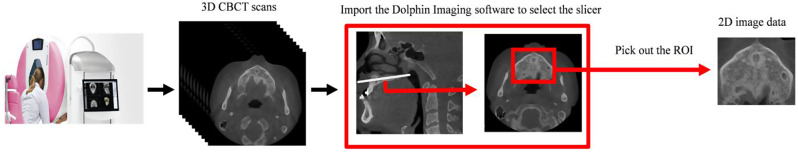
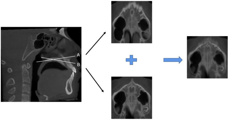
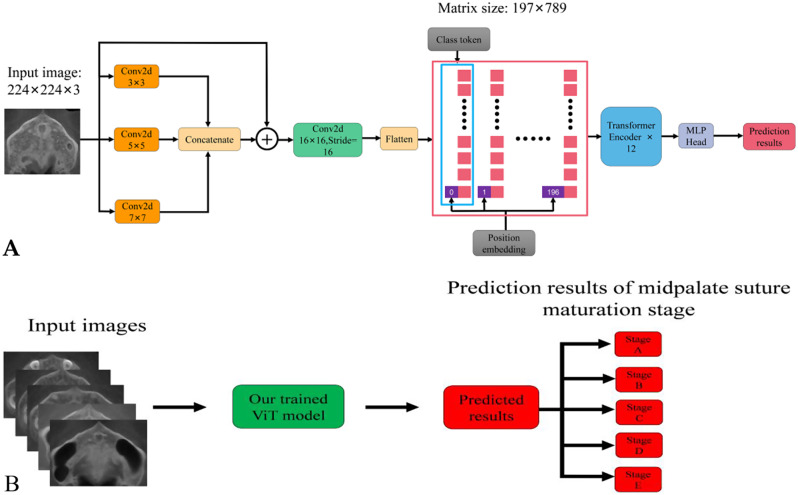
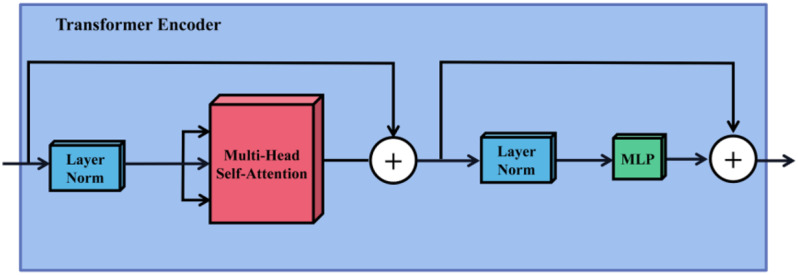
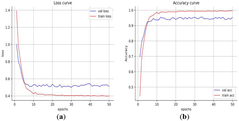
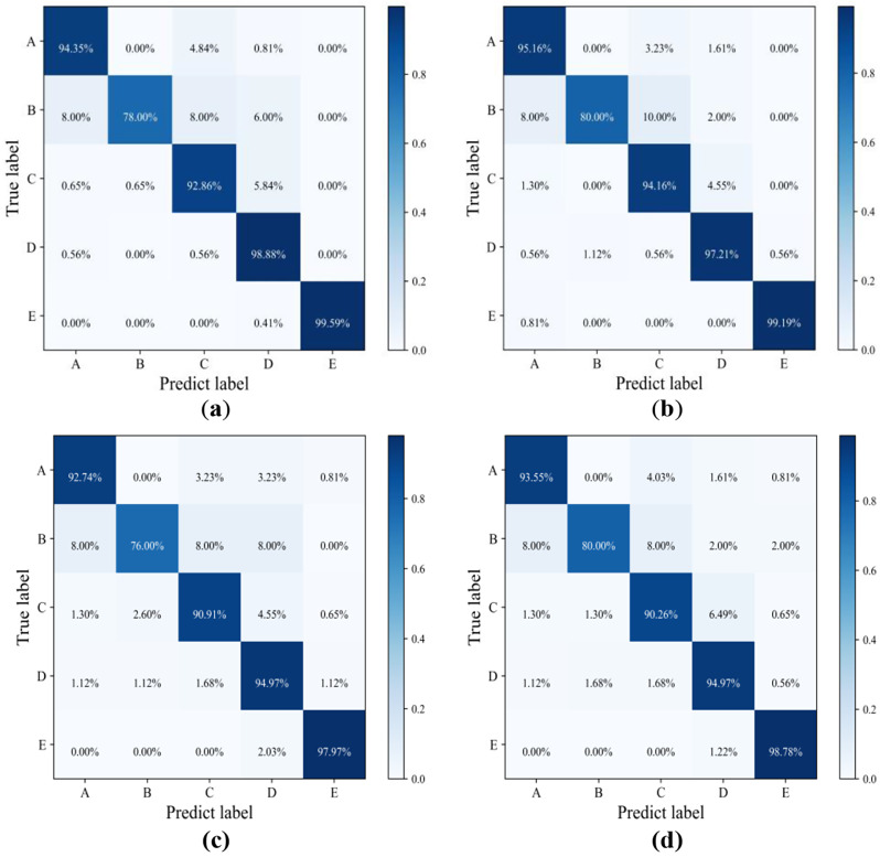
Methods: In recent years, the use of convolutional neural network (CNN) to classify images of midpalatal suture with different maturation stages has brought positive significance to the decision of the clinical maxillary expansion method. However, CNN cannot adequately learn the long-distance dependencies between images and features, which are also required for global recognition of midpalatal suture CBCT images. The Self-Attention of ViT has the function of capturing the relationship between long-distance pixels of the image. However, it lacks the inductive bias of CNN and needs more data training. To solve this problem, a CNN-enhanced ViT model based on transfer learning is proposed to classify midpalatal suture CBCT images. In this study, 2518 CBCT images of the palate plane are collected, and the images are divided into 1259 images as the training set, 506 images as the verification set, and 753 images as the test set. After the training set image preprocessing, the CNN-enhanced ViT model is trained and adjusted, and the generalization ability of the model is tested on the test set.
Results: The classification accuracy of our proposed ViT model is 95.75%, and its Macro-averaging Area under the receiver operating characteristic Curve (AUC) and Micro-averaging AUC are 97.89% and 98.36% respectively on our data test set. The classification accuracy of the best performing CNN model EfficientnetV2_S was 93.76% on our data test set. The classification accuracy of the clinician is 89.10% on our data test set.
Conclusions: The experimental results show that this method can effectively complete CBCT images classification of midpalatal suture maturation stages, and the performance is better than a clinician. Therefore, the model can provide a valuable reference for orthodontists and assist them in making correct a diagnosis.
Keywords: Cone beam computed tomography images; Midpalatal suture maturation stages; Self-attention; Vision transformer.
© 2024. The Author(s).
Conflict of interest statement
The authors declare no competing interests.
Figures


Similar articles
-
Convolutional neural network-assisted diagnosis of midpalatal suture maturation stage in cone-beam computed tomography.J Dent. 2024 Feb;141:104808. doi: 10.1016/j.jdent.2023.104808. Epub 2023 Dec 13. J Dent. 2024. PMID: 38101505
-
Performance of dental students, orthodontic residents, and orthodontists for classification of midpalatal suture maturation stages on cone-beam computed tomography scans - a preliminary study.BMC Oral Health. 2024 Mar 22;24(1):373. doi: 10.1186/s12903-024-04163-3. BMC Oral Health. 2024. PMID: 38519965 Free PMC article.
-
Cone beam computed tomography evaluation of midpalatal suture maturation in adults.Int J Oral Maxillofac Surg. 2017 Dec;46(12):1557-1561. doi: 10.1016/j.ijom.2017.06.021. Epub 2017 Jul 14. Int J Oral Maxillofac Surg. 2017. PMID: 28716474
-
Cone Beam Computed Tomography evaluation of midpalatal suture maturation according to age and sex: A systematic review.Eur J Paediatr Dent. 2022 Mar;23(1):44-50. doi: 10.23804/ejpd.2022.23.01.08. Eur J Paediatr Dent. 2022. PMID: 35274542
-
Midpalatal Suture Maturation Method for the Assessment of Maturation before Maxillary Expansion: A Systematic Review.Diagnostics (Basel). 2022 Nov 13;12(11):2774. doi: 10.3390/diagnostics12112774. Diagnostics (Basel). 2022. PMID: 36428834 Free PMC article. Review.
Cited by
-
Classification of Intraoral Photographs with Deep Learning Algorithms Trained According to Cephalometric Measurements.Diagnostics (Basel). 2025 Apr 22;15(9):1059. doi: 10.3390/diagnostics15091059. Diagnostics (Basel). 2025. PMID: 40361877 Free PMC article.
-
Preoperative assessment in lymph node metastasis of pancreatic ductal adenocarcinoma: a transformer model based on dual-energy CT.World J Surg Oncol. 2025 Apr 9;23(1):135. doi: 10.1186/s12957-025-03774-6. World J Surg Oncol. 2025. PMID: 40205450 Free PMC article.
References
MeSH terms
LinkOut - more resources
Full Text Sources
