. 2024 Aug 23;22(1):410.
doi: 10.1186/s12964-024-01788-3.

Characterization of two melanoma cell lines resistant to BRAF/MEK inhibitors (vemurafenib and cobimetinib)

Affiliations

Characterization of two melanoma cell lines resistant to BRAF/MEK inhibitors (vemurafenib and cobimetinib)

Magdalena Kot et al. Cell Commun Signal. .

Abstract

Background: BRAF (v-raf murine sarcoma viral oncogene homolog B1)/MEK (mitogen-activated protein kinase kinase) inhibitors are used for melanoma treatment. Unfortunately, patients treated with this combined therapy develop resistance to treatment quite quickly, but the mechanisms underlying this phenomenon are not yet fully understood. Here, we report and characterize two melanoma cell lines (WM9 and Hs294T) resistant to BRAF (vemurafenib) and MEK (cobimetinib) inhibitors.

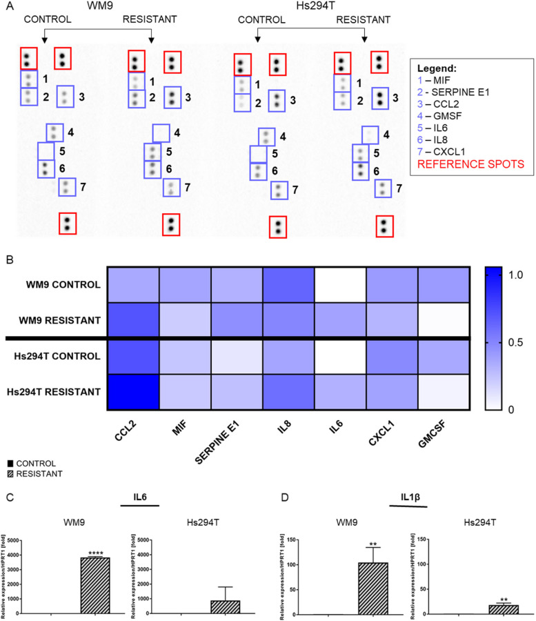
Methods: Cell viability was assessed via the XTT test. The level of selected proteins as well as activation of signaling pathways were evaluated using Western blotting. The expression of the chosen genes was assessed by RT-PCR. The distribution of cell cycle phases was analyzed by flow cytometry, and confocal microscopy was used to take photos of spheroids. The composition of cytokines secreted by cells was determined using a human cytokine array.

Results: The resistant cells had increased survival and activation of ERK kinase in the presence of BRAF/MEK inhibitors. The IC50 values for these cells were over 1000 times higher than for controls. Resistant cells also exhibited elevated activation of AKT, p38, and JNK signaling pathways with increased expression of EGFR, ErbB2, MET, and PDGFRβ receptors as well as reduced expression of ErbB3 receptor. Furthermore, these cells demonstrated increased expression of genes encoding proteins involved in drug transport and metabolism. Resistant cells also exhibited features of epithelial-mesenchymal transition and cancer stem cells as well as reduced proliferation rate and elevated cytokine secretion.

Conclusions: In summary, this work describes BRAF/MEK-inhibitor-resistant melanoma cells, allowing for better understanding the underlying mechanisms of resistance. The results may thus contribute to the development of new, more effective therapeutic strategies.

Keywords: BRAFi/MEKi; Cobimetinib; Drug resistance; Melanoma; Targeted therapy; Vemurafenib.


Conflict of interest statement

The authors declare no competing interests.

Figures

Fig. 1
Fig. 1
Sensitivity of control and resistant melanoma cell lines to the vemurafenib and cobimetinib. A The viability of the control and resistant melanoma cells after 48 h of treatment with BRAF/MEK inhibitors was measured using the XTT assay. B Based on the viability measurement and the XTT test, IC50 values were determined for control and resistant cell lines and then calculated using GraphPad Prism 7 program. C The level of phosphorylated ERK1/2 kinase after 24 h of incubation with BRAF/MEK inhibitors in control (CTRL) and resistant (R) melanoma cell lines was verified by Western Blotting in cell lysates. Control (CTRL) constitutes WM9 and Hs294T cells treated with regular media with DMSO at the concentration used for drug delivery. Representative blotting membranes of three independent experiments are shown. The loading control was the total protein content assessed by Ponceau S staining. The graphs show the average values from at least three independent experiments ± SD. Asterisks in the graphs indicate statistical significance (p) at the level of *** ≤ 0.001, **** ≤ 0.0001
Fig. 2
Fig. 2
Activation of signaling pathways in resistant and control melanoma cell lines. The level of total or phosphorylated (A) AKT, (B) p38, and (C) JNK in cell lysates was determined using Western blotting analysis. The signal was normalized to the total protein content assessed by Ponceau S staining. Representative blotting membranes of at least three biological repetitions are shown. The expression level of the (DNRAS and (E) MITF genes was estimated by real-time PCR and with the use of designed primers. HPRT1 constituted a reference gene. Control (CTRL) constitutes WM9 and Hs294T cells treated with regular media with DMSO at the concentration used for drug delivery. The graphs show average values from at least three independent experiments ± SD. Asterisks indicate statistically important differences between tested and control cells. The significance level was set at p ≤ 0.05 (*), p ≤ 0.01 (**), and p ≤ 0.001 (***)
Fig. 3
Fig. 3
Expression of selected tyrosine kinase receptors in resistant melanoma cell lines. A EGFR, (B) HER2, (CHER3, (D) MET, and (E) PDGFRβ receptor expression was assessed in control and resistant melanoma WM9 and Hs294T cells using real-time PCR. Taqman probes were used to evaluate the expression level of EGFR, HER2, HER3, and MET receptors, and GAPDH served as a housekeeping gene. The expression level of the PDGFRβ receptor was determined by utilizing the designed primers. HPRT1 constituted a reference gene. Control (CTRL) constitutes WM9 and Hs294T cells treated with regular media with DMSO at the concentration used for drug delivery. The graphs show average values from at least three independent experiments ± SD. The asterisks indicate the significance level at p ≤ 0.05 (*), p ≤ 0.01 (**) and p ≤ 0.0001 (****)
Fig. 4
Fig. 4
Expression of genes encoding proteins involved in drug transport and metabolism in resistant melanoma cells. A ABCA1, (BABCC2, and (CABCG2 transporter expression in control and resistant melanoma WM9 and Hs294T cell lines. Real-time PCR was performed with HPRT1 as a reference gene. D The level of CYP1A1 protein in cell lysates was determined using Western blotting. The signal was normalized to the total protein content assessed by Ponceau S staining. Representative blotting membranes of three independent experiments are shown. Control (CTRL) constitutes WM9 and Hs294T cells treated with regular media with DMSO at the concentration used for drug delivery. The graphs show average values from at least three independent experiments ± SD. Asterisks in the graphs indicate statistical significance (p) at the level of * ≤ 0.05, ** ≤ 0.01, and *** ≤ 0.001
Fig. 5
Fig. 5
Changes in proliferation rate and cell cycle phases distribution in resistant melanoma cell lines. A Melanoma WM9 and Hs294T cells were seeded on a 96-well plate, and their proliferation rate was calculated as a ratio of the spectrophotometric signal after 48 h divided by the signal at t0. B Cell cycle analysis in WM9 and Hs294T melanoma control cells and cells resistant to treatment with BRAFi/MEKi. Western blot analysis of the level of proteins involved in cell cycle regulation: (C) CDK6, (D) p18, (E) p21, and (F) p27 in cell lysates obtained from control and resistant melanoma cells. The signal was normalized to the total protein content assessed by Ponceau S staining. Representative blotting membranes of three independent experiments are shown. Control (CTRL) constitutes WM9 and Hs294T cells treated with regular media with DMSO at the concentration used for drug delivery. Cells for cell cycle analysis as well as those used for Western blotting analysis were synchronized to obtain reliable results for cells dividing at widely varying rates as described in the ‘Materials and Methods’ section. The graphs show average values from at least three independent experiments ± SD. Asterisks in the graphs indicate statistical significance (p) at the level of * ≤ 0.05, ** ≤ 0.01, *** ≤ 0.001, and **** ≤ 0.0001
Fig. 6
Fig. 6
Features characteristic of cancer stem cells (CSC) in resistant and control melanoma cell lines. Elevated expression of CSC markers like (A) CD24 and (BNANOG accompanied by (C) increased level of another CSC protein, ALCAM. HPRT1 is a reference gene. The Western blotting signal was normalized to the total protein content assessed by Ponceau S staining. Representative blotting membranes of three independent experiments are shown. The graphs show average values from at least three independent experiments ± SD. Asterisks in the graphs indicate statistical significance (p) at the level of * ≤ 0.05 and ** ≤ 0.01. Spheroids from control and resistant melanoma cells were obtained and then fixed and stained using phalloidin-Alexa Fluor 568 to visualize F-actin (red). Representative images taken with a fluorescence microscope are shown. Scale bar is 100 μm. Control (CTRL) constitutes WM9 and Hs294T cells treated with regular media with DMSO at the concentration used for drug delivery
Fig. 7
Fig. 7
Resistant melanoma cells display spindle-like shape and features characteristic of epithelial-mesenchymal transition (EMT). A Photographs revealing the morphology of resistant and control melanoma cells taken with a light microscope. Scale bar 300 μm. Western Blotting analysis of (B) TRGβR I, (C) TRGβR III and (D) SOX2 coupled with expression level analysis of (ESOX10 and (FSLUG genes. HPRT1 is a reference gene. The Western blotting signal was normalized to the total protein content assessed by Ponceau S staining. Representative blotting membranes of three independent experiments are shown. Control (CTRL) constitutes WM9 and Hs294T cells treated with regular media with DMSO at the concentration used for drug delivery. The graphs show the average values from at least three independent experiments ± SD. Asterisks in the graphs indicate statistical significance (p) at the level of * ≤ 0.05, ** ≤ 0.01, *** ≤ 0.001 and **** ≤ 0.0001
Fig. 8
Fig. 8
Cytokine secretion by control and resistant melanoma cell lines. Cell culture media collected from resistant and control cells were analyzed for cytokines via (A) signal (B) quantitative analysis. The results were normalized to reference spots and are shown in the form of a heatmap where darker blue indicates a higher signal intensity. Expression level of interleukins: (C) IL6 and (D) IL1β in resistant and control melanoma WM9 and Hs294T cell lines. Real-time PCR was performed, and HPRT1 constituted a reference gene. Control (CTRL) constitutes WM9 and Hs294T cells treated with regular media with DMSO at the concentration used for drug delivery. The graphs show average values from at least three independent experiments ± SD. Asterisks in the graphs indicate statistical significance (p) at the level of ** ≤ 0.01 and **** ≤ 0.0001. Abbreviations: MIF - macrophage migration inhibitory factor, CCL2 - C-C motif chemokine ligand 2, GMCSF - granulocyte/macrophage colony-stimulating factor, IL6 - interleukin 6; IL8 - interleukin 8, and CXCL1 - C-X-C motif chemokine ligand 1

Similar articles

References

    1. Dhanyamraju PK, Patel TN. Melanoma therapeutics: a literature review. J Biomed Res Educ Department Jiangsu Province. 2022;36:77. - PMC - PubMed
    1. Berk-Krauss J, Stein JA, Weber J, Polsky D, Geller AC. New systematic therapies and trends in cutaneous melanoma deaths among US whites, 1986–2016. Am J Public Health. 2020;110:731–3. 10.2105/AJPH.2020.305567 - DOI - PMC - PubMed
    1. Hartman RI, Lin JY. Cutaneous Melanoma-A Review in Detection, Staging, and Management. Hematol Oncol Clin North Am. 2019;33:25–38. https://pubmed.ncbi.nlm.nih.gov/30497675/. Cited 2024 May 29. - PubMed
    1. Leonardi GC, Falzone L, Salemi R, Zanghì A, Spandidos DA, Mccubrey JA et al. Cutaneous melanoma: From pathogenesis to therapy (Review). Int J Oncol; 2018;52:1071–80. https://pubmed.ncbi.nlm.nih.gov/29532857/. Cited 2024 May 29. - PMC - PubMed
    1. Subbiah V, Baik C, Kirkwood JM. Clinical Development of BRAF plus MEK Inhibitor Combinations. Trends Cancer; 2020;6:797–810. https://pubmed.ncbi.nlm.nih.gov/32540454/. Cited 2024 May 29. - PubMed

MeSH terms