. 2024 Aug 22;29(1):431.
doi: 10.1186/s40001-024-02017-w.

Predicting tumor invasion depth in gastric cancer: developing and validating multivariate models incorporating preoperative IVIM-DWI parameters and MRI morphological characteristics

Affiliations

Predicting tumor invasion depth in gastric cancer: developing and validating multivariate models incorporating preoperative IVIM-DWI parameters and MRI morphological characteristics

Yanling Hong et al. Eur J Med Res. .

Abstract

Introduction: Accurate assessment of the depth of tumor invasion in gastric cancer (GC) is vital for the selection of suitable patients for neoadjuvant chemotherapy (NAC). Current problem is that preoperative differentiation between T1-2 and T3-4 stage cases in GC is always highly challenging for radiologists.

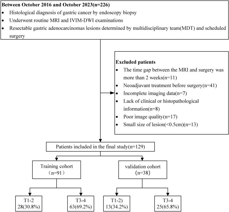
Methods: A total of 129 GC patients were divided into training (91 cases) and validation (38 cases) cohorts. Pathology from surgical specimens categorized patients into T1-2 and T3-4 stages. IVIM-DWI and MRI morphological characteristics were evaluated, and a multimodal nomogram was developed. The MRI morphological model, IVIM-DWI model, and combined model were constructed using logistic regression. Their effectiveness was assessed using receiver operating characteristic (ROC) curves, calibration curves, decision curve analysis (DCA), and clinical impact curves (CIC).

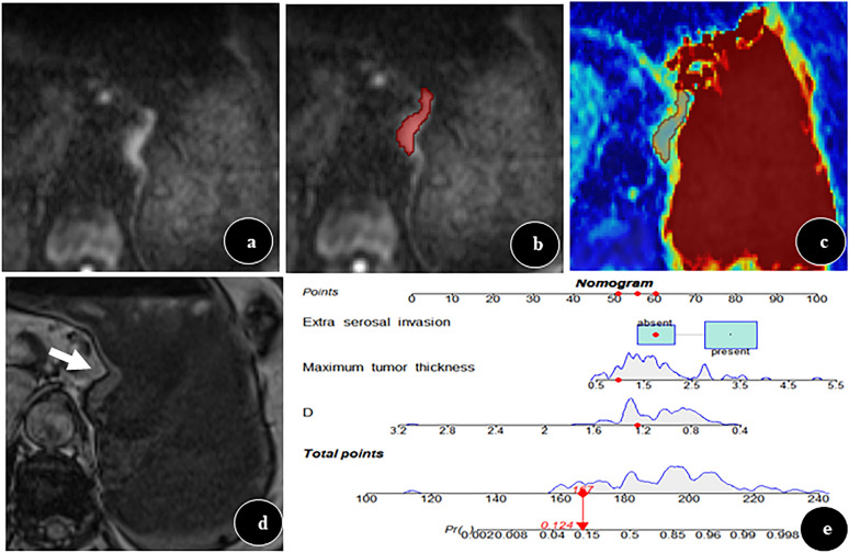
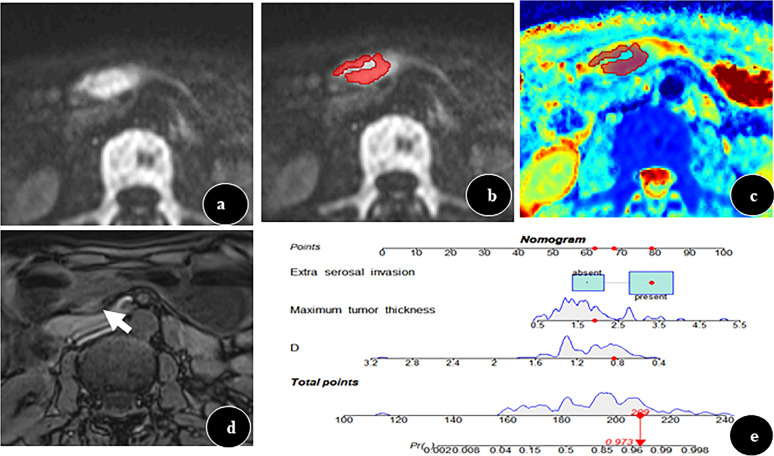
Results: The combined nomogram, integrating preoperative IVIM-DWI parameters (D value) and MRI morphological characteristics (maximum tumor thickness, extra-serosal invasion), achieved the highest area under the curve (AUC) values of 0.901 and 0.883 in the training and validation cohorts, respectively. No significant difference was observed between the AUCs of the IVIM-DWI and MRI morphological models in either cohort (training: 0.796 vs. 0.835, p = 0.593; validation: 0.794 vs. 0.766, p = 0.79).

Conclusion: The multimodal nomogram, combining IVIM-DWI parameters and MRI morphological characteristics, emerges as a promising tool for assessing tumor invasion depth in GC, potentially guiding the selection of suitable candidates for neoadjuvant chemotherapy (NAC) treatment.

Keywords: Diffusion weighted imaging; Intravoxel incoherent motion; Neoplasm staging; Nomogram; Stomach neoplasms.


Conflict of interest statement

The authors declare that they have no competing interests.

Figures

Fig. 1
Fig. 1
Flowchart Illustrating Patient Inclusion and Exclusion Criteria
Fig. 2
Fig. 2
MRT staging diagram
Fig. 3
Fig. 3
a Preoperative Nomogram for the Combined Model, Utilized for Distinguishing Between T1-2 and T3-4 Stage Tumors in Gastric Cancer Patients. b Training Cohort ROC Curves for the MRI Morphological Model, IVIM-DWI Model, and Combined Model. c Validation Cohort ROC Curves for the MRI Morphological Model, IVIM-DWI Model, and Combined Model
Fig. 4
Fig. 4
Multi-parametric Maps in a T1-2 Stage Gastric Cancer Case. a DWI map (b = 800 s/mm2) showing an irregular mass at the esophagus-cardia junction in a 66-year-old woman with histopathologically confirmed gastric cancer. b Freehand ROI drawn along the tumor's edge. The maximum thickness was 0.96 cm. c True diffusion coefficient (D) image with a D value of 1.24 × 10–3 s/mm2. d Dixon out-of-phase image showing a smooth and clear low-signal band around the lesion (white arrow), indicating no invasion of the subserosa and serosa. e Calculated scores: D value (56 points), maximum tumor thickness (51 points), and Dixon sequence evaluation (60 points), totaling 167 points. This corresponds to a predicted invasion depth of T1-2 with a probability of 0.124. Postoperative pathology confirmed a T2 stage
Fig. 5
Fig. 5
Multi-parametric Maps in a T3-4 Stage Gastric Cancer Case. a DWI map (b = 800 s/mm2) showing an irregular mass in the gastric antrum of a 71-year-old man with histopathologically confirmed gastric cancer. b Freehand ROI drawn along the tumor’s edge. The maximum thickness was 1.91 cm. c True diffusion coefficient (D) map with a D value of 0.84 × 10–3 s/mm2. d Dixon inverse-phase image showing a disrupted and interrupted low-signal band around the lesion (white arrow), indicative of subserosa and serosa invasion. e Calculated scores: D value (67.5 points), maximum tumor thickness (61.5 points), and Dixon sequence evaluation (80 points), totaling 209 points. This corresponds to a predicted invasion depth of T3-4 with a probability of 0.973. Postoperative pathology confirmed a T4a stage
Fig. 6
Fig. 6
a Calibration Curve Analysis for the Nomogram in the Training Cohort. b Calibration Curve Analysis for the Nomogram in the Validation Cohort. c Decision Curve Analysis for the MRI Morphological Model, IVIM-DWI Model, and Combined Model in the Training Cohort. d Decision Curve Analysis for the MRI Morphological Model, IVIM-DWI Model, and Combined Model in the Validation Cohort. e Clinical Impact Curve Analysis for the Nomogram in the Training Cohort. f Clinical Impact Curve Analysis for the Nomogram in the Validation Cohort

Similar articles

Cited by

References

    1. Sung H, Ferlay J, Siegel RL, Laversanne M, Soerjomataram I, Jemal A, Global cancer statistics, et al. GLOBOCAN estimates of incidence and mortality worldwide for 36 cancers in 185 countries. CA Cancer J Clin. 2020. 10.3322/caac.21660.10.3322/caac.21660 - DOI
    1. Biondi A, Lirosi MC, D’Ugo D, Fico V, Ricci R, Santullo F, et al. Neo-adjuvant chemo(radio)therapy in gastric cancer: Current status and future perspectives. World J Gastrointest Oncol. 2015;7(12):389–400. 10.4251/wjgo.v7.i12.389 - DOI - PMC - PubMed
    1. Fukagawa T, Katai H, Mizusawa J, Nakamura K, Sano T, Terashima M, et al. A prospective multi-institutional validity study to evaluate the accuracy of clinical diagnosis of pathological stage III gastric cancer (JCOG1302A). Gastric Cancer. 2018;21(1):68–73. 10.1007/s10120-017-0701-1 - DOI - PubMed
    1. Mortensen MB. Novel imaging strategies for upper gastrointestinal tract cancers. Expert Rev Gastroenterol Hepatol. 2015;9(3):295–303. 10.1586/17474124.2015.959928 - DOI - PubMed
    1. Seevaratnam R, Cardoso R, McGregor C, Lourenco L, Mahar A, Sutradhar R, et al. How useful is preoperative imaging for tumor, node, metastasis (TNM) staging of gastric cancer? A meta-analysis Gastric Cancer. 2012;15(Suppl 1):S3-18. 10.1007/s10120-011-0069-6 - DOI - PubMed

LinkOut - more resources