. 2024 Aug 23;57(1):57.
doi: 10.1186/s40659-024-00535-9.

Inhibition of forward and reverse transport of Ca2+ via Na+/Ca2+ exchangers (NCX) prevents sperm capacitation

Affiliations

Inhibition of forward and reverse transport of Ca2+ via Na+/Ca2+ exchangers (NCX) prevents sperm capacitation

Marc Yeste et al. Biol Res. .

Abstract

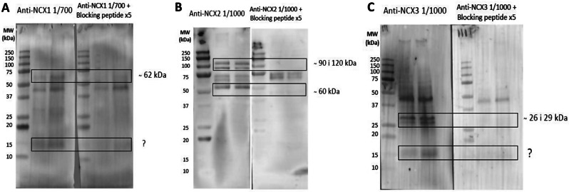
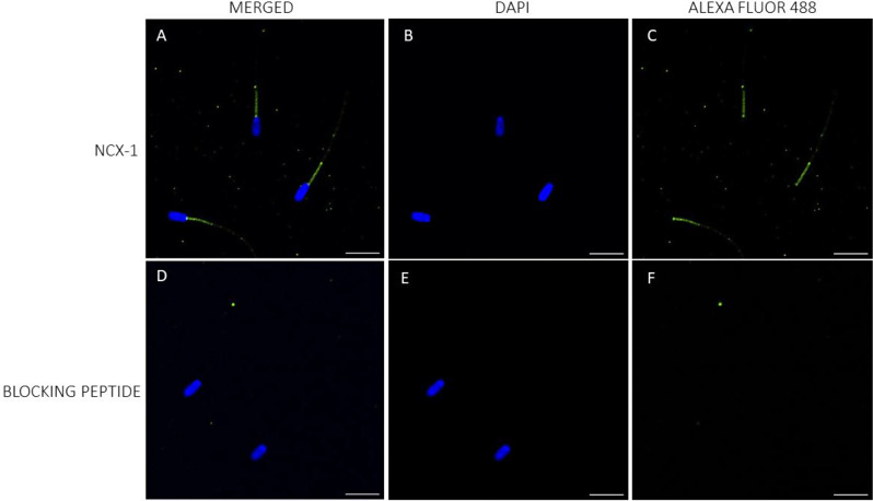
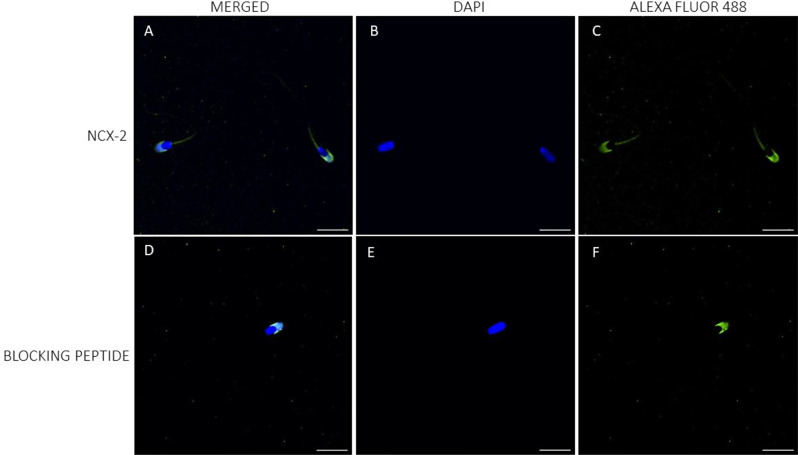
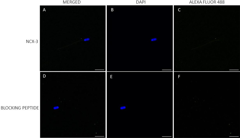
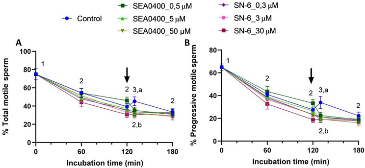
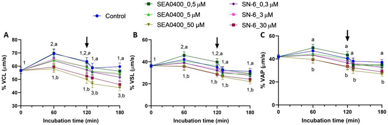
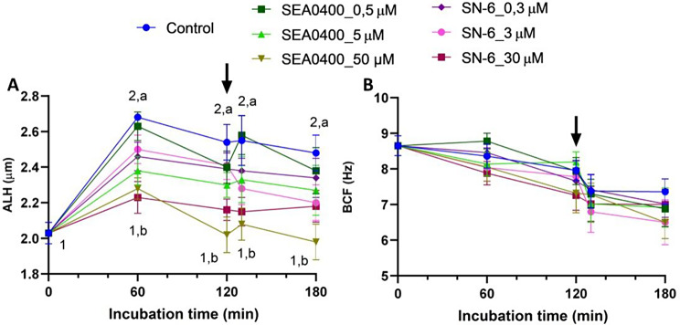
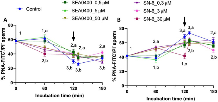
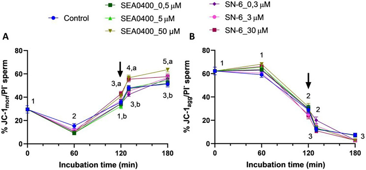
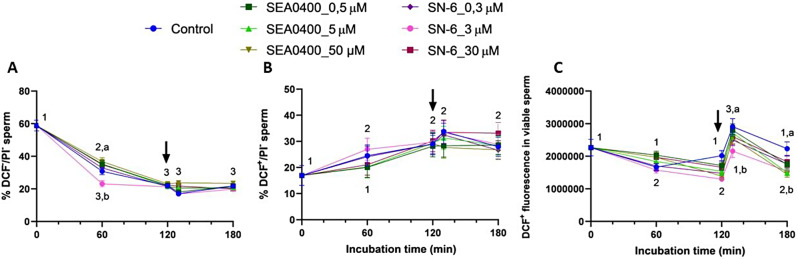
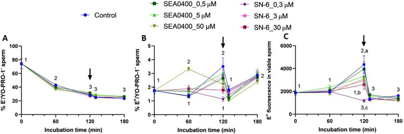
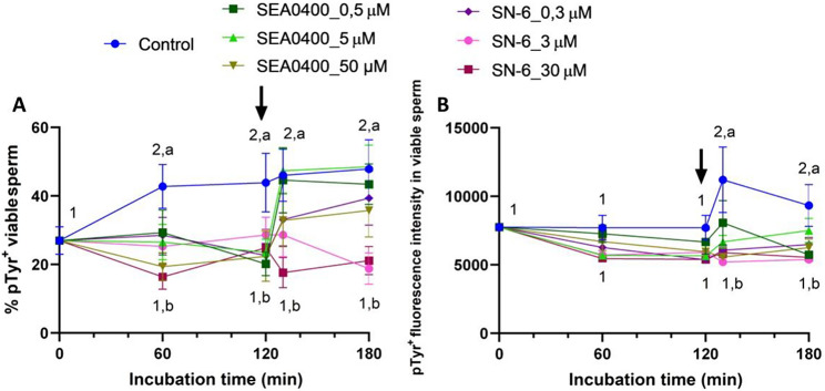
Background: While calcium is known to play a crucial role in mammalian sperm physiology, how it flows in and out of the male gamete is not completely understood. Herein, we investigated the involvement of Na+/Ca2+ exchangers (NCX) in mammalian sperm capacitation. Using the pig as an animal model, we first confirmed the presence of NCX1 and NCX2 isoforms in the sperm midpiece. Next, we partially or totally blocked Ca2+ outflux (forward transport) via NCX1/NCX2 with different concentrations of SEA0400 (2-[4-[(2,5-difluorophenyl)methoxy]phenoxy]-5-ethoxyaniline; 0, 0.5, 5 and 50 µM) and Ca2+ influx (reverse transport) with SN6 (ethyl 2-[[4-[(4-nitrophenyl)methoxy]phenyl]methyl]-1,3-thiazolidine-4-carboxylate; 0, 0.3, 3 or 30 µM). Sperm were incubated under capacitating conditions for 180 min; after 120 min, progesterone was added to induce the acrosome reaction. At 0, 60, 120, 130, and 180 min, sperm motility, membrane lipid disorder, acrosome integrity, mitochondrial membrane potential (MMP), tyrosine phosphorylation of sperm proteins, and intracellular levels of Ca2+, reactive oxygen species (ROS) and superoxides were evaluated.

Results: Partial and complete blockage of Ca2+ outflux and influx via NCX induced a significant reduction of sperm motility after progesterone addition. Early alterations on sperm kinematics were also observed, the effects being more obvious in totally blocked than in partially blocked samples. Decreased sperm motility and kinematics were related to both defective tyrosine phosphorylation and mitochondrial activity, the latter being associated to diminished MMP and ROS levels. As NCX blockage did not affect the lipid disorder of plasma membrane, the impaired acrosome integrity could result from reduced tyrosine phosphorylation.

Conclusions: Inhibition of outflux and influx of Ca2+ triggered similar effects, thus indicating that both forward and reverse Ca2+ transport through NCX exchangers are essential for sperm capacitation.

Keywords: In vitro capacitation and Ca2+ transport; Na+/Ca2+ exchangers (NCXs); Sperm.


Conflict of interest statement

Not applicable.

Figures

Fig. 1
Fig. 1
Identification of NCX isoforms. Identification of NCX1, NCX2 and NCX3 in porcine sperm. Representative immunoblots using sperm samples from different boars with A) anti-NCX1, (B) anti-NCX2 and (C) anti-NCX3 antibodies, and their respective blocking peptides
Fig. 2
Fig. 2
Immunolocalization of NCX1 isoform. Localization of NCX1 in the plasma membrane of porcine sperm (AC), and after the peptide competition assay (DF). NCX1 appears stained in green (Alexa Fluor 488) and nuclei in blue (DAPI; 4′6′-diamidion-2-phenylindole). Scale bar: 15 μm
Fig. 3
Fig. 3
Immunolocalization of NCX2 isoform. Localization of NCX2 in the plasma membrane of porcine sperm (AC), and after the peptide competition assay (DF). NCX2 appears stained in green (Alexa Fluor 488) and nuclei in blue (DAPI; 4′6′-diamidion-2-phenylindole). Scale bar: 15 μm
Fig. 4
Fig. 4
Immunolocalization of NCX3 isoform. Localization of NCX3 in the plasma membrane of porcine sperm (AC), and after the peptide competition assay (DF). NCX3 appears stained in green (Alexa Fluor 488) and nuclei in blue (DAPI; 4′6′-diamidion-2-phenylindole). Scale bar: 15 μm
Fig. 5
Fig. 5
Sperm motility. Percentages of total (A) and progressively (B) motile sperm during in vitro capacitation of control samples and samples blocked with either SEA0400 (0.5, 5, and 50 µM) or SN-6 (0.3, 3, and 30 µM). Different superscript letters indicate significant differences between control and blocked samples within a single time point (P < 0.05). Different numeral superscripts indicate significant differences between time points within a treatment (P < 0.05). The arrow indicates the addition of 10 µg/mL of progesterone at 120 min of incubation. Results are expressed as the mean ± SEM (n = 10)
Fig. 6
Fig. 6
Sperm kinematics (I). Sperm velocity parameters of VCL (A), VSL (B), and VAP (C) during in vitro capacitation of control samples and samples blocked with either SEA0400 (0.5, 5, and 50 µM) or SN-6 (0.3, 3, and 30 µM). Different superscript letters indicate significant differences between control and blocked samples within a single time point (P < 0.05). Different superscript numbers indicate significant differences between time points within a treatment (P < 0.05). The arrow indicates the addition of 10 µg/mL of progesterone at 120 min of incubation. Results are expressed as the mean ± SEM (n = 10)
Fig. 7
Fig. 7
Sperm kinematics (II). Amplitude of lateral head displacement (ALH, A) and beat cross frequency (BCF, B) during in vitro capacitation of control samples and samples blocked with either SEA0400 (0.5, 5, and 50 µM) or SN-6 (0.3, 3, and 30 µM). Different superscript letters indicate significant differences between control and blocked samples within a single time point (P < 0.05). Different superscript numbers indicate significant differences between time points within a treatment (P < 0.05). The arrow indicates the addition of 10 µg/mL of progesterone at 120 min of incubation. Results are expressed as the mean ± SEM (n = 10)
Fig. 8
Fig. 8
Acrosome integrity. Percentages of viable sperm with an intact acrosome (PNA-FITC+/EthD-1-, A) and with an exocytosed acrosome (PNA-FITC-/EthD-1-, B) during in vitro capacitation of control samples and samples blocked with either SEA0400 (0.5, 5, and 50 µM) or SN-6 (0.3, 3, and 30 µM). Different superscript letters indicate significant differences between control and blocked samples within a single time point (P < 0.05). Different superscript numbers indicate significant differences between time points within a treatment (P < 0.05). The arrow indicates the addition of 10 µg/mL of progesterone at 120 min of incubation. Results are expressed as the mean ± SEM (n = 10)
Fig. 9
Fig. 9
Intracellular Ca2+ levels. Percentages of viable sperm with high intracellular Ca2+ levels (Fluo4+/PI, A) and fluorescence intensity of Fluo4+ in viable sperm (B) of control samples and samples blocked with either SEA0400 (0.5, 5, and 50 µM) or SN-6 (0.3, 3, and 30 µM). Different superscript letters indicate significant differences between control and blocked samples within a single time point (P < 0.05). Different superscript numbers indicate significant differences between time points within a treatment (P < 0.05). The arrow indicates the addition of 10 µg/mL of progesterone at 120 min of incubation. Results are expressed as the mean ± SEM (n = 10)
Fig. 10
Fig. 10
Mitochondrial membrane potential. Percentages of viable sperm with low (JC-1mon/PI-, A) and high (JC-1agg/PI-, B) mitochondrial membrane potential during in vitro capacitation of control samples and samples blocked with either SEA0400 (0.5, 5, and 50 µM) or SN-6 (0.3, 3, and 30 µM). Different superscript letters indicate significant differences between control and blocked samples within a single time point (P < 0.05). Different superscript numbers indicate significant differences between time points within a treatment (P < 0.05). The arrow indicates the addition of 10 µg/mL of progesterone at 120 min of incubation. Results are expressed as the mean ± SEM (n = 10)
Fig. 11
Fig. 11
Total ROS levels. Percentages of viable sperm with low (DCF/PI, A) and high (DCF+/PI, B) ROS levels and fluorescence intensity of DCF+ in viable sperm (C) during in vitro capacitation of control samples and samples blocked with either SEA0400 (0.5, 5, and 50 µM) or SN-6 (0.3, 3, and 30 µM). Different superscript letters indicate significant differences between control and blocked samples within a single time point (P < 0.05). Different superscript numbers indicate significant differences between time points within a treatment (P < 0.05). The arrow indicates the addition of 10 µg/mL of progesterone at 120 min of incubation. Results are expressed as the mean ± SEM (n = 10)
Fig. 12
Fig. 12
Superoxide levels. Percentages of viable sperm with low (E/YO-PRO-1, A) and high (E+/YO-PRO-1, B) superoxide levels and fluorescence intensity of E+ in viable sperm (C) during in vitro capacitation of control samples and samples blocked with either SEA0400 (0.5, 5, and 50 µM) or SN-6 (0.3, 3, and 30 µM). Different superscript letters indicate significant differences between control and blocked samples within a single time point (P < 0.05). Different superscript numbers indicate significant differences between time points within a treatment (P < 0.05). The arrow indicates the addition of 10 µg/mL of progesterone at 120 min of incubation. Results are expressed as the mean ± SEM (n = 10)
Fig. 13
Fig. 13
Tyrosine phosphorylation of sperm proteins. Percentages of viable sperm with phosphorylated tyrosines (pTyr+, A) and fluorescence intensity of pTyr+ in viable sperm (B) of control samples and samples blocked with either SEA0400 (0.5, 5, and 50 µM) or SN-6 (0.3, 3, and 30 µM). Different superscript letters indicate significant differences between control and blocked samples within a single time point (P < 0.05). Different superscript numbers indicate significant differences between time points within a treatment (P < 0.05). The arrow indicates the addition of 10 µg/mL of progesterone at 120 min of incubation. Results are expressed as the mean ± SEM (n = 10)

References

    1. Takei GL, Ogura Y, Ujihara Y, Toyama F, Hayashi K, Fujita T. Hamster sperm possess functional Na+/Ca2+-exchanger 1: its implication in hyperactivation. Int J Mol Sci. 2023;24:8905. 10.3390/ijms24108905 - DOI - PMC - PubMed
    1. Delgado-Bermúdez A, Yeste M, Bonet S, Pinart E. A review on the role of bicarbonate and Proton Transporters during sperm capacitation in mammals. Int J Mol Sci. 2022;23:6333. 10.3390/ijms23116333 - DOI - PMC - PubMed
    1. Krasznai Z, Krasznai ZT, Morisawa M, Bazsáné ZK, Hernádi Z, Fazekas Z, et al. Role of the Na+/Ca2 + exchanger in calcium homeostasis and human sperm motility regulation. Cell Motil. 2006;63:66–76.10.1002/cm.20108 - DOI - PubMed
    1. Yoshida K, Shiba K, Sakamoto A, Ikenaga J, Matsunaga S, Inaba K, et al. Ca2 + efflux via plasma membrane Ca2+-ATPase mediates chemotaxis in ascidian sperm. Sci Rep. 2018;8:16622. 10.1038/s41598-018-35013-2 - DOI - PMC - PubMed
    1. Lishko PV, Mannowetz N. CatSper: a unique calcium channel of the sperm flagellum. Curr Opin Physiol. 2018;2:109–13. 10.1016/j.cophys.2018.02.004 - DOI - PMC - PubMed

MeSH terms

LinkOut - more resources