

[Effect of accordion technique and deferoxamine on promoting bone regeneration in distraction osteogenesis]
- PMID: 39175324
- PMCID: PMC11335587
- DOI: 10.7507/1002-1892.202404073
[Effect of accordion technique and deferoxamine on promoting bone regeneration in distraction osteogenesis]
Abstract
Objective: To compare the effects of hypoxia-inducible drugs using deferoxamine (DFO) and accordion technique (AT) on activating the hypoxia-inducible factor 1α (HIF-1α)/vascular endothelial growth factor (VEGF) signaling pathway to promote bone regeneration and remodelling during consolidation phase of distraction osteogenesis (DO).
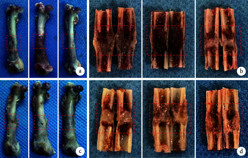
Methods: Forty-five specific-pathogen-free adult male Sprague-Dawley (SD) rats were randomly divided into the control group, DFO group, and AT group, with 15 rats in each group. All rats underwent osteotomy to establish a right femur DO model. Then, continuous distraction was started for 10 days after 5 days of latency in each group. During the consolidation phase after distraction, no intervention was performed in the control group; DFO was locally perfused into the distraction area in the DFO group starting at the 3rd week of consolidation phase; cyclic stress stimulation was given in the AT group starting at the 3rd week of consolidation phase. The general condition of rats in each group was observed. X-ray films were conducted at the end of the distraction phase and at the 2nd, 4th, and 6th weeks of the consolidation phase to observe the calcification in the distraction area. At the 4th and 6th weeks of the consolidation phase, peripheral blood was taken for ELISA detection (HIF-1α, VEGF, CD31, and Osterix), femoral specimens were harvested for gross observation, histological staining (HE staining), and immunohistochemical staining [HIF-1α, VEGF, osteopontin (OPN), osteocalcin (OCN)]. At the 6th week of the consolidation phase, Micro-CT was used to observe the new bone mineral density (BMD), bone volume/tissue volume (BV/TV), trabecular separation (Tb.Sp), trabecular number (Tb.N), and trabecular thickness (Tb.Th) in the distraction area, and biomechanical test (ultimate load, elastic modulus, energy to failure, and stiffness) to detect bone regeneration in the distraction area.
Results: The rats in all groups survived until the termination of the experiment. ELISA showed that the contents of HIF-1α, VEGF, CD31, and Osterix in the serum of the AT group were significantly higher than those of the DFO group and control group at the 4th and 6th weeks of the consolidation phase ( P<0.05). General observation, X-ray films, Micro-CT, and biomechanical test showed that bone formation in the femoral distraction area was significantly better in the DFO group and AT group than in the control group, and complete recanalization of the medullary cavity was achieved in the AT group, and BMD, BV/TV, Tb.Sp, Tb.N, and Tb.Th, as well as ultimate load, elastic modulus, energy to failure, and stiffness in the distraction area, were better in the AT group than in the DFO group and control group, and the differences were significant ( P<0.05). HE staining showed that trabecular bone formation and maturation in the distraction area were better in the AT group than in the DFO group and control group. Immunohistochemical staining showed that at the 4th week of consolidation phase, the expression levels of HIF-1α, VEGF, OCN, and OPN in the distraction area of the AT group were significantly higher than those of the DFO group and control group ( P<0.05); however, at 6th week of consolidation phase, the above indicators were lower in the AT group than in the DFO group and control group, but there was no significant difference between groups ( P>0.05).
Conclusion: Both continuous local perfusion of DFO in the distraction area and AT during the consolidation phase can activate the HIF-1α/VEGF signaling pathway. However, AT is more effective than local perfusion of DFO in promoting the process of angiogenesis, osteogenesis, and bone remodelling.
目的: 比较牵张成骨(distraction osteogenesis,DO)矿化期给予牵张区灌注低氧诱导药物去铁胺(deferoxamine,DFO)和“手风琴”技术(accordion technique,AT)循环应力,激活缺氧诱导因子1α(hypoxia-inducible factor 1α,HIF-1α)/VEGF信号通路,促进骨再生与重建的效果。.
方法: 取45只SPF级成年雄性SD大鼠,随机分为对照组、DFO组及AT组,每组15只。首先,所有大鼠截骨建立右侧股骨DO模型,截骨5 d后开始持续牵拉延长10 d;牵张完成后在矿化期内,对照组不作干预,DFO组于矿化第3周开始牵张区局部灌注DFO,AT组于矿化第3周开始给予循环应力刺激。观察各组大鼠一般情况;于牵张期结束及矿化第2、4、6周摄X线片,观察牵张区钙化情况;矿化第4、6周,取外周血行ELISA检测 HIF-1α、VEGF、CD31及成骨相关转录因子(Osterix)表达,取股骨标本行大体观察以及组织学(HE染色)及免疫组织化学染色 [HIF-1α、VEGF、骨桥蛋白(osteopontin,OPN)、骨钙素(osteocalcin,OCN)] 观测;矿化第6周,取股骨标本行Micro-CT扫描,观测牵张区新生骨骨密度(bone mineral density,BMD)、骨体积/组织体积(bone volume/tissue volume,BV/TV)、骨小梁疏密度(trabecular separation,Tb.Sp)、骨小梁数量(trabecular number,Tb.N)及骨小梁厚度(trabecular thickness,Tb.Th),以及生物力学测试极限载荷、弹性模量、断裂能量及刚度,检测牵张区骨再生情况。.
结果: 术后3组大鼠均存活至实验完成。ELISA检测示,矿化第4、6周AT组血清中HIF-1α、VEGF、CD31及Osterix含量均高于DFO组和对照组( P<0.05)。大体观察、X线片、Micro-CT及生物力学试验示,DFO组和AT组股骨牵张区内骨形成显著优于对照组,且AT组髓腔实现完全再通,AT组牵张区新生骨BMD、BV/TV、Tb.Sp、Tb.N、Tb.Th以及极限载荷、弹性模量、断裂能量、刚度均优于DFO组、对照组,差异有统计学意义( P<0.05)。HE染色示AT组牵张区骨小梁形成、成熟程度优于DFO组、对照组。免疫组织化学染色示,矿化第4周AT组牵张区HIF-1α、VEGF、OCN、OPN表达量均高于DFO组、对照组,差异有统计学意义( P<0.05);而矿化第6周时,AT组上述指标均低于DFO组、对照组,但组间差异均无统计学意义( P>0.05)。.
结论: DO矿化中期于牵张区局部持续灌注DFO或采用AT循环应力刺激均可激活HIF-1α/VEGF信号通路,但后者加速成血管-成骨耦联作用及骨重建效果优于前者。.
Keywords: Distraction osteogenesis; accordion technique; bone regeneration; bone remodelling; hypoxia-inducible drug; rat.
Conflict of interest statement
利益冲突 在课题研究和文章撰写过程中不存在利益冲突;经费支持没有影响文章观点和对研究数据客观结果的统计分析及其报道
Figures


Similar articles
-
[Effect of "accordion" technique on bone consolidation during distraction osteogenesis].Zhongguo Xiu Fu Chong Jian Wai Ke Za Zhi. 2018 May 15;32(5):558-567. doi: 10.7507/1002-1892.201712094. Zhongguo Xiu Fu Chong Jian Wai Ke Za Zhi. 2018. PMID: 29806343 Free PMC article. Chinese.
-
Enhancement of bone regeneration with the accordion technique via HIF-1α/VEGF activation in a rat distraction osteogenesis model.J Tissue Eng Regen Med. 2018 Feb;12(2):e1268-e1276. doi: 10.1002/term.2534. Epub 2017 Nov 17. J Tissue Eng Regen Med. 2018. PMID: 28763580
-
Percutaneous CO2 Treatment Accelerates Bone Generation During Distraction Osteogenesis in Rabbits.Clin Orthop Relat Res. 2020 Aug;478(8):1922-1935. doi: 10.1097/CORR.0000000000001288. Clin Orthop Relat Res. 2020. PMID: 32732577 Free PMC article.
-
H Vessel Formation as a Marker for Enhanced Bone Healing in Irradiated Distraction Osteogenesis.Semin Plast Surg. 2024 Jan 19;38(1):31-38. doi: 10.1055/s-0043-1778039. eCollection 2024 Feb. Semin Plast Surg. 2024. PMID: 38495069 Free PMC article. Review.
-
The effect of Hypoxia-Inducible Factor-1a stabilization on bone regeneration during distraction osteogenesis: A systematic review of animal studies.Arch Oral Biol. 2025 Apr;172:106184. doi: 10.1016/j.archoralbio.2025.106184. Epub 2025 Jan 23. Arch Oral Biol. 2025. PMID: 39893997
Cited by
-
Radial Periosteal Distraction as a Novel Intervention for Raynaud Syndrome with Gangrene: A Case Report.Am J Case Rep. 2025 Aug 29;26:e948422. doi: 10.12659/AJCR.948422. Am J Case Rep. 2025. PMID: 40878060 Free PMC article.
References
Publication types
MeSH terms
Substances
LinkOut - more resources
Full Text Sources
Research Materials