

A deep learning radiomics model based on CT images for predicting the biological activity of hepatic cystic echinococcosis
- PMID: 39175611
- PMCID: PMC11338923
- DOI: 10.3389/fphys.2024.1426468
A deep learning radiomics model based on CT images for predicting the biological activity of hepatic cystic echinococcosis
Abstract
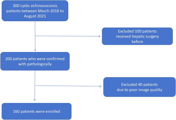
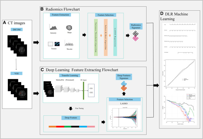
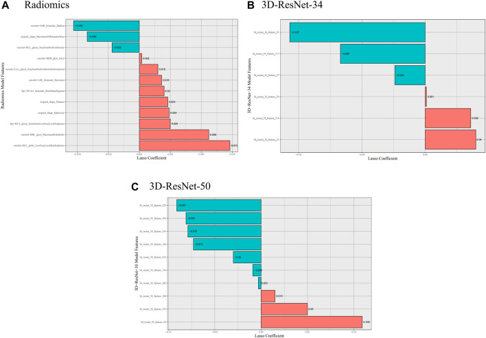
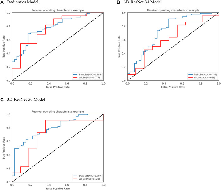
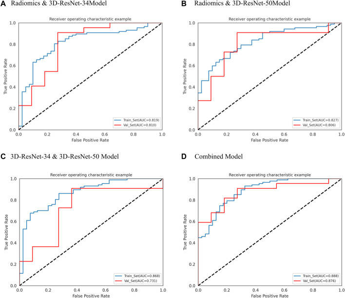
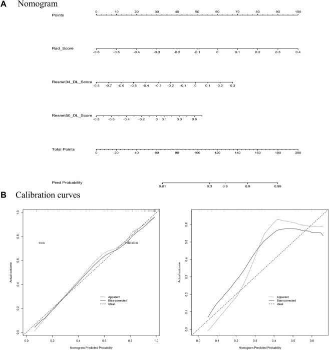
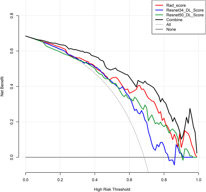
Introduction: Hepatic cystic echinococcosis (HCE) is a widely seen parasitic infection. Biological activity is crucial for treatment planning. This work aims to explore the potential applications of a deep learning radiomics (DLR) model, based on CT images, in predicting the biological activity grading of hepatic cystic echinococcosis. Methods: A retrospective analysis of 160 patients with hepatic echinococcosis was performed (127 and 33 in training and validation sets). Volume of interests (VOIs) were drawn, and radiomics features and deep neural network features were extracted. Feature selection was performed on the training set, and radiomics score (Rad Score) and deep learning score (Deep Score) were calculated. Seven diagnostics models (based on logistic regression algorithm) for the biological activity grading were constructed using the selected radiomics features and two deep model features respectively. All models were evaluated using the receiver operating characteristic curve, and the area under the curve (AUC) was calculated. A nomogram was constructed using the combined model, and its calibration, discriminatory ability, and clinical utility were assessed. Results: 12, 6 and 10 optimal radiomics features, deep learning features were selected from two deep learning network (DLN) features, respectively. For biological activity grading of hepatic cystic echinococcosis, the combined model demonstrated strong diagnostic performance, with an AUC value of 0.888 (95% CI: 0.837-0.936) in the training set and 0.876 (0.761-0.964) in the validation set. The clinical decision analysis curve indicated promising results, while the calibration curve revealed that the nomogram's prediction result was highly compatible with the actual result. Conclusion: The DLR model can be used for predicting the biological activity grading of hepatic echinococcosis.
Keywords: 3D-ResNet; biological activity grading; deep learning; hepatic cystic echinococcosis; radiomics.
Copyright © 2024 Nijiati, Tuerdi, Damola, Yimit, Yang, Abulaiti, Mutailifu, Aihait, Wang and Zou.
Conflict of interest statement
The authors declare that the research was conducted in the absence of any commercial or financial relationships that could be construed as a potential conflict of interest.
Figures
Similar articles
-
Identification of lesion bioactivity in hepatic cystic echinococcosis using a transformer-based fusion model.J Infect. 2025 Apr;90(4):106455. doi: 10.1016/j.jinf.2025.106455. Epub 2025 Mar 4. J Infect. 2025. PMID: 40049526
-
Constructing a Deep Learning Radiomics Model Based on X-ray Images and Clinical Data for Predicting and Distinguishing Acute and Chronic Osteoporotic Vertebral Fractures: A Multicenter Study.Acad Radiol. 2024 May;31(5):2011-2026. doi: 10.1016/j.acra.2023.10.061. Epub 2023 Nov 27. Acad Radiol. 2024. PMID: 38016821
-
Predicting N2 lymph node metastasis in presurgical stage I-II non-small cell lung cancer using multiview radiomics and deep learning method.Med Phys. 2023 Apr;50(4):2049-2060. doi: 10.1002/mp.16177. Epub 2023 Jan 6. Med Phys. 2023. PMID: 36563341 Clinical Trial.
-
One 3D VOI-based deep learning radiomics strategy, clinical model and radiologists for predicting lymph node metastases in pancreatic ductal adenocarcinoma based on multiphasic contrast-enhanced computer tomography.Front Oncol. 2022 Sep 9;12:990156. doi: 10.3389/fonc.2022.990156. eCollection 2022. Front Oncol. 2022. PMID: 36158647 Free PMC article.
-
A CT-based radiomics model for predicting lymph node metastasis in hepatic alveolar echinococcosis patients to support lymph node dissection.Eur J Med Res. 2024 Aug 7;29(1):409. doi: 10.1186/s40001-024-01999-x. Eur J Med Res. 2024. PMID: 39113113 Free PMC article.
Cited by
-
CT-based radiomics models using intralesional and different perilesional signatures in predicting the microvascular density of hepatic alveolar echinococcosis.BMC Med Imaging. 2025 Mar 10;25(1):84. doi: 10.1186/s12880-025-01612-5. BMC Med Imaging. 2025. PMID: 40065220 Free PMC article.
-
Advances in Novel Diagnostic Techniques for Alveolar Echinococcosis.Diagnostics (Basel). 2025 Feb 27;15(5):585. doi: 10.3390/diagnostics15050585. Diagnostics (Basel). 2025. PMID: 40075832 Free PMC article. Review.
-
Current considerations for the management of liver echinococcosis.World J Gastroenterol. 2025 Mar 14;31(10):103973. doi: 10.3748/wjg.v31.i10.103973. World J Gastroenterol. 2025. PMID: 40093668 Free PMC article. Review.
References
LinkOut - more resources
Full Text Sources