Case Reports
. 2024 Jul 26:36:100469.
doi: 10.1016/j.jctube.2024.100469. eCollection 2024 Aug.

Pilot use of a mycolactone-specific lateral flow assay for Buruli ulcer: A case report from Japan

Affiliations
Case Reports

Pilot use of a mycolactone-specific lateral flow assay for Buruli ulcer: A case report from Japan

Yasuhisa Sakakibara et al. J Clin Tuberc Other Mycobact Dis. .

Abstract

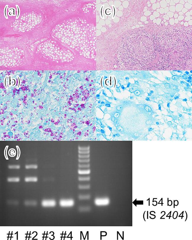
Buruli ulcer, caused by Mycobacterium (M.) ulcerans, is a neglected tropical disease (NTD) characterized by necrosis of the cutaneous tissue, predominantly affecting the limbs. The pathogenesis of this disease is mainly attributed to mycolactone, a lipid toxin produced by M. ulcerans. Here, we report the case of a 7-year-old Japanese girl who presented with worsening ulceration on her left forearm, extending to the elbow, following antimicrobial treatment. To evaluate disease progression, we used a mycolactone-specific lateral flow assay. The test yielded positive results in the advancing necrotic area, aiding in determining the extent of necessary debridement. After undergoing two debridement surgeries and receiving 38 weeks of antimicrobial treatment followed by skin grafting, the patient achieved cure. Timely diagnosis is imperative in avoiding prolonged treatment, highlighting the importance of readily available diagnostic point-of-care tests for Buruli ulcer. Moreover, detection of mycolactone not only can serve as a diagnostic tool for Buruli ulcer but also enables prediction of lesion spread and assessment of cure.

Keywords: Japan; Mycobacterium ulcerans; Paradoxical reaction; Pediatrics; Point-of-care test; Rapid diagnostic test.


Conflict of interest statement

The authors declare the following financial interests/personal relationships which may be considered as potential competing interests: Dziedzom K. de Souza, Marco Biamonte, Gerd Pluschke, Rie R. Yotsu reports financial support was provided by Global Health Innovative Technology (GHIT) Fund (G2020-202). Yuji Miyamoto, Manabu Ato reports financial support was provided by Japan Agency for Medical Research and Development. Rie Yotsu is one of the guest editors for the Special Issue on Buruli Ulcer in the Journal of Clinical Tuberculosis and Other Mycobacterial Diseases. If there are other authors, they declare that they have no known competing financial interests or personal relationships that could have appeared to influence the work reported in this paper.

Figures

Fig. 1
Fig. 1
Clinical course and symptomatic changes of Buruli ulcer on the left forearm. (a) At the time of discharge, the ulcer measured approximately 1 cm, accompanied by redness and edema around the ulcer, extending to the forearm. (b) Four weeks after initiation of antimicrobials, the ulcer enlarged, and the area with redness turned dark red. (c) Following the first debridement surgery, skin necrosis, which was previously dark red and edematous at the peripherally ulcerated with thick, brownish necrotic tissue. # Numbers indicate the sites where swab samples were obtained for the mycolactone-specific lateral flow assay. (d) At the end of treatment. After confirming the epithelialization of the grafted skin, antimycobacterial drugs were terminated. RFP: rifampicin, CAM: clarithromycin, TFLX: tosufloxacin. (For interpretation of the references to color in this figure legend, the reader is referred to the web version of this article.)
Fig. 2
Fig. 2
Histological findings and PCR testing. At the first debridement surgery, (a) Necrosis of the dermis and subcutaneous tissue with an absence of inflammatory cell infiltrate (hematoxylin-eosin staining, original magnification × 100), (b) Numerous clusters of acid-fast bacilli in the subcutaneous tissue (Ziehl–Neelsen staining, original magnification × 400). At the second debridement surgery, (c) Necrosis of dermis and subcutaneous tissue with an absence of inflammatory cells. Adjacent to it, a granuloma formation in the subcutaneous tissue with a moderate inflammatory cell infiltrate, predominantly lymphocytes, surrounding the granulomas (hematoxylin-eosin staining, original magnification × 200). (d) No acid-fast bacilli in the subcutaneous tissue (Ziehl–Neelsen staining, original magnification × 1000). (e) PCR amplification of IS2404 gene of M. ulcerans in each swab. M, 100 bp ladder marker; P, positive control; N, negative control.
Fig. 3
Fig. 3
Mycolactone-specific lateral flow assay results. The control line verifies proper sample flow and reagent function by its intensity. The test line detects mycolactone presence through a competitive immunoassay, with a weaker intensity indicating a higher concentration of mycolactone. C, control line; T, test line; T/C, T line peak value divided by C line peak value; M. shinshuense, M. ulcerans subsp. shinshuense. Sample #4 tested positive, indicated by a lower value compared to the positive control, mycolactone, at T/C. The remaining samples #1,2,3 tested negative as indicated by higher values than the positive control.

References

    1. Yotsu R.R., Suzuki K., Simmonds R.E., Bedimo R., Ablordey A., Yeboah-Manu D., et al. Buruli ulcer: a review of the current knowledge. Curr Trop Med Rep. 2018;5(4):247–256. doi: 10.1007/s40475-018-0166-2. - DOI - PMC - PubMed
    1. Omansen T.F., Erbowor-Becksen A., Yotsu R., van der Werf T.S., Tiendrebeogo A., Grout L., et al. Global epidemiology of Buruli ulcer, 2010–2017, and analysis of 2014 WHO programmatic targets. Emerg Infect Dis. 2019;25(12):2183–2190. doi: 10.3201/eid2512.190427. - DOI - PMC - PubMed
    1. George K.M., Chatterjee D., Gunawardana G., Welty D., Hayman J., Lee R., et al. Mycolactone: a polyketide toxin from Mycobacterium ulcerans required for virulence. Science. 1999;283(5403):854–857. doi: 10.1126/science.283.5403.854. - DOI - PubMed
    1. Sarfo F.S., Phillips R., Wansbrough-Jones M., Simmonds R.E. Recent advances: role of mycolactone in the pathogenesis and monitoring of Mycobacterium ulcerans infection/Buruli ulcer disease. Cell Microbiol. 2016;18(1):17–29. doi: 10.1111/cmi.12547. - DOI - PMC - PubMed
    1. Guarner J., Bartlett J., Whitney E.A.S., Raghunathan P.L., Stienstra Y., Asamoa K., et al. Histopathologic features of Mycobacterium ulcerans infection. Emerg Infect Dis. 2003;9(6):651–656. doi: 10.3201/eid0906.020485. - DOI - PMC - PubMed

Publication types

LinkOut - more resources