

Canine primary ureteral leiomyosarcoma treated with unilateral left ureteronephrectomy
- PMID: 39175966
- PMCID: PMC11338613
- DOI: 10.5455/OVJ.2024.v14.i7.20
Canine primary ureteral leiomyosarcoma treated with unilateral left ureteronephrectomy
Abstract
Background: Primary ureteral neoplasms are extremely rare in dogs, and ureteral involvement usually occurs owing to the invasion of renal and bladder tumors.
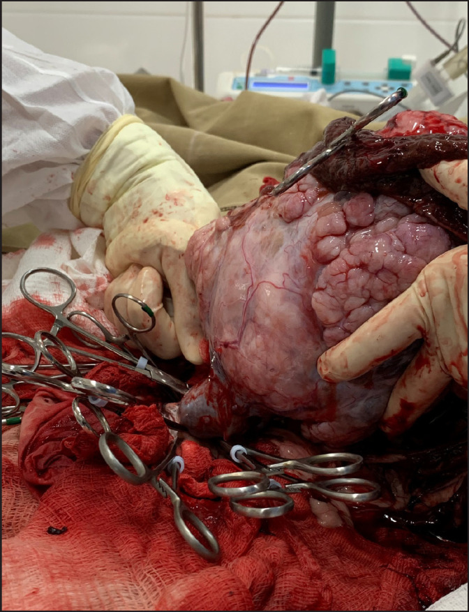
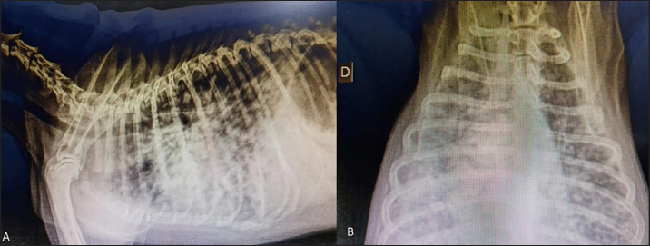
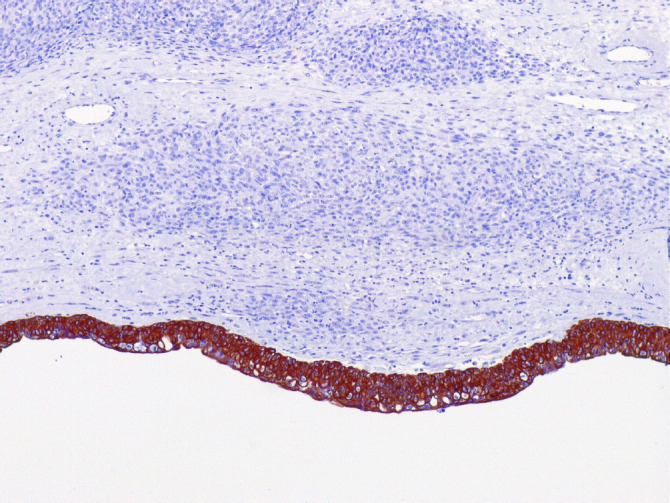
Case description: This case report describes a 12-year-old intact male mixed-breed dog referred to a private clinic with a six-month history of abdominal distention. A physical examination revealed mild abdominal pain. Hematological tests detected normocytic-normochromic anemia (hematocrit 33.6% [reference interval-RI: 37%-55%], red blood cells 4.93 M/µl [RI: 5.5-8.5 M/µl], and hemoglobin 12.4 g/dl [RI: 12-18.0 g/dl]). The results from the leukogram, thrombogram, renal, and hepatic panels were within the reference intervals for dogs. Abdominal ultrasonography revealed a cavitary mass measuring approximately 12 cm in diameter as the largest tumor in the left abdominal region over the left hepatic lobe or mesenteric site. Chest radiography did not reveal any metastasis. Therefore, the patient underwent exploratory laparotomy, during which the left ureter was found to be affected by a 12-cm mass that adhered to the left kidney. A unilateral left ureteronephrectomy was performed, and histology and immunohistochemistry (IHC) confirmed well-differentiated primary ureteral leiomyosarcoma. The patient survived for 130 days but died of lung metastasis.
Conclusion: Ureteral leiomyosarcoma should be investigated and included in the list of differential diagnoses for primary ureteral neoplasms. Regardless of the therapeutic modality, the prognosis of ureteral leiomyosarcoma may be unfavorable, as shown in this report.
Keywords: Dogs; Leiomyosarcoma; Ureteral neoplasm; Ureteronephrectomy.
Conflict of interest statement
The authors declare no conflicts of interest.
Figures


References
-
- Berent A.C, Weisse C, Bagley D. Ureteral stenting for benign and malignant disease in dogs and cats. Vet. Surg. 2007;36:E1–E29.
-
- Berzon J.L. Primary leiomyosarcoma of the ureter in a dog. J. Am. Vet. Med. Assoc. 1979;175:374–376. - PubMed
-
- Burton C.A, Day M.J, Hoston-Moore A, Holt P.E. Ureteric fibroepithelial polyps in two dogs. J. Small Anim. Pract. 1994;35:593–596.
-
- Chen L.V, Chen N, Zhu X, Zhang X, Zhong Z. Primary leiomyosarcoma of the ureter. Asian J. Surg. 2008;31:191–194. - PubMed
-
- Crow S.E. Urinary tract neoplasms in dogs and cats. Comp. Cont. Ed. Pract. Vet. 1985;7:607–618.
Publication types
MeSH terms
LinkOut - more resources
Full Text Sources