. 2025 Aug;50(8):1005.e1-1005.e8.
doi: 10.1016/j.jhsa.2024.07.008. Epub 2024 Aug 23.

The Three-Dimensional Relationship of the Axes of the Capitate and Third Metacarpal

Affiliations

The Three-Dimensional Relationship of the Axes of the Capitate and Third Metacarpal

Amy M Morton et al. J Hand Surg Am. 2025 Aug.

Abstract

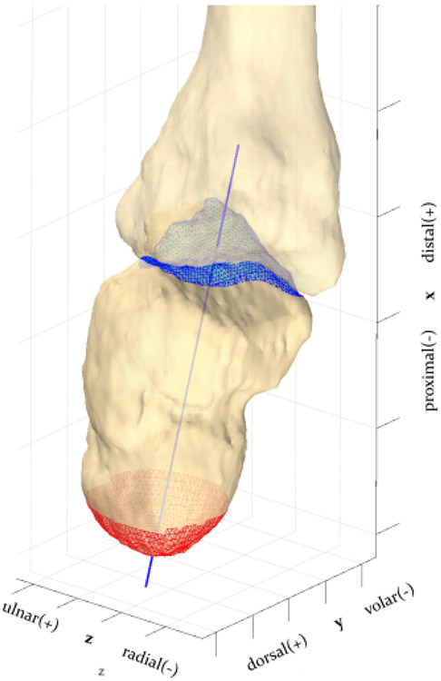
Purpose: We quantified the morphology and angulation of the third metacarpal (MC3) relative to the capitate using three-dimensional computed tomography data to inform surgical procedures such as total wrist arthroplasty and wrist arthrodesis. Specifically, we report the three-dimensional location of the intersections of the long axis of MC3 axis with the capitate cortical surface, the sagittal and coronal angles between the MC3 and capitate axes, and the MC3 shaft angle in the sagittal plane. We tested the hypothesis that these metrics did not differ between women and men.

Methods: Three-dimensional bone models of the capitate and MC3 were analyzed in 130 subjects (61M and 69F). Long axes of the MC3 and capitate were computed. The intersection of the metacarpal long axis with the cortical surface of the capitate, the angle between the metacarpal-capitate axes, and metacarpal shaft angle were calculated and compared between men and women.

Results: The long axis of the MC3 intersected the capitate at two locations on the outer cortical surface of the capitate. The proximal intersection was located near the midportion of the capitate, whereas the distal intersection was typically located within the capitate-MC3 articulation. The angle between the axes of the capitate and MC3 in the sagittal plane was a mean of 15°, ranging from 5° to 23°. The mean sagittal MC3 shaft angle was 166° and ranged from 158° to 173°.There were only subtle differences in these metrics between the sexes.

Conclusions: The long axis of the MC3 penetrates the dorsal surface of the capitate about its midportion, but there is notable variation in this location as well as in the angular relationships.

Clinical relevance: Three-dimensional measurements of the relationships between the third metacarpal and the capitate may serve as an important reference for the placement of intramedullary wires, plates, devices, and prosthetics.

Keywords: Alignment; capitate; third metacarpal; three-dimensional; total wrist arthroplasty.


Conflict of interest statement

Conflicts of Interest No benefits in any form have been received or will be received related directly to this article.

Figures

Figure 1.
Figure 1.
The principal axes of inertia of the MC3 was used to define its coordinate system (MCS). Long axis orients distal-proximal (X, red), volar-dorsal (Y, green), and radial-ulnar (Z, blue). Sagittal plane (XY, Z=0) and axial plane (YZ) density data cross sections are also shown.
Figure 2.
Figure 2.
The long axis of the capitate defined by the centroid of the proximal lobe (red mesh) and distal articular capitate surface (blue mesh).
Figure 3.
Figure 3.
(A) Sagittal (left) and coronal (right) angles between the capitate axis (blue) and the MC3 long axis computed from principal axes of inertia (black). Ulnar view (left) and volar view (right) of a right hand MC3 and capitate with respective dorsal/volar ulnar/radial left/right indications at the top of page. (B) Mean (box) sagittal and coronal angles for women (W) and men (M).
Figure 4.
Figure 4.
(A) MC3 shaft angle (brown) was the angle between proximal (pink) and distal (lilac) portions of the MC3, which were assumed to be linear segments. (B) Mean (box) MC3 shaft angle for women (W) and men (M).
Figure 5.
Figure 5.
The distal (blue) and proximal (red) intersections of the MC3 long axis with the dorsal surface of the capitate in an oblique view (A), radial view (B), dorsal view (C). The black line represents the mean MC3 axis for all subjects. Units are in the percentage of the capitate dimensions in proximal-distal (X, red axis), volar-dorsal (Y, green axis), and radial-ulnar (Z, blue) directions, illustrated with the capitate from one representative subject of mean size.

References

    1. Johnson NA, Dias R, Dias JJ. Scaphoid alignment in dorsally displaced distal radial fracture: a radiographic study. J Hand Surg Eur Vol. 2021;46(6):600–606. doi: 10.1177/1753193420983696 - DOI - PubMed
    1. Venter RG, Burger MC, Ikram A, Lamberts RP. Bony anatomy of the third metacarpal and relationship with the capitate: a computed tomography study. Surg Radiol Anat SRA. Published online June 27, 2019. doi: 10.1007/s00276-019-02272-1 - DOI - PubMed
    1. Larsen CF, Stigsby B, Lindequist S, Bellstrøm T, Mathiesen FK, Ipsen T. Observer variability in measurements of carpal bone angles on lateral wrist radiographs. J Hand Surg. 1991;16(5):893–898. doi: 10.1016/s0363-5023(10)80157-1 - DOI - PubMed
    1. Rosengren J, Brodin N. Validity and reliability of the Swedish version of the Patient Specific Functional Scale in patients treated surgically for carpometacarpal joint osteoarthritis. J Hand Ther Off J Am Soc Hand Ther. 2013;26(1):53–60; quiz 61. doi: 10.1016/j.jht.2012.10.007 - DOI - PubMed
    1. Roh YH, Noh JH, Lee BK, Baek JR, Oh JH, Gong HS, Baek GH. Reliability and validity of carpal alignment measurements in evaluating deformities of scaphoid fractures. Arch Orthop Trauma Surg. 2014;134(6):887–893. doi: 10.1007/s00402-014-1998-4 - DOI - PubMed

LinkOut - more resources