. 2024 Sep 24;43(9):114664.
doi: 10.1016/j.celrep.2024.114664. Epub 2024 Aug 22.

Trained immunity is regulated by T cell-induced CD40-TRAF6 signaling

Affiliations

Trained immunity is regulated by T cell-induced CD40-TRAF6 signaling

Maaike M E Jacobs et al. Cell Rep. .

Abstract

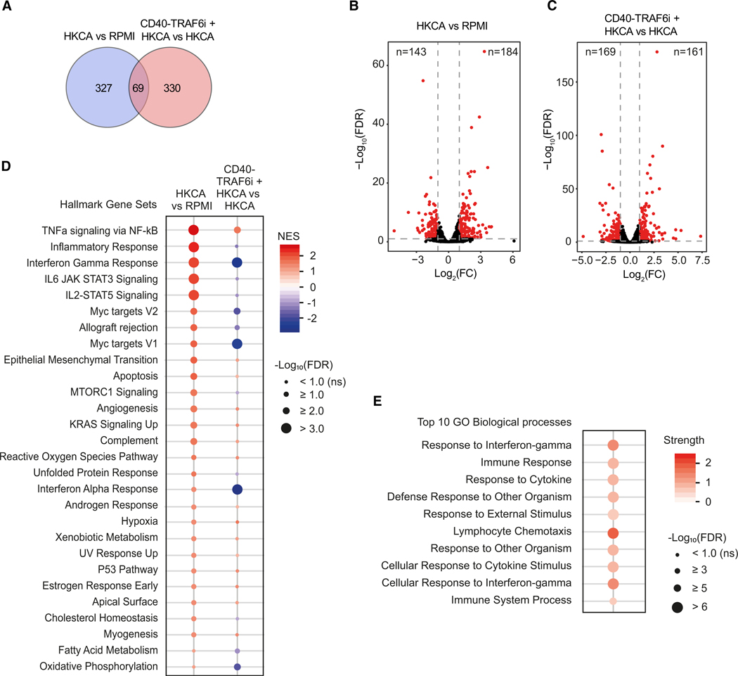
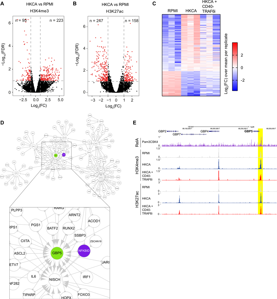
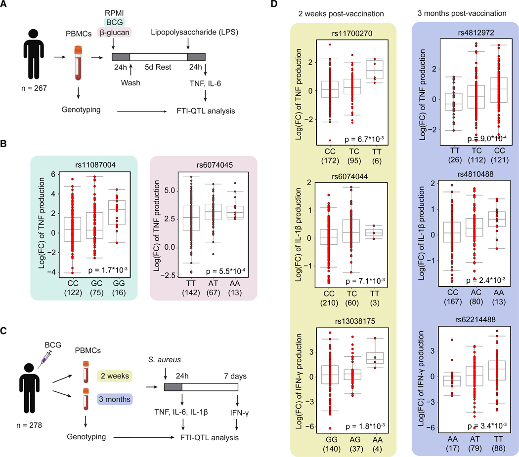
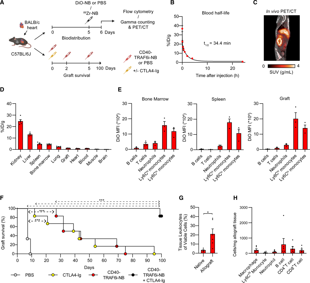
Trained immunity is characterized by histone modifications and metabolic changes in innate immune cells following exposure to inflammatory signals, leading to heightened responsiveness to secondary stimuli. Although our understanding of the molecular regulation of trained immunity has increased, the role of adaptive immune cells herein remains largely unknown. Here, we show that T cells modulate trained immunity via cluster of differentiation 40-tissue necrosis factor receptor-associated factor 6 (CD40-TRAF6) signaling. CD40-TRAF6 inhibition modulates functional, transcriptomic, and metabolic reprogramming and modifies histone 3 lysine 4 trimethylation associated with trained immunity. Besides in vitro studies, we reveal that single-nucleotide polymorphisms in the proximity of CD40 are linked to trained immunity responses in vivo and that combining CD40-TRAF6 inhibition with cytotoxic T lymphocyte antigen 4-immunoglobulin (CTLA4-Ig)-mediated co-stimulatory blockade induces long-term graft acceptance in a murine heart transplantation model. Combined, our results reveal that trained immunity is modulated by CD40-TRAF6 signaling between myeloid and adaptive immune cells and that this can be leveraged for therapeutic purposes.

Keywords: CD40-CD40L; CP: Immunology; T cells; innate immunity; monocytes; nanobiologics; trained immunity.


Conflict of interest statement

Declaration of interests J.O., L.A.B.J., M.G.N., and W.J.M.M. declare that they are scientific founders of Trained Therapeutics Discovery.

Figures

Figure 1.
Figure 1.. T cells modulate trained immunity responses in monocytes
(A) Schematic representation of the in vitro assays for evaluating trained immunity induction. (B) TNF and IL-6 production in adherent PBMCs and purified monocytes not treated or treated with heat-killed Candida albicans (HKCA) upon RPMI, lipopolysaccharide (LPS), or Pam3CSK4 (P3C) restimulation (n = 7 donors). (C) TNF and IL-6 production in adherent PBMCs and purified monocytes not treated or treated with Bacille Calmette Guérin (BCG) vaccine after RPMI, LPS, or P3C restimulation (n = 7 donors). (D) TNF and IL-6 production in HKCA-treated or untreated autologous monocytes:T cells co-cultured at different ratios after restimulation with LPS (n = 6 donors). Mean ± SEM are shown, *p < 0.05 and **p < 0.01. Paired t tests were used. See also Figures S1 and S2.
Figure 2.
Figure 2.. Trained immunity induction is mediated via CD40-TRAF6 signaling in monocytes
(A) TNF and IL-6 production in autologous monocyte:T cell co-cultures and Transwell co-cultures not treated or treated with HKCA after restimulation with LPS (n = 6 donors). (B) Volcano plot indicating RNA expression of genes encoding co-stimulatory and co-inhibitory receptors in HKCA-stimulated adherent PBMCs versus unstimulated PBMCs 24 h post-stimulation. Fold changes (FCs) and p values were calculated for each gene using pairwise analysis with DESeq2 (n = 3 donors per group). Dotted lines indicate an FC of >2 or <0.5, and Bonferroni-adjusted p value < 0.05. (C) TNF and IL-6 production in HKCA-treated and untreated adherent PBMCs in the presence or absence of CD40-TRAF6i after restimulation with LPS (n = 9 donors) or P3C (n = 10 donors). (D) TNF and IL-6 production in CD40 ligand (CD40L)-treated and untreated adherent PBMCs after restimulation with LPS (n = 15 donors) or P3C (n = 13 donors). (E) TNF and IL-6 production in CD40L-treated and untreated monocytes after restimulation with LPS (n = 15 donors) or P3C (n = 13 donors). Mean ± SEM are shown, *p < 0.05, **p < 0.01, and ***p < 0.001. Paired t tests and paired two-way ANOVA with Šidák’s post-test were used. See also Figures S3–S6 and Table S1.
Figure 3.
Figure 3.. Effect of CD40-TRAF6 inhibition on transcriptional profiles in HKCA-trained monocytes
(A) Number of differentially expressed genes (DEGs) in monocytes treated for 24 h with HKCA versus RPMI and HKCA plus CD40-TRAF6i versus HKCA only (FC > 2 or < 0.5, false discovery rate (FDR) < 0.1 (n = 3 donors per group). (B) Volcano plot indicating RNA expression in HKCA-stimulated monocytes versus unstimulated monocytes (RPMI) 24 h post-stimulation. FC and FDR were calculated for each gene using pairwise analysis with DESeq2 (n = 3 donors per group). Dotted lines indicate an FC of >2 or <0.5, and FDR < 0.1. (C) Volcano plot indicating RNA expression in HKCA-stimulated monocytes treated with CD40-TRAF6i versus HKCA-stimulated monocytes 24 h post-stimulation. FC and FDR were calculated for each gene using pairwise analysis with DESeq2 (n = 3 donors per group). Dotted lines indicate an FC of >2 or <0.5, and FDR < 0.1. (D) Significantly altered gene sets of the HALLMARK database in monocytes treated for 24 h with HKCA compared to RPMI or monocytes treated for 24 h with HKCA in the presence of CD40-TRAF6i compared to monocytes treated with HKCA alone (FDR < 0.1) (n = 3 donors per group). (E) Top 10 enriched Gene Ontology biological processes among 33 DEGs in HKCA-treated monocytes compared to RPMI-treated monocytes, for which expression is significantly reversed upon CD40-TRAF6i treatment, sorted on FDR (n = 3 donors per group). NES, normalized enrichment score. See also Figures S7 and S8.
Figure 4.
Figure 4.. CD40-TRAF6 inhibition alters oxidative phosphorylation and glycolysis in HKCA-trained PBMCs
(A) Spider plot showing metabolic parameters 6 days after stimulation in PBMCs that were not treated or treated with HKCA in the presence or absence of CD40-TRAF6i for 24 h (n = 5 donors). Values were normalized to untrained PBMCs. The asterisk (*) indicates a significant difference between HKCA+CD40-TRAF6i versus HKCA-treated PBMCs. (B) Extracellular acidification rate (ECAR) upon injection of glucose, oligomycin, and 2-deoxyglucose (2-DG) at indicated time points in PBMCs not treated or treated with HKCA in the presence or absence of CD40-TRAF6i, measured 6 days after treatment using Seahorse technology (n = 5 donors). (C) Glycolysis rate analyzed with Seahorse technology in PBMCs not treated or treated with HKCA in the presence or absence of CD40-TRAF6i for 24 h, 6 days after treatment (n = 5 donors). (D) Oxygen consumption rate (OCR) upon injection of oligomycin, carbonyl cyanide-4-(trifluoromethoxy)phenylhydrazone) (FCCP), and antimycin A + rotenone at indicated time points in PBMCs not treated or treated with HKCA in the presence or absence of CD40-TRAF6i, measured 6 days after treatment using Seahorse technology (n = 5 donors). (E) Basal respiration, ATP-linked respiration, and non-mitochondrial respiration in PBMCs not treated or HKCA-treated in the presence or absence of CD40-TRAF6i for 24 h, 6 days after treatment (n = 5 donors). Mean ± SEM are shown. *p < 0.05 and **p < 0.01. Paired t tests and paired two-way ANOVA with Šidák’s post-test were used. See also Figure S9.
Figure 5.
Figure 5.. Effect of CD40-TRAF6 inhibition on H3K4me3 modifications of HKCA-trained monocytes
(A) Volcano plot showing genomic regions with significantly altered H3K4me3 peak intensity (FC > 2 or < 0.5, FDR < 0.1) in HKCA-stimulated versus unstimulated monocytes (RPMI) 5 days post-stimulation (n = 3 donors), analyzed with DESeq2. (B) Volcano plot showing genomic regions with significantly altered H3K27ac peak intensity (FC > 2 or < 0.5, FDR < 0.1) in HKCA-stimulated versus unstimulated monocytes (RPMI) 6 days post-stimulation (n = 3 donors), analyzed with DESeq2. (C) Heatmap showing the intensity of H3K4me3 peaks in unstimulated monocytes (RPMI) and monocytes stimulated with HKCA in the presence or absence of CD40-TRAF6 inhibitor, for 318 genomic regions with significantly altered H3K4me3 peak intensity in HKCA-stimulated versus RPMI-stimulated monocytes (FC > 2 or < 0.5, FDR < 0.1) (n = 3 donors). (D) Gene regulatory network inferred from bulk RNA-seq showing genes where HKCA-induced expression was diminished by CD40-TRAF6 inhibitor. Boxed image on the bottom zooms in at NFKBID-GBP5. (E) Genomic view around GBP5. Tracks indicates RelA binding in P3C-treated THP-1 cells (available from ENCODE3), H3K4me3 and H3K27ac binding in monocytes treated with RPMI, or HKCA in the presence or absence of CD40-TRAF6 inhibitor. The yellow highlighted region is the GBP5 promoter. See also Figure S10.
Figure 6.
Figure 6.. SNPs in the proximity of CD40 associate with ex vivo and in vivo trained immunity responses
(A) Experimental design for the FTI-QTL analysis with in vitro stimulation. (B) Associations of SNPs rs11087004 (n = 213, β = −0.58 [C versus G], p = 1.7 × 10⁻³) and rs6074045 (n = 222, β = −0.54 [T versus A], p = 5.5 × 10⁻⁴) in the proximity of CD40 with TNF production for BCG- and β-glucan-induced trained immunity, respectively. Boxplots show genotype-stratified FCs of stimulated versus unstimulated cells. β-Values indicate effect size and direction. (C) Experimental design for the FTI-QTL analysis with in vivo BCG vaccination. (D) Associations of SNPs in CD40 rs11700270 (n = 273, β = 0.32 [T versus C], p = 6.7 × 10⁻³), rs6074044 (n = 273, β = 0.21 [T versus C], p = 7.1 × 10⁻³), and rs13038175 (n = 181, β = −0.68 [G versus A], p = 1.8 × 10⁻³) with cytokine production of PBMCs collected 2 weeks after trained immunity induction with BCG vaccination, and SNPs in the proximity of CD40 rs4812972 (n = 259, β = 0.34 [C versus T], p = 9.0 × 10⁻⁴), rs4810488 (n = 260, β = 0.20 [A versus C], p = 2.4 × 10⁻³), and rs62214488 (n = 184, β = −0.41 [A versus T], p = 3.4 × 10⁻³) with cytokine production of PBMCs collected 3 months after trained immunity induction using BCG vaccination, upon stimulation with Staphylococcus aureus (S. aureus). Boxplots show genotype-stratified FCs of cytokine production compared to PBMCs collected from the same individual before BCG vaccination. β-Values indicate effect size and direction. Production of TNF and IL-1β was measured 24 h after S. aureus stimulation, and production of IFN-γ was quantified 7 days after S. aureus stimulation.
Figure 7.
Figure 7.. Myeloid-specific CD40-TRAF6 inhibition combined with CTLA4-Ig prolongs allograft survival in a heart transplant mouse model
(A) Schematic of the experimental setup. (B) Determination of blood half-life time of 89Zr-NB (n = 5 mice/group). (C) Representative whole-body 2D positron emission tomography with computed tomography (PET/CT) image of a C57BL/6J mouse heterotopically transplanted with a BALB/c heart 24 h after injection of 89Zr-NBs. (D) Gamma counting of organs from C57BL/6J mice heterotopically transplanted with a BALB/c heart 24 h after 89Zr-NBs injection (n = 5 mice/group). (E) Flow cytometry of DiO-NB uptake in bone marrow, spleen, and graft 24 h after DiO-NB administration (n = 3 mice/group). (F) Kaplan-Meier curve with log-rank test for graft survival (n = 6 mice/group). ***p < 0.001. (G) Leukocytes infiltrated in native hearts and allografts of CTLA4-Ig plus CD40-TRAF6i-NB-treated mice 100 days post-transplantation (n = 6 mice). *p < 0.05, paired t test. (H) Immune cell subsets in allografts of CTLA4-Ig plus CD40-TRAF6i-NB-treated recipient mice 100 days post-transplantation (n = 6 mice). Mean ± SEM are shown. See also Figures S11 and S12.

Similar articles

Cited by

  • The immune system in cardiovascular diseases: from basic mechanisms to therapeutic implications.
    Wang X, Chen L, Wei J, Zheng H, Zhou N, Xu X, Deng X, Liu T, Zou Y. Wang X, et al. Signal Transduct Target Ther. 2025 May 23;10(1):166. doi: 10.1038/s41392-025-02220-z. Signal Transduct Target Ther. 2025. PMID: 40404619 Free PMC article. Review.
  • C1q reprograms innate immune memory.
    Jonkman I, Jacobs MME, Negishi Y, van Heck JIP, Matzaraki V, Martens JHA, Baltissen M, Vermeulen M, Fayad ZA, Teunissen AJP, Mulder WJM, Hilbrands LB, Joosten LAB, Netea MG, Mhlanga MM, Rother N, Duivenvoorden R. Jonkman I, et al. Front Immunol. 2025 May 23;16:1515127. doi: 10.3389/fimmu.2025.1515127. eCollection 2025. Front Immunol. 2025. PMID: 40486519 Free PMC article.

References

    1. Netea MG, Quintin J, and van der Meer JWM (2011). Trained immunity: a memory for innate host defense. Cell Host Microbe 9, 355–361. 10.1016/j.chom.2011.04.006. - DOI - PubMed
    1. Mitroulis I, Ruppova K, Wang B, Chen L-S, Grzybek M, Grinenko T, Eugster A, Troullinaki M, Palladini A, Kourtzelis I, et al. (2018). Modulation of myelopoiesis progenitors is an integral component of trained immunity. Cell 172, 147–161.e12. 10.1016/j.cell.2017.11.034. - DOI - PMC - PubMed
    1. Netea MG, Domínguez-Andrés J, Barreiro LB, Chavakis T, Divangahi M, Fuchs E, Joosten LAB, van der Meer JWM, Mhlanga MM, Mulder WJM, et al. (2020). Defining trained immunity and its role in health and disease. Nat. Rev. Immunol 20, 375–388. 10.1038/s41577-020-0285-6. - DOI - PMC - PubMed
    1. Quintin J, Saeed S, Martens JHA, Giamarellos-Bourboulis EJ, Ifrim DC, Logie C, Jacobs L, Jansen T, Kullberg BJ, Wijmenga C, et al. (2012). Candida albicans infection affords protection against reinfection via functional reprogramming of monocytes. Cell Host Microbe 12, 223–232. 10.1016/j.chom.2012.06.006. - DOI - PMC - PubMed
    1. Jentho E, and Weis S. (2021). DAMPs and innate immune training. Front. Immunol 12, 699563. 10.3389/fimmu.2021.699563. - DOI - PMC - PubMed

Substances