. 2024 Aug 27;121(35):e2322422121.
doi: 10.1073/pnas.2322422121. Epub 2024 Aug 23.

Phosphatidylinositol-3-phosphate mediates Arc capsid secretion through the multivesicular body pathway

Affiliations

Phosphatidylinositol-3-phosphate mediates Arc capsid secretion through the multivesicular body pathway

Kritika Mehta et al. Proc Natl Acad Sci U S A. .

Erratum in

Abstract

Activity-regulated cytoskeleton-associated protein (Arc/Arg3.1) is an immediate early gene that plays a vital role in learning and memory. Arc protein has structural and functional properties similar to viral Group-specific antigen (Gag) protein and mediates the intercellular RNA transfer through virus-like capsids. However, the regulators and secretion pathway through which Arc capsids maneuver cargos are unclear. Here, we identified that phosphatidylinositol-3-phosphate (PI3P) mediates Arc capsid assembly and secretion through the endosomal-multivesicular body (MVB) pathway. Indeed, reconstituted Arc protein preferably binds to PI3P. In HEK293T cells, Arc forms puncta that colocalize with FYVE, an endosomal PI3P marker, as well as Rab5 and CD63, early endosomal and MVB markers, respectively. Superresolution imaging resolves Arc accumulates within the intraluminal vesicles of MVB. CRISPR double knockout of RalA and RalB, crucial GTPases for MVB biogenesis and exocytosis, severely reduces the Arc-mediated RNA transfer efficiency. RalA/B double knockdown in cultured rat cortical neurons increases the percentage of mature dendritic spines. Intake of extracellular vesicles purified from Arc-expressing wild-type, but not RalA/B double knockdown, cells in mouse cortical neurons reduces their surface GlutA1 levels. These results suggest that unlike the HIV Gag, whose membrane targeting requires interaction with plasma-membrane-specific phosphatidyl inositol (4,5) bisphosphate (PI(4,5)P2), the assembly of Arc capsids is mediated by PI3P at endocytic membranes. Understanding Arc's secretion pathway helps gain insights into its role in intercellular cargo transfer and highlights the commonality and distinction of trafficking mechanisms between structurally resembled capsid proteins.

Keywords: activity-regulated cytoskeleton-associated protein; intercellular RNA transfer; multivesicular body; phospholipids; virus-like capsid.


Conflict of interest statement

Competing interests statement:The authors declare no competing interest.

Figures

Fig. 1.
Fig. 1.
Halo-Arc can transfer between HEK293T cells. (A) Workflow of Arc-mediated intercellular RNA transfer. (B) Donor-recipient assay using nontransfected (Top) or HaloArc-expressing (Bottom) cells. (B′) HaloArc expression in recipient cells. (Scale bar, 50 µm.) (C) Epifluorescence imaging of EVs isolated from the conditioned medium of untransfected (Top) or HaloArc-transfected (Bottom) cells labeled with 5 μM DiO (green) and 100 nM JF549 Halo ligand (magenta). (Scale bar, 2 µm.) (D) Representative 2D-STED image of an EV stained with DiO (green, confocal mode) and JF-646 halo ligand (magenta, STED mode) capturing HaloArc resides inside the EVs. (Scale bar, 500 nm.) (E) Western blot analysis of HaloArc expression in cell lysates (Left) or EVs (Right). (F) Reverse transcription PCR analysis of HaloArc mRNA inside the EV fraction with and without RNase treatment.
Fig. 2.
Fig. 2.
Halo-Arc assembles into membrane-associated clusters in live cells. (A) (i) A representative HILO image of a HEK293T cell expressing HaloArc stained in JF549 halo ligand (arrows indicate the HaloArc clusters). (ii) Histogram for the diffusion coefficient for tracked halo-Arc particles (n = 624 particles; N = 3 experiments). (B) Same as A but for HaloTag only (n = 1,770 particles; N = 3 experiments). (C) Scatter plot of the diffusion coefficients of HaloArc and HaloTag particles (Mann–Whitney test, ***P < 0.001). (D) (i and ii) Representative convolved epifluorescence image of transfected HEK293T cells stained with DiO (Left, green) and JF549 HaloTag ligand for HaloArc (Right, magenta). (iii) Merged image of (i and ii). Arrowheads indicate HaloArc clusters localized within DiO-stained vesicles. (iv) Intensity profile across the ROI in (iii). (Scale bar, 5 µm.) (E) (i) Representative epifluorescence image of a HEK293T cell expressing sfGFP-Arc (arrows indicate Arc clusters). (ii) Intensity profile across the ROI in (i). (F) Representative epifluorescence (Left) and convolved (Right) images of a stained NIH3T3 fibroblast expressing HaloArc. Inset: HaloArc cluster in cells. (G) Same as F but in SH-SY5Y cells. (H) Immunofluorescence staining of MAP2 (neuronal marker) overlaid with HaloArc (magenta) and DAPI (nucleus).
Fig. 3.
Fig. 3.
Arc protein interacts with PI3P. (A) Results of PIP-strip lipid-overlay assay showing preferable Arc binding to monophosphorylated phosphoinositides (highlighted in yellow). Arc protein is probed using the Arc antibody. (B) Quantification of PIP-strip signal intensity in A. (C) Representative 2-color HILO image of a HEK293T cell coexpressing EGFP-2×FYVE (green; a PI3P marker) and HaloArc (magenta). Arrowheads mark HaloArc clusters colocalized with the Fyve domain (dual). (Scale bar, 5 μm.) (D) Profile of fluorescence intensity along the line in C. Colocalized clusters are shown in yellow. (E) Quantification of the count of HaloArc clusters per cell under the HILO field with (pink; n = 34 cells) or without (green; n = 35 cells) FYVE cotransfection. (F) Percentages of HaloArc-positive EVs produced by cells expressing HaloArc or HaloArc+MTMR1, normalized by the number of all EVs stained by DiO. (Mann–Whitney test; *P < 0.05 and **P < 0.001).
Fig. 4.
Fig. 4.
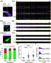
Arc clusters exist inside different endosomal species. (A) (i) A representative 2-color HILO image of HEK293T cells cotransfected with HaloArc (magenta) and EGFP-Rab5 (green). (ii) Time-lapse images of the selected ROI in (i). Arrowheads indicate HaloArc colocalized with Rab5+ vesicles. (B) (i) A representative 2-color HILO image of HEK293T cells cotransfected with HaloArc (magenta) and sfGFP-CD63 (green). (ii) Time-lapse images of the selected ROI in (i). Arrowheads indicate HaloArc colocalized with CD63+ vesicles. (C) (i) A representative 2-color HILO image of a HEK293T cell cotransfected with HaloArc (magenta) and EGFP-Rab7 (green). (ii) Time-lapse images of the selected ROI in (i). Arrowheads indicate HaloArc colocalized with Rab7+ vesicles. (D) Quantification for the frequency of overlap between HaloArc clusters and each vesicle marker (Rab5: early endosome, n = 104 clusters; CD63: multivesicular body, n = 99 clusters; and Rab7: late endosome, n = 54 clusters). (E) Average integrated fluorescence intensities of HaloArc clusters overlapped with each vesicle marker (Rab5, n = 20; CD63, n = 56; Rab7, n = 47). (F) Step size histogram of HaloArc cluster overlapped with each vesicle marker. (Mann–Whitney test; *P < 0.05 and ***P < 0.001). (Scale bar, 5 μm for all images.)
Fig. 5.
Fig. 5.
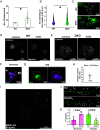
Decreased levels of RalA and RalB alters HaloArc trafficking and spine morphology. (A) Quantification of the number of HaloArc clusters in WT (pink) and DKO (green) HEK293T cells. (B) Quantification of integrated fluorescence intensity of HaloArc clusters in WT (pink) and DKO (green) HEK-293T cells. (C) A representative HILO image of WT HEK293T cell (Top) and DKO HEK293T cell (Bottom) expressing sfGFP-CD63. Representative images of HaloArc clusters inside the cytoplasm of wild-type (D) or DKO (E) HEK293T cells in confocal and STED modes. (F) A 2D-confocal image of a HEK293T cell expressing SnapArc (magenta, labeled with AF-647 SNAP-tag ligand) and sfGFP-CD63 (green). (G) Zoom-in view of an Arc-containing MVB (CD63). (H) MINFLUX imaging of SnapArc within the MVB in G. (I) Analysis of the size of SnapArc puncta resolved by MINFLUX. (J) Confocal imaging of cultured primary rat cortical neurons expressing membrane-associated EGFP cotransduced with either scrambled shRNA (Top) or RalA/B double knockdown shRNA (Bottom). Mushroom-like and filopodial spines are marked with asterisks and arrowheads, respectively. (K) Quantification of spine morphology in neurons treated with scrambled or RalA/B double knockdown shRNA (n = 21 dendrites for scrambled, n = 15 dendrites for knockdown condition) (Mann–Whitney test; *P < 0.05 and ***P < 0.001).
Fig. 6.
Fig. 6.
Effects of reduced levels of RalA and RalB on HaloArc-mediated intercellular RNA transfer and function in recipient cells. Comparison of EV concentration (A) and size (B) distribution of WT and DKO HEK293T cells with and without HaloArc expression. (C) Representative epifluorescence images of recipient cells treated with the conditioned medium from HaloArc-expressing wild-type cells. (D) Same as C except using the conditioned medium harvested from DKO cells. (Scale bars in C and D, 100 µm.) (E) Quantification of the RNA transfer efficiency from HaloArc-expressing wild-type (n = 29 images) or DKO cells (n = 27 images) (Mann–Whitney test; ***P < 0.001). (F) Immunocytochemistry of surface GluA1 subunit of AMPAR in untreated cultured mouse cortical neurons (Left) or those treated with EVs isolated from HaloArc-expressing wild-type (Middle) or RalA/B double knockdown (Right). (G) Quantification of the surface GluA1 level normalized to the average intensity from untreated neurons (n = 8 to 9 neurons each condition) (one-way ANOVA with the Tukey test *P < 0.05, ***P < 0.001, and n.s.: nonsignificant).
Fig. 7.
Fig. 7.
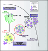
PI3P mediates Arc capsid trafficking and secretion through the endosome–MVB pathway. Arc protein is targeted to the limiting membrane of the early endosome through its interaction with PI3P (green), mediated by PI3K activity, and sorted through the axis of the late endosome and MVB. Endosomal PI3P could facilitate Arc oligomerization. Arc capsid assembly could occur either inside the cytoplasm or, more likely, within the intraluminal vesicles of MVB. Upon membrane fusion between MVB and the plasma membrane, EVs containing Arc are secreted.

Update of

References

    1. Link W., et al. , Somatodendritic expression of an immediate early gene is regulated by synaptic activity. Proc. Natl. Acad. Sci. U.S.A. 92, 5734–5738 (1995). - PMC - PubMed
    1. Korb E., Finkbeiner S., Arc in synaptic plasticity: From gene to behavior. Trends Neurosci. 34, 591–598 (2011). - PMC - PubMed
    1. Bramham C. R., et al. , The Arc of synaptic memory. Exp. Brain Res. 200, 125–140 (2010). - PMC - PubMed
    1. Griffiths D. J., Endogenous retroviruses in the human genome sequence. Genome Biol. 2, reviews1017.1 (2001). - PMC - PubMed
    1. Lynch C., Tristem M., A co-opted gypsy-type LTR-retrotransposon is conserved in the genomes of humans, sheep, mice, and rats. Curr. Biol. 13, 1518–1523 (2003). - PubMed

Substances

LinkOut - more resources