. 2024 Aug 27;121(35):e2406748121.
doi: 10.1073/pnas.2406748121. Epub 2024 Aug 23.

Platelet-activating factor (PAF) promotes immunosuppressive neutrophil differentiation within tumors

Affiliations

Platelet-activating factor (PAF) promotes immunosuppressive neutrophil differentiation within tumors

Ankit Dahal et al. Proc Natl Acad Sci U S A. .

Abstract

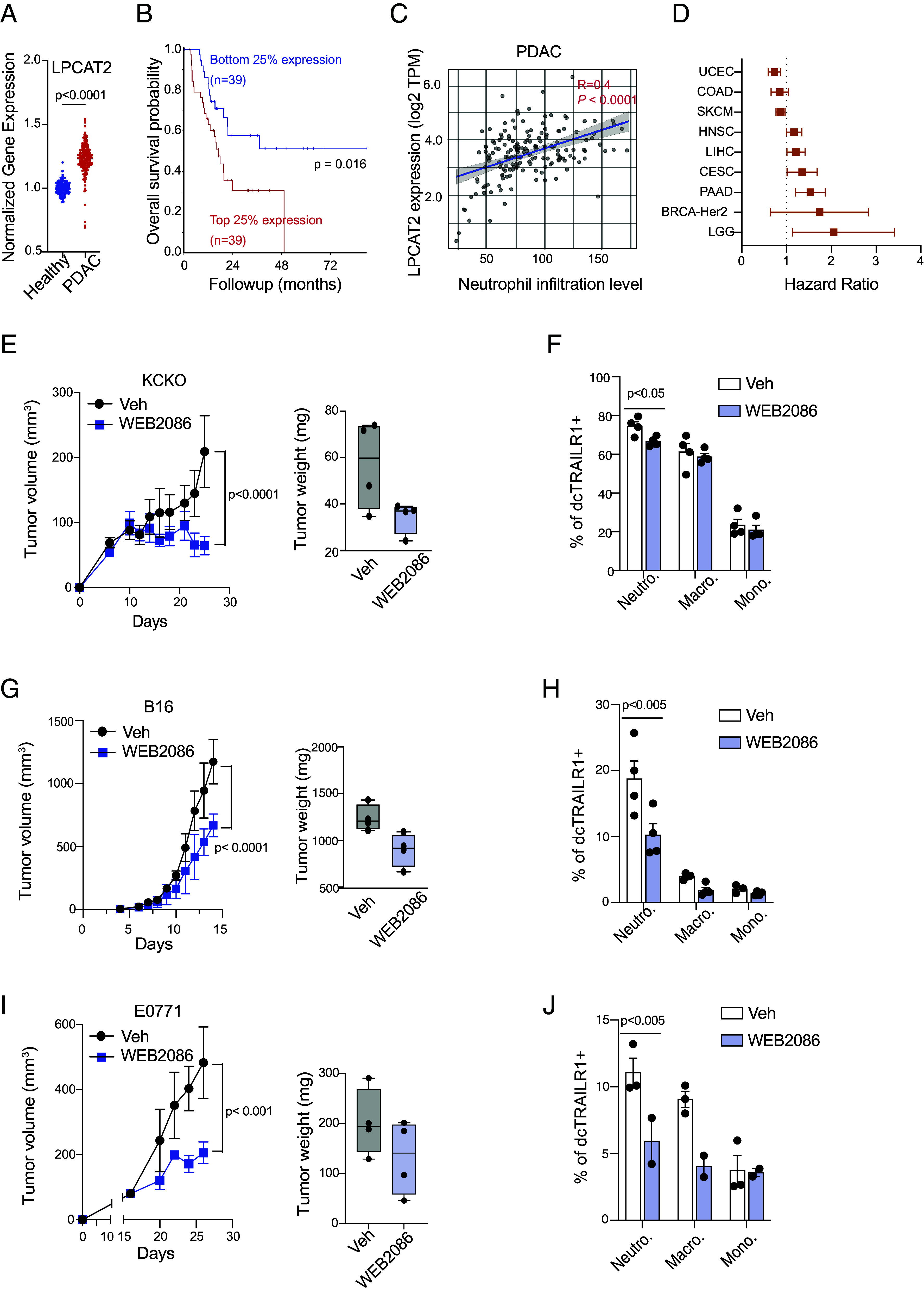
Chronic inflammatory milieu in the tumor microenvironment (TME) leads to the recruitment and differentiation of myeloid-derived suppressor cells (MDSCs). Polymorphonuclear (PMN)-MDSCs, which are phenotypically and morphologically defined as a subset of neutrophils, cause major immune suppression in the TME, posing a significant challenge in the development of effective immunotherapies. Despite recent advances in our understanding of PMN-MDSC functions, the mechanism that gives rise to immunosuppressive neutrophils within the TME remains elusive. Both in vivo and in vitro, newly recruited neutrophils into the tumor sites remained activated and highly motile for several days and developed immunosuppressive phenotypes, as indicated by increased arginase 1 (Arg1) and dcTrail-R1 expression and suppressed anticancer CD8 T cell cytotoxicity. The strong suppressive function was successfully recapitulated by incubating naive neutrophils with cancer cell culture supernatant in vitro. Cancer metabolite secretome analyses of the culture supernatant revealed that both murine and human cancers released lipid mediators to induce the differentiation of immunosuppressive neutrophils. Liquid chromatography-mass spectrometry (LC-MS) lipidomic analysis identified platelet-activation factor (PAF; 1-O-alkyl-2-acetyl-sn-glycero-3-phosphocholine) as a common tumor-derived lipid mediator that induces neutrophil differentiation. Lysophosphatidylcholine acyltransferase 2 (LPCAT2), the PAF biosynthetic enzyme, is up-regulated in human pancreatic ductal adenocarcinoma (PDAC) and shows an unfavorable correlation with patient survival across multiple cancer types. Our study identifies PAF as a lipid-driven mechanism of MDSC differentiation in the TME, providing a potential target for cancer immunotherapy.

Keywords: Cancer; MDSC; myeloid cells; neutrophil; tumor microenvironment.


Conflict of interest statement

Competing interests statement:The authors declare no competing interest.

Figures

Fig. 1.
Fig. 1.
Cancer cell–neutrophil interaction directly regulates PMN-MDSC differentiation. (A) Neutrophils, CD8, and CD4 T cells were isolated from orthotopic KCKO tumors, and relative abundance of each cell type was quantified using flow cytometry. (B) qPCR and flow cytometric quantification of (C) Arginase 1 and (D) dcTRAIL-R1 expression in neutrophils isolated from different organs of naïve and tumor-bearing mice. (E and F) Neutrophils isolated from different organs were cocultured with CFSE-labeled CD8 T cells to assess neutrophil-mediated suppression of T cell proliferation. (G) Schematic of neutrophil migration into KCKO tumoroid. Images from 0 h and 12 h after incubation are shown. The graph depicts the MFI of neutrophils that migrated into the KCKO tumoroid (Red). Neutrophil migration kinetics after adding Pertussis Toxin (PTX) is shown in blue. (H) The meandering index and track velocity of neutrophils cultured with cancer cells were calculated at different time points after incubation with KCKO cancer. Data from (B), (C), and (F) include n = 3, P < 0.0001, and (D) n = 3, P < 0.05 as determined by the one-way ANOVA test with Tukey’s multiple-comparison post hoc test. (H) One-way ANOVA test with Tukey MCT.
Fig. 2.
Fig. 2.
Cancer-secreted factors promote PMN-MDSC differentiation in vitro. (A) MFI of neutrophil activation markers CD11b and CD62L was quantified using flow cytometry. (B) Neutrophil survival was measured at different time points using flow cytometry to count the number of live neutrophils present in the culture at each day. (C) Arg1 expression was quantified at different time points after neutrophils were cultured in KCKO cancer supernatant using qPCR for RNA and flow cytometry for Arg1 protein expression after 24 h in culture. (D) Naïve neutrophils were cultured either in culture media or media containing pancreatic interstitial fluid or tumor interstitial fluids for 18 h. Neutrophil activation (CD11b), percent alive, and percent dcTRAIL-R1 positive are quantified. (E and F) Suppression of CD8 T cell proliferation. (G) OT1 T cells were cocultured with OVA-pulsed KCKO or E0771 cancer cells in the presence of cancer supernatant pulsed neutrophils. Cytotoxicity was quantified using flow cytometric reading for 7AAD-positive cancer cells. (A) n = 3. (B and C) Ordinary two-way ANOVA test, n = 3, P < 0.0001. (D) n = 3, one-way ANOVA, Tukey’s MCT. (F) Student’s t test, P < 0.0001. (G) One-way ANOVA test, n = 3, Tukey’s MCT, P < 0.01.
Fig. 3.
Fig. 3.
Cancer-secreted factors promote MDSC differentiation in murine and human neutrophils across multiple cancer types. (A and B) Neutrophil activation (A) and survival (B) across different cancer supernatants after 12 h in culture were quantified using MFI of CD11b expression and Annexin V staining, respectively. (C) Kinetics of Arg1 expression quantifying expression across the various murine cancer types up to 24 h in culture. (D) CD8 T cell proliferation index (the average number of divisions of proliferating cells) after overnight coculture with cancer-supernatant conditioned neutrophils. (E and F) Activation (E) and survival (F) of human neutrophils after coculturing with supernatants prepared from human cancers or cancer-associated fibroblast. (G) Human CD8 T cell proliferation after being cocultured with cancer supernatant-conditioned human neutrophils for 3 d. Data from A, B, C, D, E, F, G, and H: n = 3, one-way ANOVA, Tukey’s MCT. C: n = 3, two-way ANOVA, P < 0.001.
Fig. 4.
Fig. 4.
Cancer-secreted lipid mediators induce PMN-MDSC differentiation in both murine and human cancers. (AC) Neutrophil activation (A; CD11b MFI), Arg1 (B), and dcTrail-R1 (C) expression levels were quantified after overnight incubation in heat-inactivated KCKO cancer supernatant. (D) Mean and 95% CI of EV concentration in KCKO supernatant before and after EV depletion with ultracentrifugation. (E and F) Quantification of neutrophil activation (E) and Arg1 expression (F) after culturing with EV-depleted KCKO supernatant. (G and H) Neutrophil activation (G) and Arg1 expression (H) after incubation in lipid-depleted supernatants prepared from KCKO and E0771 murine cancers. (I) Expression of dcTrail-R1 in neutrophils after overnight coculture in lipid-depleted cancer supernatant. (AC) One-way ANOVA test with Tukey’s MCT, n = 3 (A) ns, (B) P < 0.0001, and (C) P < 0.0001. (H and I) Two-way ANOVA test, n = 3, P < 0.0001. (EG) One-way ANOVA, Tukey’s MCT, n = 3, ns.
Fig. 5.
Fig. 5.
Cancer-secreted PAF induces neutrophil differentiation. (A) LC-MS analysis of lipids present in KCKO, PDAC3, HT29, H1299, and TOV11D cancer supernatants. Lipids with FC > 2 and P-value < 0.05 are marked (red). (B) Four shared lipids are identified across murine and human cancers. (C) Arg1 expression in murine and human neutrophils after adding reconstituted lipids (1 μM) to media. (D) Arg1 expression in murine neutrophils after adding PAF to lipid-depleted KCKO and E0771 supernatant. (E) Kinetics of Arg1 and dcTrail-R1 expression in murine neutrophils in the presence of PAF. (F) CD8 T cell proliferation after overnight coculture with neutrophils conditioned with DMSO or PAF. (G) ELISA quantifying concentration of PAF in TIF of mock surgery (pancreas) vs. orthotopic KCKO tumors. (H) Quantification of Arg1 and dcTrail-R1 expression after incubation of neutrophils in KCKO and E0771 supernatants in the presence of PAFR inhibitor, WEB-2086 (5 μM). (I) Bulk RNAseq of neutrophils cultured in the presence of DMSO or PAF overnight, highlighting the expression of intratumoral MDSC and inflammatory signature genes. (C and E) One-way ANOVA, Tukey’s MCT, n = 3, P < 0.05 and <0.0001, respectively. (D) Two-way ANOVA, Šidák MCT, n = 3, P < 0.0001. (F and H) Student’s t test. n = 9 P < 0.005.
Fig. 6.
Fig. 6.
Inhibition of PAFR limits tumor growth in vivo. (A) Normalized LPCAT2 expression in healthy pancreas vs. PDAC tumors was obtained using GTEX and TCGA datasets from the UCSC Xena data analysis tool. (B) TCGA survival curve subsetting top and bottom 25% LPCAT2 expressing PDAC tumors obtained using R2 Genome Browser tool. (C) Intratumoral neutrophil infiltration compared to LPCAT2 expression across human PDAC dataset obtained from TIMER 2.0 tool. (D) TCGA data correlating LPCAT2 expression and patient survival were obtained using the R2 genome browser tool. (E) KCKO cells growth kinetics and endpoint tumor weight at endpoint with vehicle (5% ethanol: PBS) and 10 mg/kg WEB2086. The drug was injected intraperitoneally (I.P.) every day. (F) dcTrail-R1 expression of tumor-infiltrating myeloid cells in vehicle vs. WEB2086-treated mice. (G) B16F0 growth kinetics and tumor weight and (H) expression of dcTrail-R1 in myeloid cells. (I) E0771 growth kinetics and tumor weight and (J) expression of dcTrail-R1 in myeloid cells. (E, G, I) Two-way ANOVA, n = 4. (F, H, J) Multiple t test, Holm–Šidák MCT, n = 4.

References

    1. Veglia F., Perego M., Gabrilovich D., Myeloid-derived suppressor cells coming of age. Nat. Immunol. 19, 108–119 (2018). - PMC - PubMed
    1. Jin L., Kim H. S., Shi J., Neutrophil in the pancreatic tumor microenvironment. Biomolecules 11, 1170 (2021). - PMC - PubMed
    1. Gentles A. J., et al. , The prognostic landscape of genes and infiltrating immune cells across human cancers. Nat. Med. 21, 938–945 (2015). - PMC - PubMed
    1. Pekarek L. A., Starr B. A., Toledano A. Y., Schreiber H., Inhibition of tumor growth by elimination of granulocytes. J. Exp. Med. 181, 435–440 (1995). - PMC - PubMed
    1. Gabitass R. F., Annels N. E., Stocken D. D., Pandha H. A., Middleton G. W., Elevated myeloid-derived suppressor cells in pancreatic, esophageal and gastric cancer are an independent prognostic factor and are associated with significant elevation of the Th2 cytokine interleukin-13. Cancer Immunol. Immunother 60, 1419–1430 (2011). - PMC - PubMed

MeSH terms

Substances