. 2024 Aug 23;10(34):eado6566.
doi: 10.1126/sciadv.ado6566. Epub 2024 Aug 23.

The nuclear export protein XPO1 provides a peptide ligand for natural killer cells

Affiliations

The nuclear export protein XPO1 provides a peptide ligand for natural killer cells

Matthew D Blunt et al. Sci Adv. .

Abstract

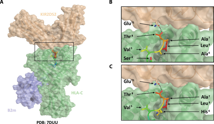
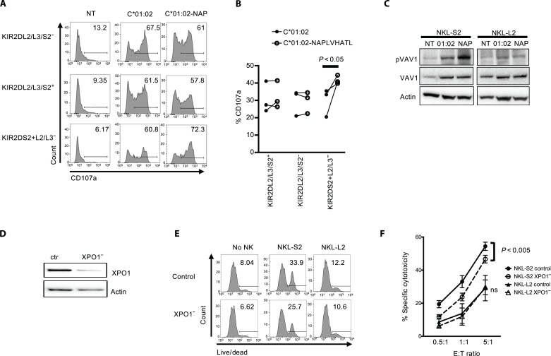
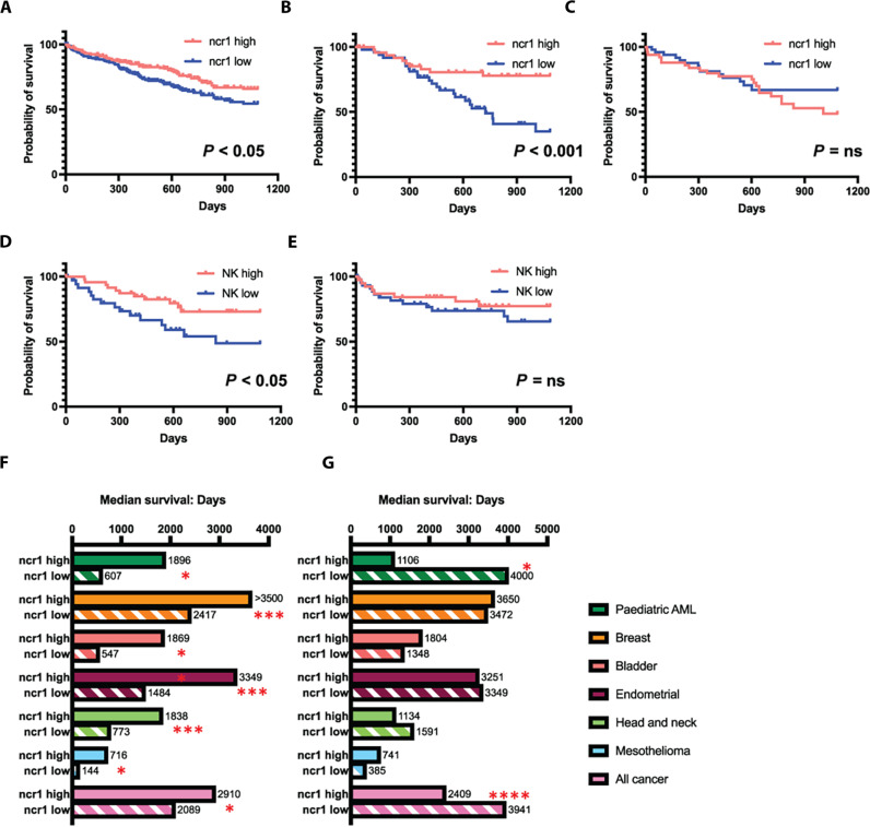
XPO1 (Exportin-1/CRM1) is a nuclear export protein that is frequently overexpressed in cancer and functions as a driver of oncogenesis. Currently small molecules that target XPO1 are being used in the clinic as anticancer agents. We identify XPO1 as a target for natural killer (NK) cells. Using immunopeptidomics, we have identified a peptide derived from XPO1 that can be recognized by the activating NK cell receptor KIR2DS2 in the context of human leukocyte antigen-C. The peptide can be endogenously processed and presented to activate NK cells specifically through this receptor. Although high XPO1 expression in cancer is commonly associated with a poor prognosis, we show that the outcome of specific cancers, such as hepatocellular carcinoma, can be substantially improved if there is concomitant evidence of NK cell infiltration. We thus identify XPO1 as a bona fide tumor antigen recognized by NK cells that offers an opportunity for a personalized approach to NK cell therapy for solid tumors.


Figures

Fig. 1.
Fig. 1.. An XPO1-derived peptide is a ligand for KIR2DS2.
(A) SeqLogo plots of peptides eluted from Huh7 and Huh7:HLA-C*01:02 cell lines. (B) Representative and annotated ms2 spectrum that has been assigned to the peptide sequence NAPLVHATL. The resulting Byonic score, the posterior error probability (PEP) and the mass/charge ratio (m/z) value of the singly protonated species is shown in the inset. (C) 721.174 cells were incubated with NAPLVHATL at the indicated concentrations, stained for HLA-C using the DT9 antibody, and analyzed by flow cytometry. The mean fluorescence intensity (MFI) of DT9 staining compared to the positive control VAPWNSFAL peptide is shown. (D) 721.174 cells were incubated with 100 μM NAPLVHATL peptide and stained with a KIR2DS2-tetramer and analyzed by flow cytometry. Histogram plots of KIR2DS2-staining compared with a no peptide control are shown, with median fluorescence intensities indicated. (E to G) 721.174 cells were incubated overnight with the indicated peptides at 200 μM and cocultured with NKL-2DS2 cells, and then the cells were lysed and analyzed for phosphorylation of Syk (Tyr323 and Tyr317) and Vav1 (Tyr174) by Western blotting. A representative Western blot is shown in (E); the ratios of pVav1 to Vav1 (F) and pSyk to Syk (G) from three independent experiments are also shown. P values indicate comparison of the NAPLVHATL peptide with negative (VAPWNSDAL) and positive (LNPSVAATL) control peptides as determined by one-way analysis of variance (ANOVA).
Fig. 2.
Fig. 2.. Modeling of HLA-C*01:02, NAPLVHATL, and KIR2DS2 binding interactions.
On the basis of the crystal structure of the HLA-KIR complex (PDB: 7DUU), a potential binding mode for the XPO1-derived peptide, NAPLVHATL, was investigated using PyMOL. In silico mutagenesis of the peptide was performed using the protein mutagenesis tool within PyMOL, with the minimization of steric clashes being used to determine the optimal rotameric state of the mutated residue. Peptide-protein interactions were analyzed using PISA. KIR2DS2 is shown as a brown transparent surface, HLA as a green transparent surface, and β2-microglobulin as a blue transparent surface. The peptide is shown as a rainbow ribbon backbone with side chains as sticks. Hydrogen bonds are shown as a black dashed line. (A) Model of KIR2DS2 binding with HLA-C*01:02 and NAPLVHATL. Conservation of the KIR2DS2 Glu71–peptide Thr8 hydrogen bond is shown in (B) for the LNPSVAATL crystal structure and in (C) for the modeled NAPLVHATL peptide.
Fig. 3.
Fig. 3.. Endogenously presented NAPLVHATL activates KIR2DS2+ NK cells.
(A and B) 721.221 cells were transfected with HLA-C*01:02 alone (C*01:02) or in combination with the peptide NAPLVHATL (C*01:02-NAP), and the cell lines were used as targets for degranulation assays with interleukin-15–activated NK cells as effectors. CD107a expression on the indicated subpopulations of CD3CD56+ NK cells was assessed by flow cytometry against these targets. (NT = no target). One representative flow cytometry plots of CD107a expression is shown from six healthy donors tested (A), and degranulation from three patients with HCC is shown in (B). P values were determined by paired t test. (C) NKL-2DS2 or NKL-2DL2 cells were incubated with either no target (NT), 721.221:HLA-C*01:02 (C*01:02) or 721.221:HLA-C*01:02+NAPLVHATL (NAP) cells for 5 min and assessed for Vav1 (Tyr174) phosphorylation by immunoblotting. A representative image from six experiments is shown. (D) Representative immunoblot of XPO1 silencing following XPO1-targeting siRNA treatment of Huh7:HLA-C*01:02 cells. (E and F) Huh7:HLA-C*01:02 cells were treated with control or XPO1-targeting siRNA and used as targets in cytotoxicity assays with NKL-S2 or NKL-L2 cells. Killing was determined using the LIVE/DEAD stain. Representative plots at an E:T ratio of 5:1 are shown in (E), and the mean specific cytotoxicity and SEM from five independent experiments are shown (F). P values were determined by two-way ANOVA.
Fig. 4.
Fig. 4.. The combination of high levels of XPO1 and ncr1 expression is associated with improved survival in cancer.
(A to C) Kaplan-Meier plots of HCC TCGA data comparing 3-year survival in individuals with highest (red lines) and lowest (blue lines) quartile levels of ncr1 expression in the whole cohort (A), individuals with levels of XPO1 above the median (B), and individuals with levels of XPO1 below the median (C). (D and E) show the Kaplan-Meier plots of the XPO1 high (D) and low (E) analyzed according to peripheral blood NK cell infiltration into the tumors as determined by Cibersort X analysis comparing the highest and lowest quartiles of NK cell infiltration. For all plots, P values were calculated using the log-rank Mantel-Cox test. (F and G) Median survival of individuals with high and low levels of ncr1 expression in different TCGA datasets comparing individual groups with XPO1 levels above (F) and below (G) the median value for each group. P values compare the ncr1high and ncr1low groups using the log-rank Mantel-Cox test. The following numbers of cases analyzed in each cohort were as follows: pediatric acute myeloid leukemia (AML) (187 cases), breast cancer (1098 cases), bladder cancer (412 cases), endometrial cancer (559 cases), head and neck cancer (528 cases), mesothelioma (86 cases), and all cancers (11,768 cases). *P < 0.05, **P < 0.01, ***P < 0.005, ****P < 0.0001.

References

    1. Chiossone L., Dumas P. Y., Vienne M., Vivier E., Natural killer cells and other innate lymphoid cells in cancer. Nat. Rev. Immunol. 18, 671–688 (2018). - PubMed
    1. Vilches C., Parham P., KIR: Diverse, rapidly evolving receptors of innate and adaptive immunity. Annu. Rev. Immunol. 20, 217–251 (2002). - PubMed
    1. Leone P., De Re V., Vacca A., Dammacco F., Racanelli V., Cancer treatment and the KIR-HLA system: An overview. Clin. Exp. Med. 17, 419–429 (2017). - PubMed
    1. Bao X., Hanson A. L., Madeleine M. M., Wang S. S., Schwartz S. M., Newell F., Pettersson-Kymmer U., Hemminki K., Tiews S., Steinberg W., Rader J. S., Castro F., Safaeian M., Franco E. L., Coutlee F., Ohlsson C., Cortes A., Marshall M., Mukhopadhyay P., Cremin K., Johnson L. G., Garland S. M., Tabrizi S. N., Wentzensen N., Sitas F., Trimble C., Little J., Cruickshank M., Frazer I. H., Hildesheim A., Brown M. A., Duncan E. L., Sun Y. P., Leo P. J., HLA and KIR associations of cervical neoplasia. J. Infect. Dis. 218, 2006–2015 (2018). - PMC - PubMed
    1. Cariani E., Pilli M., Zerbini A., Rota C., Olivani A., Zanelli P., Zanetti A., Trenti T., Ferrari C., Missale G., HLA and killer immunoglobulin-like receptor genes as outcome predictors of hepatitis C virus-related hepatocellular carcinoma. Clin. Cancer Res. 19, 5465–5473 (2013). - PubMed

LinkOut - more resources