. 2024 Aug 23;20(8):e1012498.
doi: 10.1371/journal.ppat.1012498. eCollection 2024 Aug.

Cell-intrinsic regulation of phagocyte function by interferon lambda during pulmonary viral, bacterial super-infection

Affiliations

Cell-intrinsic regulation of phagocyte function by interferon lambda during pulmonary viral, bacterial super-infection

Danielle Antos et al. PLoS Pathog. .

Abstract

Influenza infections result in a significant number of severe illnesses annually, many of which are complicated by secondary bacterial super-infection. Primary influenza infection has been shown to increase susceptibility to secondary methicillin-resistant Staphylococcus aureus (MRSA) infection by altering the host immune response, leading to significant immunopathology. Type III interferons (IFNs), or IFNλs, have gained traction as potential antiviral therapeutics due to their restriction of viral replication without damaging inflammation. The role of IFNλ in regulating epithelial biology in super-infection has recently been established; however, the impact of IFNλ on immune cells is less defined. In this study, we infected wild-type and IFNLR1-/- mice with influenza A/PR/8/34 followed by S. aureus USA300. We demonstrated that global IFNLR1-/- mice have enhanced bacterial clearance through increased uptake by phagocytes, which was shown to be cell-intrinsic specifically in myeloid cells in mixed bone marrow chimeras. We also showed that depletion of IFNLR1 on CX3CR1 expressing myeloid immune cells, but not neutrophils, was sufficient to significantly reduce bacterial burden compared to mice with intact IFNLR1. These findings provide insight into how IFNλ in an influenza-infected lung impedes bacterial clearance during super-infection and show a direct cell intrinsic role for IFNλ signaling on myeloid cells.


Conflict of interest statement

The authors have declared that no competing interests exist.

Figures
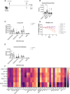
Fig 1
Fig 1. Global IFNLR1-/- mice have increased bacterial clearance compared with wild type (WT) mice during pulmonary super-infection.
A. Mice were infected with 900 PFU A/PR/8/34 on day 0 and 5 x 10⁷ colony forming units (CFU) Staphylococcus aureus strain USA300 on day 6, followed by harvest on day 7. Figure created on Biorender.com. B. Global IFNLR1-/- mice have reduced bacterial burden compared to WT mice 24 hours post bacterial infection (IFNLR1-/-: F/S n = 18, -/S n = 12; WT: F/S n = 17, -/S n = 16). C. Protein levels in the airways of infected mice are not different between IFNLR1-/- and WT mice during super-infection (IFNLR1-/- n = 13, WT n = 13), single bacterial (IFNLR1-/- n = 12, WT = 11) or viral infection (IFNLR1-/- n = 8, WT n = 9), or at basal levels (IFNLR1-/- n = 7, WT n = 10). D. Mouse weight loss was monitored daily during infection time-course (data from at least 3 replicates). E. Immune cell infiltration into the airways was assessed by quantification of cells in bronchoalveolar lavage (BAL) fluid during super-infection (IFNLR1-/- n = 13, WT n = 11), single bacterial (IFNLR1-/- n = 12, WT = 12) or viral infection (IFNLR1-/- n = 8, WT n = 8), or at basal levels (IFNLR1-/- n = 7, WT n = 8). F. Global cytokine levels were detected using Bio-Plex assays in mice on day 7 of the model during super-infection (IFNLR1-/- n = 12, WT n = 12), single infections (Bacteria: IFNLR1-/- n = 5, WT n = 6; Virus: (IFNLR1-/- n = 4, WT n = 4), and at basal levels (IFNLR1-/- n = 2, WT n = 2). Statistics compare IFNLR1-/- to WT mice within each infection type. p values: *<0.05, **<0.01, ***<0.001, ****<0.0001.
Fig 2
Fig 2. IFNλ modulates chemokine production to influence antibacterial type 17 immunity in the lung.
A. IL-17 and IL-22 were measured in the BAL fluid by ELISA, and both were increased in super-infected IFNLR1-/- mice (F/S: IFNLR1-/- n = 9, WT n = 9; -/S: IFNLR1-/- n = 7, WT n = 7; data from 3 replicates). B. Antibody blockade of IL-17A increased bacterial burden in super-infected IFNLR1-/- mice (Ad-IL-17 n = 11, Ad-eGFP n = 11). C. Neutrophil recruitment to the lung was measured and quantified by flow cytometry in total cell counts (left) and frequency (right; representative data from 2 replicates, n = 7). D. Levels of IL-1β (left; IFNLR1-/- n = 12, WT n = 13), IL-23 (middle; IFNLR1-/- n = 10, WT n = 10), and IL-6 (right; IFNLR1-/- n = 8, WT n = 7) were assessed in super-infected mice by Bio-plex or ELISA (data from 3 replicates). p values: *<0.05, **<0.01.
Fig 3
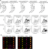
Fig 3. Disruption of IFNλ signaling in phagocytes directly increases their ability to phagocytose bacteria.
A. Global IFNLR1-/- and WT mice were super-infected as previously stated and fluorescently labeled MSSA strain RN6390 (dsRed expressing) were used to measure and quantify bacteria taken up by phagocytes. IFNLR1-/- mice had increased uptake both 6 hours post bacterial infection (hashed bars) and 24 hours post bacterial infection (open bars), the latter shown in flow plots below (data from 3 replicates). Populations of interest included inflammatory monocytes (CD45+, Siglec-F-, Ly6C++, CD11b+, MHC-II+/-), lung monocytes (CD45+, CD11b+, Siglec-F-, Ly6C+/-, CX3CR1+), interstitial macrophages (CD45+, CD11b+, Siglec-F-, MHC-II+, CX3CR1+), and neutrophils (CD45+, CD11b+, Ly6G+). B. Amnis ImageStream was used to visualize increased phagocytosis in IFNLR1-/- immune cells. IFNLR1-/- cells (left; n = 8) have increased dsRed S. aureus staining (yellow) within the cell membrane compared to WT cells (right; n = 7). Cells that stain positively for S. aureus also express CX3CR1 highly. p values: *<0.05.
Fig 4
Fig 4. IFNλ acts directly on immune cells to impede bacterial phagocytosis.
A. Weight loss is equal between WT host mice and IFNLR1-/- host mice. B. Mixed bone marrow chimeras have comparable bacterial burden (left), immune cell infiltration (middle), and lung leak (right) regardless of host genotype (host: KO n = 12, WT n = 12). C. In a WT host, IFNLR1-/- immune cells (inflammatory monocytes, lung monocytes, and interstitial macrophages) take up more bacteria, visualized by MFI, than WT immune cells. Uptake in the IFNLR1-/- host is not significantly altered (host: KO n = 8, WT n = 8). D. In WT hosts, IFNLR1-/- immune cells have increased frequencies of dsRed+ cells compared to WT immune cells (host: KO n = 8, WT n = 8). These trends are consistent but not significant in KO hosts. E. Visualization of increased dsRed+ populations in IFNLR1-/- immune cells compared to WT immune cells within the same WT host. p values: *<0.05, **<0.01.
Fig 5
Fig 5. IFNλ does not directly act on neutrophils to decrease bacterial clearance during super-infection.
A. S100A8-Cre-IFNLR1fl/fl mice constitutively express Cre, and thus were super-infected as previously stated with harvest on day 7. Figure created on Biorender.com. B. S100A8-Cre-IFNLR1fl/fl (cKO) mice do not have significantly altered bacterial burden compared to Cre- littermate controls (cKO n = 10, Cre- n = 12). C. Neutrophil uptake of dsRed-labeled bacteria was not altered by frequency (left; cKO n = 10, Cre- n = 12) or total number (right; cKO n = 6, Cre- n = 6) of dsRed+ cells in S100A8-Cre-IFNLR1fl/fl versus Cre- controls. D. Neutrophil recruitment to the lung, measured by flow cytometry, was not altered by frequency (left; cKO n = 10, Cre- n = 12) or total number (right; cKO n = 6, Cre- n = 6) in S100A8-Cre-IFNLR1fl/fl versus Cre- controls. E. Mortality curve showed no difference in S100A8-Cre-IFNLR1fl/fl or Cre- control survival during lethal super-infection (cKO n = 7, Cre- n = 9).
Fig 6
Fig 6. Depletion of IFNLR1 specifically on CX3CR1 cells is sufficient to increase bacterial clearance during super-infection.
A. CX3CR1-Cre-IFNLR1fl/fl mice or Cre- littermate controls were injected with 2mg of Tamoxifen two weeks and one week before influenza infection on day 0. Super-infection proceeded as previously, with additional injections given on days 0 and 5 of the model. Figure created on Biorender.com. B. CX3CR1-Cre-IFNLR1fl/fl (cKO) mice had significantly reduced bacterial burden during super-infection (open bars; cKO n = 6, Cre- n = 10, data from 3 replicates) compared to littermate controls, but there are no differences during single bacterial infection (hashed bars; cKO n = 5, Cre- n = 5, data from 2 replicates). C. CX3CR1 cKO mice have greater and longer survival after lethal super-infection compared to littermate controls (p = 0.05; cKO n = 29, Cre- n = 27). D-E. Bacterial uptake by immune cells was largely unchanged in super-infected CX3CR1 cKO mice and littermate controls 24h post bacterial infection (D, cKO n = 8, Cre- n = 11), also shown by representative flow plots below (E). p values: *<0.05, **<0.01.
Fig 7
Fig 7. Specific deletion of IFNLR1 on lung myeloid cells increases localization of bacteria within phagolysosomes.
A. CX3CR1-Cre-IFNLR1fl/fl mice or littermate controls were super-infected as previously with pHrodo red-labeled S. aureus and pHrodo red+ cells were quantified by flow cytometry 6h post bacterial infection (cKO n = 7, Cre- n = 8). B. ImageStream analysis shows high colocalization of dsRed S. aureus and endosomal marker CD63 in global IFNLR1-/- and WT mice (IFNLR1-/- n = 8, WT n = 8). p values: *<0.05, **<0.01.
Fig 8
Fig 8. Deletion of IFNLR1 in CX3CR1+ immune cells impacts many immune pathways.
A. CX3CR1-Cre-eYFP-IFNLR1fl/fl mice were treated with 2 mg Tamoxifen or corn oil controls as previously and infected with influenza for 6 days. After sacrifice, whole lung was processed into single cell suspension and sorted for eYFP+ cells before RNA isolation and bulk sequencing. In tamoxifen-treated mice, eYFP+ cells had deletion of IFNLR1 while the receptor was intact in eYFP+ cells from untreated mice (cKO: treated n = 5, untreated n = 4). Figure created on Biorender.com. B. All differentially expressed genes (DEGs) in treated mice as compared to untreated were plotted as a volcano plot. C. DEGs within the interferome were specifically analyzed, and those upregulated in treated mice were plotted as a heatmap. D. GO analysis was performed using clusterprofiler (Bioconductor) in R and significantly altered pathways in treated mice compared to controls are shown.

References

    1. Kotenko S V., Gallagher G, Baurin V V, Lewis-Antes A, Shen M, Shah NK, et al. IFN-λs mediate antiviral protection through a distinct class II cytokine receptor complex. Nat Immunol. 2003;4: 69–77. doi: 10.1038/ni875 - DOI - PubMed
    1. Sheppard P, Kindsvogel W, Xu W, Henderson K, Schlutsmeyer S, Whitmore TE, et al. IL-28, IL-29 and their class II cytokine receptor IL-28R. Nat Immunol. 2003;4: 63–68. doi: 10.1038/ni873 - DOI - PubMed
    1. Galani IE, Triantafyllia V, Eleminiadou EE, Koltsida O, Stavropoulos A, Manioudaki M, et al. Interferon-λ Mediates Non-redundant Front-Line Antiviral Protection against Influenza Virus Infection without Compromising Host Fitness. Immunity. 2017;46: 875–890.e6. doi: 10.1016/j.immuni.2017.04.025 - DOI - PubMed
    1. Davidson S, McCabe TM, Crotta S, Gad HH, Hessel EM, Beinke S, et al. IFN λ is a potent anti-influenza therapeutic without the inflammatory side effects of IFN α treatment. EMBO Mol Med. 2016;8: 1099–1112. doi: 10.15252/emmm.201606413 - DOI - PMC - PubMed
    1. Crotta S, Davidson S, Mahlakoiv T, Desmet CJ, Buckwalter MR, Albert ML, et al. Type I and Type III Interferons Drive Redundant Amplification Loops to Induce a Transcriptional Signature in Influenza-Infected Airway Epithelia. PLoS Pathog. 2013;9. doi: 10.1371/journal.ppat.1003773 - DOI - PMC - PubMed

MeSH terms