. 2024 Sep:107:105274.
doi: 10.1016/j.ebiom.2024.105274. Epub 2024 Aug 22.

Altered memory CCR6+ Th17-polarised T-cell function and biology in people with HIV under successful antiretroviral therapy and HIV elite controllers

Affiliations

Altered memory CCR6+ Th17-polarised T-cell function and biology in people with HIV under successful antiretroviral therapy and HIV elite controllers

Alexis Yero et al. EBioMedicine. 2024 Sep.

Abstract

Background: Despite successful antiretroviral therapy (ART), frequencies and immunological functions of memory CCR6+ Th17-polarised CD4+ T-cells are not fully restored in people with HIV (PWH). Moreover, long-lived Th17 cells contribute to HIV persistence under ART. However, the molecular mechanisms underlying these observations remain understudied.

Methods: mRNA-sequencing was performed using Illumina technology on freshly FACS-sorted memory CCR6+CD4+ T-cells from successfully ART-treated (ST), elite controllers (EC), and uninfected donors (HD). Gene expression validation was performed by RT-PCR, flow cytometry, and in vitro functional assays.

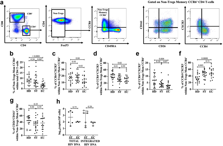
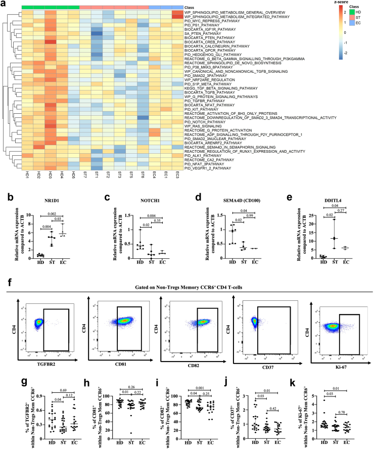
Findings: Decreased Th17 cell frequencies in STs and ECs versus HDs coincided with reduced Th17-lineage cytokine production in vitro. Accordingly, the RORγt/RORC2 repressor NR1D1 was upregulated, while the RORγt/RORC2 inducer Semaphorin 4D was decreased in memory CCR6+ T-cells of STs and ECs versus HDs. The presence of HIV-DNA in memory CCR6+ T-cells of ST and EC corresponded with the downregulation of HIV restriction factors (SERINC3, KLF3, and RNF125) and HIV inhibitors (tetraspanins), along with increased expression of the HIV-dependency factor MRE11, indicative of higher susceptibility/permissiveness to HIV-1 infection. Furthermore, markers of DNA damage/modification were elevated in memory CCR6+ T-cells of STs and ECs versus HDs, in line with their increased activation (CD38/HLA-DR), senescence/exhaustion phenotype (CTLA-4/PD-1/CD57) and their decreased expression of proliferation marker Ki-67.

Interpretation: These results reveal new molecular mechanisms of Th17 cell deficit in ST and EC PWH despite a successful control of HIV-1 replication. This knowledge points to potential therapeutic interventions to limit HIV-1 infection and restore frequencies, effector functions, and senescence/exhaustion in Th17 cells.

Funding: This study was funded by the Canadian Institutes of Health Research (CIHR, operating grant MOP 142294, and the Canadian HIV Cure Enterprise [CanCURE 2.0] Team Grant HB2 164064), and in part, by the Réseau SIDA et maladies infectieuses du Fonds de recherche du Québec-Santé (FRQ-S).

Keywords: CCR6; DNA damage; DNA repair; HIV infection; HIV persistence; Th17 cells; Transcriptomics.


Conflict of interest statement

Declaration of interests We have no competing interest to declare.

Figures

Fig. 1
Fig. 1
Differential gene expression in memory CCR6+CD4+ T-cells from PWH under successful ART and elite controllers compared to HIV-uninfected individuals. a) Before cell-sorting, total CD4+ T-cells were first isolated from PBMCs of HIV healthy donors (HD, n = 5), successfully ART-Treated HIV+ individuals (ST, n = 6), and HIV elite controllers (EC, n = 3) by negative selection using magnetic beads. Gating strategy used for the FACS-sorting of memory CD4+CD25CD45RACCR6+ T-cells. Total RNA from FACS-sorted cells was used for genome-wide transcriptomics analysis. The differential expression analysis was performed using a linear model with moderated t-statistics as implemented in the Bioconductor package limma (version 3.58.1). b) Heatmap showing differential gene expression in sorted non-Tregs memory CCR6+ cells in the 3 study groups related to different T-helpers (Th1, Th2, and Th17) and Tregs cells. c) Representation of the number of commonly and differentially expressed genes when comparing EC versus HD, EC versus ST, and ST versus HD individuals. d) Principal component analysis plots. Scores plot of PC1 versus PC2 from PCA are shown for mRNA-seq results. Each dot represents one sample. Colours are assigned based on the known of specimen assignment. e) Volcano plots showing differentially expressed genes when comparing EC versus HD, EC versus ST, and ST versus HD individuals. f) Top-modulated 50 genes in ST and EC individuals compared to HD subjects. Highlighted genes correspond to genes that were previously associated with (1) Th17 cell differentiation, stability, and functions; (2) susceptibility to HIV infection; (3) HIV latency and persistence; and (4) DNA replication, DNA damage repair, cell cycle and cell proliferation.
Fig. 2
Fig. 2
Decreased frequencies of memory CCR6+CD4+T-cell sub-populations in PWH under successful ART and elite controllers compared to HIV-uninfected controls. a) Gating strategy in flow cytometry to determine non-Treg (FoxP3-) memory CCR6+ sub-populations. b) Frequencies of non-Tregs memory CCR6+ T-cells within total CD4 T-cells determined by flow cytometry. Percentages determined in flow cytometry of CCR4CXCR3+ (c), CCR4+CXCR3 (d), CCR4+CXCR3+ (e), CCR4CXCR3 (f), and CD26+CD161+ (g) within memory CCR6+ cells. Quantification of total HIV DNA and integrated HIV DNA (h) in memory CCR6+ CD4+ T-cells from successfully ART-Treated HIV+ individuals (ST) (n = 7) and HIV elite controllers (EC) (n = 4). Horizontal lines refer to the median. After the Kruskal–Wallis analysis, the differences among the three study groups were determined by a nonparametric Mann–Whitney rank test for unpaired variables. Sample size in flow cytometry analysis: HD (n = 20), ST (n = 23), and ED (n = 18).
Fig. 3
Fig. 3
Diminished Th17 cell-specific cytokine production by memory CCR6+CD4+T-cells in PWH under successful ART and elite controllers compared to HIV-uninfected controls. a) Schematic representation of in vitro stimulation protocol with PMA/Ionomycin. Cells were stimulated for 4 h with phorbol myristate acetate (PMA) at 100 ng/ml and ionomycin (1 μg/ml), followed by an additional 4 h in the presence of brefeldin A and monensin. b) Gating strategy in flow cytometry to determine IL-17A, IL-17F, IL-22, IFN-γ, and TGF-β1 production by PMA/Ionomycin-stimulated memory CCR6+ CD4+ T-cells. Flow cytometry analysis was used to determine the percentages of expression of IL-17A (c), IL-17F (d), IL-22 (e), IFN-γ (f), IFN-γ+IL-17A+ (g), IFN-γ+IL-17F+ (h), IFN-γ+IL-22+ (i), and TGF-β1 (j) within memory CCR6+ CD4+ T-cells.
Fig. 4
Fig. 4
The differentiation, stability, proliferation, and functions of memory CCR6+ CD4+ T-cells are dysregulated in successfully ART-Treated HIV+individuals and elite controllers. a) Top-regulated canonical pathways (C2) related to Th17 cell differentiation, stability, proliferation, and functions were identified using gene set enrichment analysis in FACS-sorted memory CCR6+CD4+ T-cells from HIV healthy donors (HD, n = 5), successfully ART-Treated HIV+ individuals (ST, n = 6), and HIV elite controllers (EC, n = 3). Relative expression of NR1D1 (b), NOTCH1 (c), Semaphorin 4D (CD100) (d), and DDIT4L (e) RNA relative to the ACTB housekeeping gene in FACS-sorted memory CCR6+CD4+ T-cells from HIV healthy donors (HD, n = 9), successfully ART-Treated HIV+ individuals (ST, n = 7), and HIV elite controllers (EC, n = 4). f) Gating strategy in flow cytometry to determine TGFBR2+, CD81+, CD82+, CD37+, and Ki-67+ cells within CCR6+CD4+ T-cells. Percentages determined in flow cytometry of TGFBR2+ (g), CD81+ (h), CD82+ (i), CD37 (j), and Ki-67 (k) within CCR6+ CD4+ T-cells. Following Kruskal–Wallis analysis, the differences among the three study groups were determined by nonparametric Mann–Whitney rank test for unpaired variables. Sample size in flow cytometry analysis: HD, n = 20; ST, n = 23; and EC, n = 18.
Fig. 5
Fig. 5
Gene expression of memory CCR6+CD4+T-cells from successfully ART-Treated HIV+individuals and elite controllers is associated with higher susceptibility to HIV infection. a) Top-regulated canonical pathways (C2) related to HIV susceptibility were identified using gene set enrichment analysis in FACS-sorted memory CCR6+CD4+ T-cells from HIV healthy donors (HD, n = 5), successfully ART-Treated HIV+ individuals (ST, n = 6), and HIV elite controllers (EC, n = 3). Relative expression of KLF3 (b), SERINC3 (c), RNF125 (d), MRE11 (e), IFIT2 (f), and CDK6 (g) RNA relative to the ACTB housekeeping gene from HIV-uninfected individuals (HD) (n = 9), successfully ART-Treated HIV+ individuals (ST) (n = 7), and HIV elite controllers (EC) (n = 4). Following Kruskal–Wallis analysis, the differences among the three study groups was determined by nonparametric Mann–Whitney rank test for unpaired variables.
Fig. 6
Fig. 6
Pathways and genes associated with HIV persistence are dysregulated in memory CCR6+CD4+T-cells from successfully ART-Treated HIV+individuals and elite controllers. a) Top-regulated canonical pathways (C2) related to HIV persistence were identified using gene set enrichment analysis in FACS-sorted memory CCR6+CD4+ T-cells from HIV healthy donors (HD, n = 5), successfully ART-Treated HIV+ individuals (ST, n = 6), and HIV elite controllers (EC, n = 3). Relative expression of EED (b), TP53BP1 (c), and TFAP4 (d) RNA relative to the ACTB housekeeping gene in FACS-sorted memory CCR6+CD4+ T-cells from HIV healthy donors (HD, n = 9), successfully ART-Treated HIV+ individuals (ST, n = 7), and HIV elite controllers (EC, n = 4). Following Kruskal–Wallis analysis, the differences among the three study groups was determined by nonparametric Mann–Whitney rank test for unpaired variables.
Fig. 7
Fig. 7
Dysregulation of markers associated with DNA damage, DNA modifications, and DNA repair in memory CCR6+CD4+T-cells from successfully ART-Treated HIV+individuals and elite controllers. a) Top-regulated canonical pathways (C2) related to DNA damage, DNA modifications, and DNA repair mechanisms were identified using gene set enrichment analysis in FACS-sorted memory CCR6+CD4+ T-cells from HIV healthy donors (HD, n = 5), successfully ART-Treated HIV+ individuals (ST, n = 6), and HIV elite controllers (EC, n = 3). Relative expression of RPA2 (b), OGG1 (c), NASP (d), DNMT1 (e), and DNMT3a (f) RNA relative to the ACTB housekeeping gene in FACS-sorted memory CCR6+CD4+ T-cells from HIV healthy donors (HD, n = 9), successfully ART-Treated HIV+ individuals (ST, n = 7), and HIV elite controllers (EC, n = 4). g) Gating strategy in flow cytometry to determine PARP/H2AX expression within memory CCR6+ cells. Percentages determined in flow cytometry of PARP+ (h), H2AX+ (i), and H2AX+PARP+ (j) cells within memory CCR6+ cells. After Kruskal–Wallis analysis, the differences among the three study groups were determined by nonparametric Mann–Whitney rank test for unpaired variables. Sample size in flow cytometry analysis: HD, n = 20; ST, n = 23; and EC, n = 18.
Fig. 8
Fig. 8
Higher expression of cellular markers of immune activation, exhaustion, and senescence in memory CCR6+CD4+T-cells from successfully ART-Treated HIV+individuals and elite controllers. a) Gating strategy in flow cytometry to determine CD38/HLA-DR, PD-1/CTLA-4, PD-1/CD57, and CD28/CD57 expression within memory CCR6+ cells. Percentages determined in flow cytometry of CD38+ (b), HLA-DR+ (c), CD38+HLA-DR+ (d), PD-1+ (e), CTLA-4+ (f), PD-1+CTLA-4+ (g), PD-1+CD57+ (h), and CD28-CD57+ (i) cells within memory CCR6+ cells. After Kruskal–Wallis analysis, the differences among the three study groups was determined by nonparametric Mann–Whitney rank test for unpaired variables. Sample size in flow cytometry analysis: HD, n = 20; ST, n = 23; and EC, n = 18.
Supplementary Figure S1
Supplementary Figure S2
Supplementary Figure S3

References

    1. Langrish C.L., Chen Y., Blumenschein W.M., et al. IL-23 drives a pathogenic T cell population that induces autoimmune inflammation. J Exp Med. 2005;201(2):233–240. - PMC - PubMed
    1. Park H., Li Z., Yang X.O., et al. A distinct lineage of CD4 T cells regulates tissue inflammation by producing interleukin 17. Nat Immunol. 2005;6(11):1133–1141. - PMC - PubMed
    1. Harrington L.E., Hatton R.D., Mangan P.R., et al. Interleukin 17-producing CD4+ effector T cells develop via a lineage distinct from the T helper type 1 and 2 lineages. Nat Immunol. 2005;6(11):1123–1132. - PubMed
    1. Wacleche V.S., Landay A., Routy J.P., Ancuta P. The Th17 lineage: from barrier surfaces homeostasis to autoimmunity, cancer, and HIV-1 pathogenesis. Viruses. 2017;9(10) - PMC - PubMed
    1. Yero A., Bouassa R.M., Ancuta P., Estaquier J., Jenabian M.A. Immuno-metabolic control of the balance Th17-polarized and regulatory T-cells during HIV infection. Cytokine Growth Factor Rev. 2023;69:1–13. - PubMed

MeSH terms