Review
. 2024 Sep:107:105294.
doi: 10.1016/j.ebiom.2024.105294. Epub 2024 Aug 22.

Renal tubular epithelial cells response to injury in acute kidney injury

Affiliations
Review

Renal tubular epithelial cells response to injury in acute kidney injury

Zuo-Lin Li et al. EBioMedicine. 2024 Sep.

Abstract

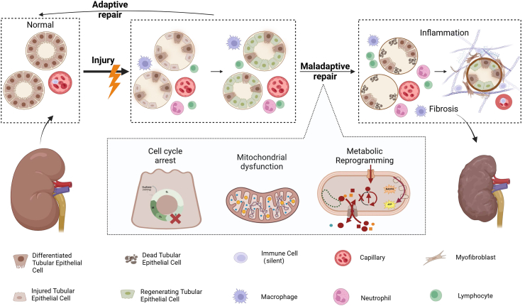
Acute kidney injury (AKI) is a clinical syndrome characterized by a rapid and significant decrease in renal function that can arise from various etiologies, and is associated with high morbidity and mortality. The renal tubular epithelial cells (TECs) represent the central cell type affected by AKI, and their notable regenerative capacity is critical for the recovery of renal function in afflicted patients. The adaptive repair process initiated by surviving TECs following mild AKI facilitates full renal recovery. Conversely, when injury is severe or persistent, it allows the TECs to undergo pathological responses, abnormal adaptive repair and phenotypic transformation, which will lead to the development of renal fibrosis. Given the implications of TECs fate after injury in renal outcomes, a deeper understanding of these mechanisms is necessary to identify promising therapeutic targets and biomarkers of the repair process in the human kidney.

Keywords: Acute kidney injury; Adaptive repair; Phenotypic transformation; Renal tubular epithelial cells.


Conflict of interest statement

Declaration of interests The authors confirm that there are no conflicts of interest.

Figures

Fig. 1
Fig. 1
Origin of proliferating tubule cells. A. The first hypothesis suggests that renal tubular repair originates from a pre-existing population of fixed progenitor cells that exhibit MSC-like characteristics, possess self-renewal capacity, and are induced to give rise to multiple clonal cell lines. B. The second hypothesis suggests that renal tubular repair originates from all surviving TECs, which are all capable of temporarily adopting a scattered tubular cell (STC) phenotype and re-entering the cell cycle, thus having the potential to differentiate.
Fig. 2
Fig. 2
The shematic diagram of major cell state transitions in TEC after AKI. After AKI, dead TECs release pro-inflammatory and pro-fibrotic factors that chemotaxis immune cells to the site of injury to engulf themselves and repair injured TECs. In less severe injuries, TEC cells undergo adaptive repair and kidney function is restored. However, in the presence of non-adaptive repair such as persistent metabolic reprogramming, mitochondrial dysfunction, and cell cycle blockage, renal fibrosis will eventually result.
Fig. 3
Fig. 3
Injury response and prognosis of adaptive versus maladaptive repair. Two scenarios that may occur in the kidney after AKI with varying degrees of injury are depicted. In the first scenario, mild injury activates the innate immune response in time, leading to adaptive tubular repair and kidney regeneration. In the second scenario, sustained or severe injury leads to phenotypic changes in TEC and maladaptive tubular repair.
Fig. 4
Fig. 4
Crosstalk of TEC with other cells in renal repair after AKI. Interactions between TECs, immune cells and mesenchymal fibroblasts play an important role in renal repair. After AKI, dead TECs trigger inflammation by releasing DAMPs, which activate PRRs on macrophages and recruit immune cells to the site of injury. Surviving TECs undergo dedifferentiation and proliferation and repopulate the tubular basement membrane, where they may also communicate with immune cells via exosomes containing signaling molecules. During renal repair, pro-inflammatory (M1) macrophages change phenotype to anti-inflammatory (M2) macrophages. Pro-fibrotic factors produced by injured TECs also contribute to fibroblast activation into myofibroblasts. In addition to supporting tubular remodeling by providing mechanical support through ECM synthesis, myofibroblasts promote the repair and proliferation of TECs through intercellular contact signaling and paracrine signaling and even the secretion of pro-regenerative factors. The interaction of TECs with endothelial cells is also a key aspect of renal repair, with TECs releasing a wide range of angiogenic factors to promote endothelial cell proliferation and migration, and endothelial cells being able to participate in renal vascular repair and regeneration by regulating chronic inflammatory responses. Disruption of communication between TECs and endothelial cells leads to maladaptive repair and fibrosis. Abbreviations: TEC, tubular epithelial cell; DAMP, damage-associated molecular pattern; PRRs, pattern recognition receptor; ECM, extra cellular matrix; IL, interleukin; TGF, transforming growth factor; PDGF, platelet-derived growth factor; ICAM, Intercellular adhesion molecule; VCAM, vascular cell adhesion molecule; VEGF, vascular endothelial growth factor.

References

    1. Matsuura R., Doi K., Rabb H. Acute kidney injury and distant organ dysfunction-network system analysis. Kidney Int. 2023;103(6):1041–1055. - PubMed
    1. Ronco C., Bellomo R., Kellum J.A. Acute kidney injury. Lancet. 2019;394(10212):1949–1964. - PubMed
    1. Rana R., Breyer M.D. Tackling AKI. J Am Soc Nephrol. 2023;34(6):935–936. - PMC - PubMed
    1. Li Z.L., Huang M.M., Yu M.Y., et al. Mitochondrial fumarate promotes ischemia/reperfusion-induced tubular injury. Acta Physiol (Oxf) 2024;240(4) - PubMed
    1. Bonventre J.V., Yang L. Cellular pathophysiology of ischemic acute kidney injury. J Clin Invest. 2011;121(11):4210–4221. - PMC - PubMed

LinkOut - more resources