. 2024 Aug 23;15(1):7267.
doi: 10.1038/s41467-024-51498-0.

Complementary dual-virus strategy drives synthetic target and cognate T-cell engager expression for endogenous-antigen agnostic immunotherapy

Affiliations

Complementary dual-virus strategy drives synthetic target and cognate T-cell engager expression for endogenous-antigen agnostic immunotherapy

Zaid Taha et al. Nat Commun. .

Abstract

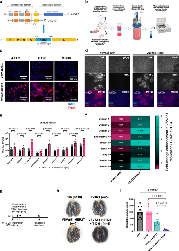
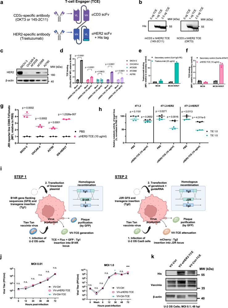
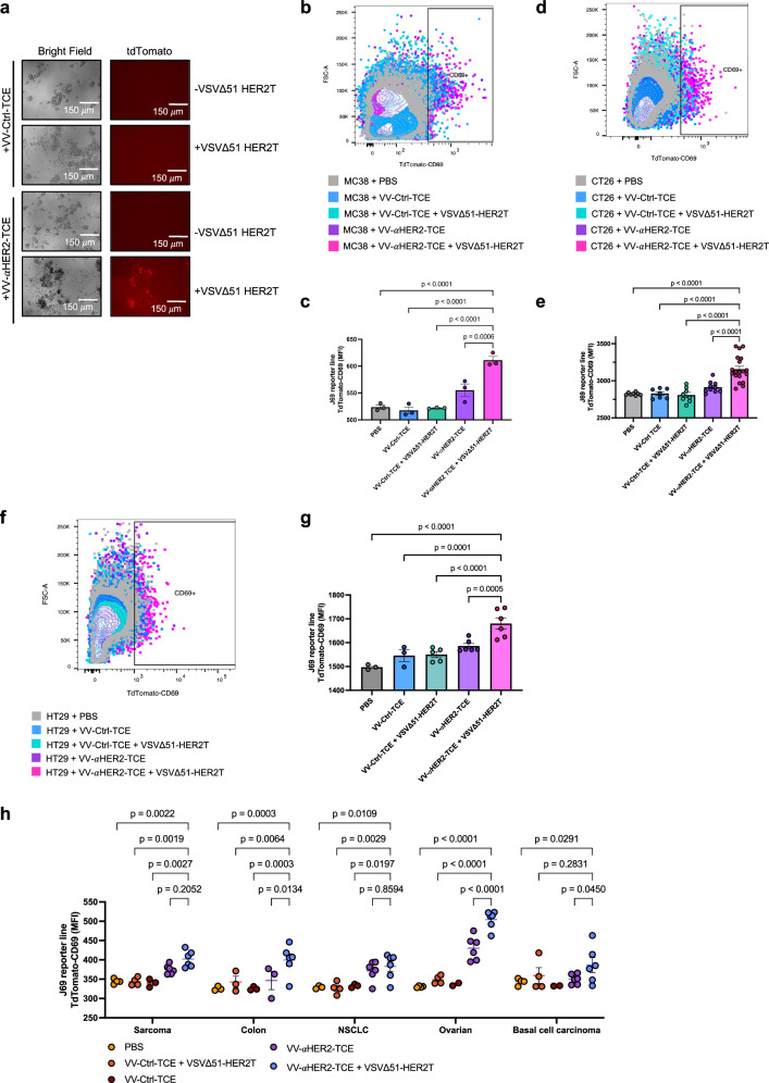
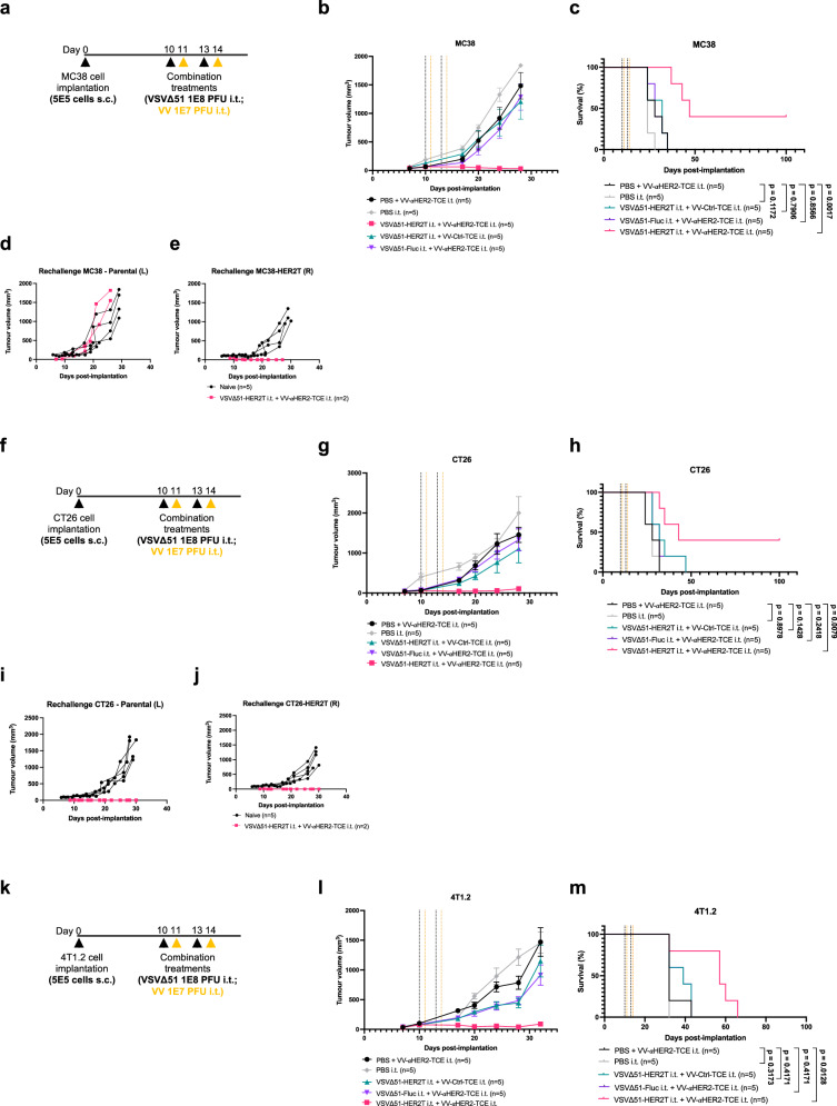
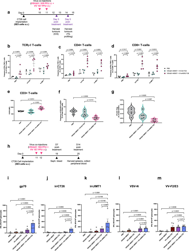
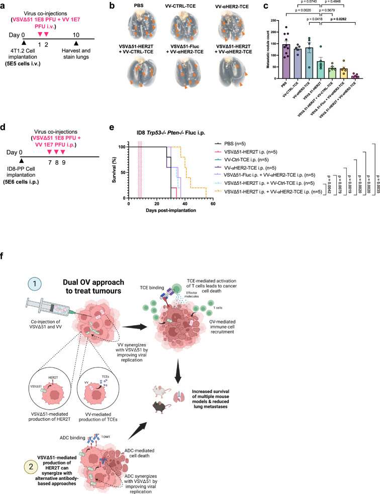
Targeted antineoplastic immunotherapies have achieved remarkable clinical outcomes. However, resistance to these therapies due to target absence or antigen shedding limits their efficacy and excludes tumours from candidacy. To address this limitation, here we engineer an oncolytic rhabdovirus, vesicular stomatitis virus (VSVΔ51), to express a truncated targeted antigen, which allows for HER2-targeting with trastuzumab. The truncated HER2 (HER2T) lacks signaling capabilities and is efficiently expressed on infected cell surfaces. VSVΔ51-mediated HER2T expression simulates HER2-positive status in tumours, enabling effective treatment with the antibody-drug conjugate trastuzumab emtansine in vitro, ex vivo, and in vivo. Additionally, we combine VSVΔ51-HER2T with an oncolytic vaccinia virus expressing a HER2-targeted T-cell engager. This dual-virus therapeutic strategy demonstrates potent curative efficacy in vivo in female mice using CD3+ infiltrate for anti-tumour immunity. Our findings showcase the ability to tailor the tumour microenvironment using oncolytic viruses, thereby enhancing compatibility with "off-the-shelf" targeted therapies.


Conflict of interest statement

Z.T., M.J.F.C, J.C.B., J-S.D. report having a pending patent directly related to this work entitled “Delivery vehicles expressing engineered antigens and uses thereof” (U.S. Provisional Patent Application No. 63/680, 672). The other authors declare no competing interests.

Figures

Fig. 1
Fig. 1. VSVΔ51-encoded HER2T is expressed in vitro and in vivo.
a Schematic of the HER2T construct encoded within VSVΔ51. b Overview of VSVΔ51 rescue, propagation, and purification. c 4T1.2, CT26, and MC38 cells were seeded on glass coverslips and infected with the indicated VSVΔ51 variants at MOI 0.1 and incubated for 20 h. Cells were fixed in 4% paraformaldehyde (PFA) and stained using trastuzumab as a primary antibody (1:1000) and goat anti-human IgG-Alexa Fluor 594 (1:300) as a secondary antibody. Cells were mounted using ProLong Gold Antifade with DAPI, and imaged at 20× magnification using a Zeiss AxioImager M1. Similar results were seen across 3 different independent experiments. d Tumour samples were stained using trastuzumab as a primary antibody (1:1000), and goat anti-human IgG-Alexa Fluor 594 (1:300) as a secondary antibody. Sections were mounted using Prolong Gold Antifade with DAPI. Sections were imaged using the Zeiss AxioImager M1 at 10× magnification. Similar results were seen across 3 different independent experiments. e, f Patient tumour cores were infected with (e) VSVΔ51-HER2T and co-treated with T-DM1 or mock (PBS) as indicated. Viral titre in supernatants was quantified 48 hpi. Shown are mean ± SEM, n = 4–14 cores, P-value calculated by multiple unpaired t-tests. (Endometrial 1 n = 4, Renal 1 n = 6, Ovarian 2 and Breast 1 n = 8, Parotid 2 n = 14, all others n = 12). f Ratios of mean viral titre of T-DM1/PBS for samples infected with VSVΔ51-HER2T or VSVΔ51-GFP were calculated and plotted as a heat map. g Experimental overview: 5 ×10⁵ 4T1.2 cells were injected i.v. by teil vein injection into wildtype BALB/c mice on D0 and then treated on D1 and D2 by i.v. administration of PBS, VSVΔ51-HER2T, T-DM1, or VSVΔ51-HER2 + T-DM1. Mice were euthanized on D10 post-implantation, and lungs were extracted, perfused, and stained for counting of metastatic lung nodules. h Following staining with black India ink, lungs were fixed and imaged. i Lung nodules from g and h were counted. Shown are mean ± SEM, n = 10 mice in PBS, n = 5 mice in treatment groups, P-value calculated by one-way ANOVA relative to PBS, with Dunnett correction for multiple comparisons. Source data are provided as a Source Data file.
Fig. 2
Fig. 2. Engineered αHER2-TCE recognizes HER2T antigen and can be encoded in VV.
a Schematic of the anti-HER2 TCE design. b TCE expression plasmids were transfected into HEK293T cells, and supernatants were resolved by SDS-PAGE 48 h post-transfection. Blots were probed with anti-His antibody to detect His-tagged TCEs. Similar results were seen across 3 different independent experiments. c Whole cell lysates of the indicated cell lines were resolved by SDS-PAGE and probed with anti-HER2 antibody. d TCE binding assay for the indicated cells lines (mean ± SEM, n = 3, P-value calculated by two-way ANOVA with Sidak’s correction for multiple comparisons). e, f trastuzumab and TCE binding assays for MC38 and MC38-HER2T. (mean ± SEM, n = 3). g J69 cells (1 × 10⁶) were co-cultured for 24 h with the indicated cell lines (5 × 10⁵) in the presence or absence of 10 μg/ml αHER2-TCE. J69 cells were then dislodged and isolated and TdTomato signal was quantified by the BioTek Cytation5 (Gen5 v2 software) at Ex555nm/Em580nm (mean ± SEM, n = 3, P-value calculated by multiple unpaired t-tests with the Holm-Sidak correction for multiple comparisons.) h Naïve BALB/c splenocytes were isolated and co-cultured with the indicated target cells at a T:E ratio of 1:0 or 1:5. Co-cultures were incubated for 72 h with or without 10 μg/ml αHER2-TCE. Cell viability was assessed by AlamarBlue and normalized to untreated target cells alone (mean ± SEM, n = 3, P-value calculated by multiple unpaired t-tests with the Holm-Sidak correction for multiple comparisons.) i Schematic of the engineering of TCEs into VV and subsequent viral rescue and purification. j Multi-step (left, MOI 0.01) and single-step (right, MOI 1.0) VV growth curves. U-2 OS cells were seeded in 6-well plates at 1 × 10⁶ cells per well and incubated overnight. Cells were infected with VV-Ctrl, VV-Ctrl-TCE, or VV-αHER2-TCE at the indicated MOI, and frozen at −80 °C at the indicated timepoints. Cells undergo a total of 3 freeze-thaw cycles to liberate intracellular VV, which was subsequently quantified by plaque assay. Shown are mean ± SEM, n = 3, P-value calculated by two-way ANOVA with Tukey’s correction for multiple comparisons. *P < 0.05, ***P < 0.001, relative to VV-αHER2-TCE. (P-values resulting from the two-way ANOVA are included in the Source Data file for this figure). k Whole cell lysates of U-2 OS cells infected with VV and resolved by SDS-PAGE. Blots were probed with anti-His IgG to quantify His-tagged TCE, and anti-Vaccinia IgG to confirm VV infection. Similar results were seen across 3 different independent experiments. Source data are provided as a Source Data file.
Fig. 3
Fig. 3. Infection of tumours with VV-αHER2-TCE ex vivo leads to T-cell activation when combined with VSVΔ51-HER2.
a Images acquired using the EVOS M5000 microscope of the co-cultured J69 cells, which were cultured with MC38 tumour cores under different treatment conditions as indicated (20× magnification) b J69 cells from a were isolated and assessed for TdTomato signal by flow cytometric analysis. c MFI of TdTomato+ J69 cells was quantified. Shown are mean ± SEM, n = 3 cores per group; P-value calculated by one-way ANOVA, with Dunnett correction for multiple comparisons. d J69 cells were isolated from co-culture with CT26 tumour cores following treatment as indicated, and TdTomato signal was quantified by flow cytometric analysis. e Quantification of TdTomato signal from d. Shown are mean ± SEM, n = 6–19 cores per group; P-value calculated by one-way ANOVA related to VSVΔ51-HER2T+VV-αHER2-TCE, with Dunnett correction for multiple comparisons. PBS n = 8, VV-Ctrl-TCE n = 7, VV-Ctrl-TCE+VSVΔ51-HER2T n = 8, VV-αHER2-TCE n = 10, VV-αHER2-TCE+VSVΔ51-HER2T n = 19). f HT29 xenografts were excised and cored by punch biopsy, then treated and co-cultured with J69 cells. J69 cells were isolated and assessed for TdTomato signal by flow cytometric analysis. g Quantification of TdTomato signal from f. Shown are mean ± SEM, n = 3–6 cores per group; P-value calculated by one-way ANOVA related to VSVΔ51-HER2T+VV-αHER2-TCE, with Dunnett correction for multiple comparisons. (PBS n = 3, VV-Ctrl-TCE n = 3, VV-Ctrl-TCE+VSVΔ51-HER2T n = 5, VV-αHER2-TCE n = 6, VV-αHER2-TCE+VSVΔ51-HER2T n = 6). h Patient tumour specimens were treated and analysed for J69 TdTomato signal. Shown are mean ± SEM, n = 2–6 cores per group (for exact n values, see Source Data); P-value calculated by one-way ANOVA related to VSVΔ51-HER2T+VV-αHER2-TCE within each specimen, calculated only for n = 3 or more cores. Source data are provided as a Source Data file.
Fig. 4
Fig. 4. Treatment of tumour-bearing mice with VV-αHER2-TCE and VSVΔ51-HER2T prolongs overall survival in localized disease models.
a Experimental overview: 5 × 10⁵ MC38 cells were implanted s.c. in C57BL/6 mice in the right flank. Mice were treated i.t. as indicated. b Tumour volumes (mean ± SEM, n = 5 per group) and (c) overall survival were monitored (d, e). Cured mice from ac were rechallenged with bilateral s.c. tumours, d parental MC38 (left flank) or e MC38-HER2T (right flank). Naïve mice were challenged with bilateral s.c. tumours in parallel as controls. f Experimental overview: 5 × 10⁵ CT26 cells were implanted s.c. in BALB/c mice in the right flank. Mice were treated i.t. as indicated. g Tumour volumes (mean ± SEM, n = 5 per group) and h overall survival were monitored. i, j Cured mice from fh were rechallenged with bilateral s.c. tumours, i parental CT26 (left flank) or j CT26-HER2T (right flank). Naïve mice were challenged with bilateral s.c. tumours in parallel as controls. k Experimental overview: 5 × 10⁵ 4T1.2 cells were implanted s.c. in BALB/c mice in the right flank. Mice were treated i.t. as indicated. l Tumour volumes (mean ± SEM, n = 5 per group) and m overall survival were monitored. c, h, m For analysis of survival data, P-values were calculated by the Kaplan–Meier method followed by log-rank test. Source data are provided as a Source Data file.
Fig. 5
Fig. 5. Intratumoural injection of VV-αHER2-TCE and VSVΔ51-HER2T induces anti-tumour immune responses.
a Experimental overview: 5 × 10⁵ CT26 cells were implanted s.c. in BALB/c mice in the right flank. Mice were treated i.t. as indicated. Tumours were harvested on day 3 for intracellular staining (ICS) of cytokines, and on day 5 for assessing the profiles of the immune infiltrating populations. bd Following tumour-dissociation, immune cells were cultured ex vivo for 24 h and Golgi plugged following stimulation with PBS control, 2 × 10⁶ irradiated CT26 cells, or 2 × 10⁶ irradiated JIMT1 cells. Immune cells were analysed by flow cytometric analysis for intracellular levels of IFNγ and TNFα in b TCRγδ T-cells, c CD4+ T-cells, and d CD8+ T-cells. (mean ± SEM, n = 3 mice in PBS and VSVΔ51-HER2T+VV-Ctrl-TCE groups, n = 5 mice in VV-αHER2-TCE+VSVΔ51-HER2T group; P-value calculated by one-way ANOVA relative to VV-αHER2-TCE+VSVΔ51-HER2T, with Dunnett correction for multiple comparisons). eh Tumours dissociated at day 5 were analysed by flow cytometric analysis. e Levels of CXCR3 (MFI) in total CD3+ T-cells are shown. Frequency of (f) PD1 + LAG3 + CD3+ T-cells and (g) CTLA4+ Tregs were determined relative to total CD45+ cells. For all violin plots, n = 5 mice per group; P-value calculated by one-way ANOVA relative to VV-αHER2-TCE+VSVΔ51-HER2T, with Dunnett correction for multiple comparison. h Experimental overview: 5 × 10⁵ CT26 cells were implanted s.c. in BALB/c mice in the right flank. Mice were treated i.t. as indicated. Following harvest and dissociation of spleens at D14 post-treatment, splenocytes were subjected to cultured in an ELISpot plate to quantify IFNγ-secreting cells following 16 h culture in the presence of the following stimulation (im): 10 µM gp70 peptide, irradiated CT26 cells, irradiated JIMT1 cells, 10 µM VSV-N peptide, 10 µM VV-F2/E3 peptides (mean ± SEM, n = 5 mice per group; P-value calculated by one-way ANOVA relative to VSVΔ51-HER2T+VV-αHER2-TCE, with Dunnett correction for multiple comparisons). Source data are provided as a Source Data file.
Fig. 6
Fig. 6. Treatment of tumour-bearing mice with VV-αHER2-TCE and VSVΔ51-HER2T prolongs overall survival and reduces lung metastases in disseminated disease models.
a Experimental overview: 5 × 10⁵ 4T1.2 cells were injected i.v. by teil vein injection into BALB/c mice, and treated as indicated, followed by harvest and staining of lungs. b Following staining with black India ink, lungs were fixed and imaged. c Metastatic lung nodules were quantified. Shown are mean ± SEM, n = 10 mice in the PBS group, n = 5 mice in all other groups; P-value calculated by one-way ANOVA relative to VSVΔ51-HER2T, with Dunnett correction for multiple comparisons. d Experimental overview: 5 × 10⁶ ID8-PP cells were implanted i.p. in C57BL/6 mice, followed by treatments as indicated. e Overall survival was monitored; P-values indicated next to treatment groups is relative to VSVΔ51-HER2T+VV-αHER2-TCE. For analysis of survival data, P-values were calculated by the Kaplan–Meier method followed by log-rank test. Source data are provided as a Source Data file. f Schematic depicting the dual oncolytic virus approach to treat tumours (indicated by the number 1), whereby VSVΔ51 and VV produce the HER2T synthetic target and its cognate TCE, respectively. These two OVs synergize since VV dampens interferon levels and increases VSVΔ51 replication. VV-produced TCEs recognize and bind to HER2T, forming a pseudo-immunological synapse that allows for the release of effector molecules (e.g. granzyme, perforin). OVs lead to the recruitment of immune cells in the tumour, and TCEs mediate the activation of T cells that trigger apoptosis of cancer cells. Ultimately, this strategy improves survival of mice bearing different tumour types and decreases lung metastases. Importantly, VSVΔ51-mediated production of HER2T on the surface of cancer cells can synergize with other alternative antibody-based approaches, such as antibody-drug conjugates like T-DM1 (indicated by the number 2). In this case, the ADC can synergize with VSVΔ51 to promote viral replication. The ADC-mediated cell death also leads to increased survival in preclinical animal models and decreased lung metastases in mice.

References

    1. Finck, A., Gill, S. I. & June, C. H. Cancer immunotherapy comes of age and looks for maturity. Nat. Commun.11, 10.1038/s41467-020-17140-5 (2020). - PMC - PubMed
    1. Arulanandam, R. et al. The strategic combination of trastuzumab emtansine with oncolytic rhabdoviruses leads to therapeutic synergy. Commun. Biol.3, 254 (2020). - PMC - PubMed
    1. von Minckwitz, G. et al. Trastuzumab Emtansine for Residual Invasive HER2-Positive Breast Cancer. N. Engl. J. Med.380, 617–628 (2019). 10.1056/NEJMoa1814017 - DOI - PubMed
    1. Cortés, J. et al. Trastuzumab Deruxtecan versus Trastuzumab Emtansine for Breast Cancer. N. Engl. J. Med.386, 1143–1154 (2022). 10.1056/NEJMoa2115022 - DOI - PubMed
    1. Swain, S. M., Shastry, M. & Hamilton, E. Targeting HER2-positive breast cancer: advances and future directions. Nat. Rev. Drug Discov.22, 101–126, (2023). - PMC - PubMed

MeSH terms

LinkOut - more resources