. 2024 Aug 23;14(1):19594.
doi: 10.1038/s41598-024-70601-5.

Regulation viral RNA transcription and replication by higher-order RNA structures within the nsp1 coding region of MERS coronavirus

Affiliations

Regulation viral RNA transcription and replication by higher-order RNA structures within the nsp1 coding region of MERS coronavirus

Yutaka Terada et al. Sci Rep. .

Abstract

Coronavirus (CoV) possesses numerous functional cis-acting elements in its positive-strand genomic RNA. Although most of these RNA structures participate in viral replication, the functions of RNA structures in the genomic RNA of CoV in viral replication remain unclear. In this study, we investigated the functions of the higher-order RNA stem-loop (SL) structures SL5B, SL5C, and SL5D in the ORF1a coding region of Middle East respiratory syndrome coronavirus (MERS-CoV) in viral replication. Our approach, using reverse genetics of a bacterial artificial chromosome system, revealed that SL5B and SL5C play essential roles in the discontinuous transcription of MERS-CoV. In silico analyses predicted that SL5C interacts with a bulged stem-loop (BSL) in the 3' untranslated region, suggesting that the RNA structure of SL5C is important for viral RNA transcription. Conversely, SL5D did not affect transcription, but mediated the synthesis of positive-strand genomic RNA. Additionally, the RNA secondary structure of SL5 in the revertant virus of the SL5D mutant was similar to that of the wild-type, indicating that the RNA structure of SL5D can finely tune RNA replication in MERS-CoV. Our data indicate novel regulatory mechanisms of viral RNA transcription and replication by higher-order RNA structures in the MERS-CoV genomic RNA.


Conflict of interest statement

The authors declare no competing interests.

Figures
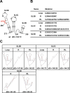
Fig. 1
Fig. 1
Construction of SL5 structure disrupting mutants. (A) RNA structures predicted by Mfold. Numbers indicate the nucleotide positions in the MERS-CoV EMC strain (DDBJ accession No. NC_019843). Circles indicate the mutation sites for each of the SL5 mutants shown in (B). ATG: Start codon of ORF1a. (B) Nucleotide mutations of SL5B, SL5C, and SL5D mutants used in this study. (C) Predicted secondary RNA structures of each mutant.
Fig. 2
Fig. 2
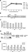
Effects of SL5B, SL5C, and SL5D RNA structures on viral propagation. (A) Viral growth of SL5 mutants. Huh7 cells were transfected with the MERS-CoV cDNA clones and cultured. Experiments were carried out in triplicate and representative results were shown. Culture supernatants were collected to harvest the virus, and viral titers were determined by TCID50 assay using Vero cells. Mock indicates the transfected Huh7 cells with backbone BAC plasmid without any viral sequence. **** P < 0.0001. (B) Real-time RT-PCR results evaluating viral RNA transcription. Huh7 cells transfected with each cDNA clone were cultured for the indicated times. Total RNA was extracted from each infected cell, and real-time RT-PCR targeting sg N mRNA was performed. The sg N mRNA levels were normalized to the GAPDH mRNA levels. **** P < 0.0001. # represents the limit of detection.  (C) Immunoblot analysis for the detection of MERS-CoV N protein. Cell lysates of Huh7 cells transfected with each cDNA clone were subjected to western blot analysis using anti-MERS-CoV N antibody (a-N) and anti-actin antibody (a-Actin). (D) Luciferase activities of replicons carrying each of the SL5 mutants. 293T cells were transfected with each cDNA replicon and cultured for 48 h. Nluc activities in cultured cells were measured using confocal microscopy. Firefly luciferase activities from pGL3-control plasmid were also measured as an internal control. Polymerase dead mutant (SAA) was used as a negative control.* P <0.05.
Fig. 3
Fig. 3
Functional step of SL5B and SL5C in MERS-CoV lifecycle. (A) Genomic RNA stabilities of viruses containing mutant SL5B or SL5C regions with disrupted secondary structures. Huh7 cells were transfected with each cDNA clone and cultured for 0, 4, 8, and 12 h. Total RNA were extracted from the cells and subjected to real-time RT-PCR targeting ORF1a region to measure the levels of genomic RNA. The levels of genomic RNAs were normalized to the GAPDH mRNA levels. (B) Schematic of pcDNA3.1-SL5-fluc plasmid. MERS-CoV sequence from nts 265–343 was inserted between the CMV promoter and the firefly luciferase gene (fluc). Gray squares indicate the nucleotide regions of SL5B, SL5C, and SL5D. (C) Effects of SL5B and SL5C structures on protein translation. 293T cells were transfected with pcDNA3.1-SL5-fluc or SL5B/SL5C mutants. pcDNA3.1-fluc without the viral sequence was used as a positive control. Renilla luciferase reporter plasmid, pRL-SV40, was used as the internal control. After 24 h of incubation, the transfected cells were collected, and luciferase activities were measured. Firefly luciferase activities were normalized to the Renilla luciferase activities. (D) Schematic diagram of the reporter assay for detecting the MERS-CoV protease, nsp5. cDNA clones of WT, SL5B mutant, or SL5C mutant were co-transfected into Huh7 cells with the pGlo-VRLQS biosensor plasmid. Translated proteins from pGlo-VRLQS do not show luciferase activity. Cleavage caused by MERS-CoV nsp5 increases luciferase activity. Transfected Huh7 cells were cultured for 24 and 48 h, and luciferase activities in cells were measured using confocal microscopy. (E) Expression of nsp5 in SL5B and SL5C mutants. The results of the MERS-CoV nsp5 biosensor assay described in (D). The Nsp5 deletion mutant, pBAC-MERS-nsp5-rpsL, was used as a negative control (Control).
Fig. 4
Fig. 4
Prediction of long RNA-RNA interaction with SL5. (A) Plot of all predicted LRI values with P < 0.005 observed in four MERS-CoV genomes. The outer circle represents the genome information of MERS-CoV. The inner circle shows all predicted interactions between all genome positions. The plot was created with Circos. (B) Predicted RNA-RNA interaction between SL5C and the BSL within the 3′UTR based on the LRIscan analysis. The nucleotide positions in MERS-CoV EMC are indicated (DDBJ accession No. NC_019843).
Fig. 5
Fig. 5
Functional step of SL5D in MERS-CoV lifecycle. (A, B) Correlative light and electron microscopy (CLEM) analysis of rMERS-ZsGreen or rMERS-ZsGreen-SL5D-R mutants. Vero cells were inoculated with rMERS-ZsGreen or rMERS-ZsGreen-SL5D-R (MOI = 0.1) and cultured for 24 h. (A) Infected cells were identified by green fluorescence and (B) subjected to transmission electron microscopy (TEM) analysis. Black arrows indicate the viral particles inside the infected cells. White arrows indicate the viral particles on the cell surface. Scale bar = 1 µm. N nucleolus. Bottom: Zoomed image of red square areas from top TEM image. (C) Viral release of rMERS-ZsGreen or rMERS-ZsGreen-SL5D-R mutants from Vero cells. Vero cells were inoculated with rMERS-ZsGreen or rMERS-ZsGreen-SL5D-R (MOI = 0.01) and cultured. At the indicated time, culture supernatants were collected. Cells were collected in fresh medium, stored frozen, and then thawed before use. Viral titers of both supernatants (Sup) and cell (Cell) samples were measured to determine the levels of released virus relative to that inside the cell. * P < 0.05  (D) Examination of the levels of strand-specific genomic RNA in wildtype or the SL5D-R mutant virus. Huh7 cells were transfected with pBAC-MERS or pBAC-MERS-SL5D-R and cultured for 72 h, and total RNAs were extracted. RNA samples were reverse-transcribed using positive- or negative-strand specific primers and then treated with RNaseH. The strand-specific cDNAs were subjected to real-time PCR targeting the ORF1a region. * P < 0.05.
Fig. 6
Fig. 6
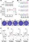
Emergence of revertant SL5D-R mutant virus in Vero cells. (A) Nucleotide sequences from 330–344 of WT and SL5D-R mutant viruses. Mutated nucleotides are shown in red. (B) Nucleotide sequences of rMERS-SL5D-R viruses passaged in Vero cells. Vero cells were inoculated with rMERS-SL5D-R virus and passaged. After ten passages in Vero cells, viral RNAs were extracted from the P1, P2, P5, and P10 virus stocks, and RT-PCR was performed to amplify the SL5D region and read the nucleotide sequences. The amplified PCR products were cloned into a Blunt vector and transformed to E. coli. The cloned vector DNA from ten clones was extracted and the nucleotide sequences were identified. (C) Growth kinetics of passaged rMERS-SL5D-R viruses in Vero cells. Vero cells were inoculated with rMERS or passaged rMERS-SL5D-R viruses (MOI = 0.01) and cultured. Viral titers of culture supernatants were determined using the TCID50 assay. (D) Plaque morphology of the passaged rMERS-SL5D-R viruses. Seeded Vero cells were inoculated with 25 TCID50 of each virus. Infected Vero cells were fixed with phosphate-buffered formaldehyde at 3 d post infection and stained using crystal violet. White arrow indicates small plaques. White arrowheads indicate large plaques. Scale bar = 1 cm. (E) RNA structures of positive-strand RNA of P10 virus predicted following Mfold analysis. (F) RNA structures of negative-strand RNA predicted following Mfold analysis. Predicted structures were constructed using the reverse-complement sequences of nts 1–460 from the WT, SL5D-R mutant, and SL5D-R P10 viruses. Numbers indicate the corresponding nucleotide position of positive-strand genomic RNA.

Similar articles

References

    1. Weiss, S. R. & Navas-Martin, S. Coronavirus pathogenesis and the emerging pathogen severe acute respiratory syndrome coronavirus. Microbiol. Mol. Biol. Rev.69, 635–664. 10.1128/MMBR.69.4.635-664.2005 (2005). 10.1128/MMBR.69.4.635-664.2005 - DOI - PMC - PubMed
    1. Bradburne, A. F., Bynoe, M. L. & Tyrrell, D. A. Effects of a “new” human respiratory virus in volunteers. Br. Med. J.3, 767–769. 10.1136/bmj.3.5568.767 (1967). 10.1136/bmj.3.5568.767 - DOI - PMC - PubMed
    1. van der Hoek, L., Pyrc, K. & Berkhout, B. Human coronavirus NL63, a new respiratory virus. FEMS Microbiol. Rev.30, 760–773. 10.1111/j.1574-6976.2006.00032.x (2006). 10.1111/j.1574-6976.2006.00032.x - DOI - PMC - PubMed
    1. van der Hoek, L. et al. Identification of a new human coronavirus. Nat. Med.10, 368–373. 10.1038/nm1024 (2004). 10.1038/nm1024 - DOI - PMC - PubMed
    1. Woo, P. C. et al. Characterization and complete genome sequence of a novel coronavirus, coronavirus HKU1, from patients with pneumonia. J. Virol.79, 884–895. 10.1128/JVI.79.2.884-895.2005 (2005). 10.1128/JVI.79.2.884-895.2005 - DOI - PMC - PubMed

MeSH terms

LinkOut - more resources