. 2024 Oct;43(41):3049-3061.
doi: 10.1038/s41388-024-03131-z. Epub 2024 Aug 23.

Targeting MERTK on tumour cells and macrophages: a potential intervention for sporadic and NF2-related meningioma and schwannoma tumours

Affiliations

Targeting MERTK on tumour cells and macrophages: a potential intervention for sporadic and NF2-related meningioma and schwannoma tumours

Foram Dave et al. Oncogene. 2024 Oct.

Erratum in

Abstract

Meningioma and schwannoma are common tumours of the nervous system. They occur sporadically or as part of the hereditary NF2-related schwannomatosis syndrome. There is an unmet need for new effective drug treatments for both tumour types. In this paper, we demonstrate overexpression/activation of TAM (TYRO3/AXL/MERTK) receptors (TAMs) and overexpression/release of ligand GAS6 in patient-derived meningioma tumour cells and tissue. For the first time, we reveal the formation of MERTK/TYRO3 heterocomplexes in meningioma and schwannoma tissue. We demonstrate the dependence of AXL and TYRO3 expression on MERTK in both tumour types, as well as interdependency of MERTK and AXL expression in meningioma. We show that MERTK and AXL contribute to increased proliferation and survival of meningioma and schwannoma cells, which we inhibited in vitro using the MERTK/FLT3 inhibitor UNC2025 and the AXL inhibitor BGB324. UNC2025 was effective in both tumour types with superior efficacy over BGB324. Finally, we found that TAMs are expressed by tumour-associated macrophages in meningioma and schwannoma tumours and that UNC2025 strongly depleted macrophages in both tumour types.


Conflict of interest statement

The authors declare no competing interests.

Figures

Fig. 1
Fig. 1. MERTK/pMERTK, AXL/pAXL, TYRO3/pTYRO3 and GAS6 are overexpressed in meningioma tissue of all grades.
A, B MERTK, AXL and TYRO3 (A) and pMERTK and pAXL (B) expression is significantly increased in grade-I (MN-GI) and grades- II/III (MN-GII/III) meningioma tissue compared to normal meningeal tissue (NMT), with no significant difference between grades. Expression of pTYRO3 is increased in all meningioma grades compared to NMT but this result is not significant. C, D Adenovirus-mediated reintroduction of wild-type Merlin into Merlin-negative MN-GI primary cells resulted in a significant partial decrease in the expression of AXL, TYRO3 and MERTK, with no significant effect on GAS6. Western blot data were normalised to the loading control GAPDH. P values were calculated by Kruskal Wallis test followed by post hoc Dunn’s test (A, B) and ANOVA with Dunnett’s multiple comparison test (D) and are represented as follows: ns (not significant) P > 0.05, * P < 0.05, ** P < 0.01 and *** P < 0.001. Mean ± SEM is indicated in all graphs.
Fig. 2
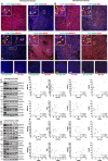
Fig. 2. TAM receptors and GAS6 are expressed by tumour-associated macrophages in meningioma and schwannoma tumours.
A, B MERTK, AXL, TYRO3 and GAS6 are expressed in CD68-negative tumour cells (white arrows) and CD68-positive macrophages (yellow arrows) in grade-I meningioma and schwannoma tissue. Omission of primary antibody or an appropriate IgG control antibody were used as controls. Images were captured at 20x magnification and scale bars in (A, B) are 50 μm.Western blot data demonstrating correlation between the expression of MERTK, AXL, TYRO3, GAS6, and CD163 and CD68 in meningioma (C, D) and schwannoma (E, F) across successive passages of cell culture (p0-p4). Data in D and F are normalised to a loading control GAPDH and presented as percentage of p0. Spearman’s rank correlation coefficient ‘r’ was calculated to describe the strength and direction of association between CD68 or CD163 and MERTK, AXL, TYRO3 or GAS6.
Fig. 3
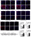
Fig. 3. AXL and MERTK are involved in the increased proliferation of meningioma and schwannoma cells.
AJ MERTK depletion using shRNA decreased proliferation of meningioma and schwannoma cells, and AXL depletion decreased proliferation of meningioma cells determined by reduced number of Ki67+ cells and decreased expression of Cyclin D1. TYRO3 depletion had no effect on proliferation in either of tumour cell type. In (F, G) data are normalised to total cell number monitored by DAPI staining and presented as percentage of scramble control (Scr). In (I, J), data are normalised to GAPDH and are presented as percentage of scramble (Scr). One-way ANOVA with post hoc Tukey’s was used with statistical values as follows: ns (not significant) P > 0.05, * P < 0.05, ** P < 0.01. Numbers on the X axis in F, G, I and J graphs refer to different shRNA constructs. In figures the mean ± SEM is given. Scale bars in (AC), are 40 μm (×20 magnification), in (D) 100 μm (×10 magnification), in (E) 50 μm in the left panel (upper ×10 magnification, and lower ×20 magnification) and 100 μm in the right panel (×10 magnification).
Fig. 4
Fig. 4. TAM receptors are interdependent in meningioma and schwannoma.
A, D, E MERTK knockdown (KD) decreases the expression of AXL and TYRO3 in meningioma (A, D) and schwannoma (A, E) cells compared to a non-silencing shRNA sequence (Scr). B, D AXL-KD reduces the expression of MERTK in meningioma cells. CE TYRO3-KD has no effect on either AXL or MERTK expression in meningioma (D) or schwannoma (E) cells. F Quantitative PCR in meningioma cells demonstrate that MERTK-shRNAs 202 and 203 have no effect on either AXL-mRNA or TYRO3-mRNA expression and AXL-shRNAs 1652 and 653 have no effect on either MERTK-mRNA or TYRO3-mRNA expression. MERTK-shRNA 1643 decreased expression of AXL-mRNA and AXL-shRNA 030 decreased expression of MERTK-mRNA. G MERTK/FLT3 inhibitor UNC2025 (3 µM, 24 hrs) reduces expression of MERTK and AXL at the cell membrane in grade-I Merlin-negative meningioma cell line BM-I. HI MERTK and TYRO3 but not MERTK and AXL or TYRO3 and AXL form a heterocomplex in meningioma (H) and schwannoma (I) tissue. J No complexes between MERTK and TYRO3, MERTK and AXL or TYRO3 and AXL are detected in BM-I cells. In (D, E) data are normalised to GAPDH and presented as percentage of scramble control (Scr). In (DF) one-way ANOVA with post hoc Tukey’s test was used. In (E) TYRO3 438 and TYRO3 476, TYRO3, Kruskal Wallis followed by post hoc Dunn’s test was used. Statistical P values are represented as follows: ns (not significant) P > 0.05, * P < 0.05, ** P < 0.01. In (G), Z-stack imaging using 20x objective on a Leica SPE microscope was used and Pearson’s Correlation Coefficient (PCC) r coloc value was calculated using ImageJ. White arrows indicate membranous MERTK and AXL colocalisation. In panels (DF) the mean ± SEM is graphed.
Fig. 5
Fig. 5. Inhibition of MERTK and AXL decrease proliferation and increase apoptosis in meningioma and schwannoma cells.
UNC2025 (A, B) and BGB324 (C, D) decrease total cell number and increase number of dead cells in meningioma and schwannoma cells after 72 hrs and 7 days. UNC2025 (E, F, I, J) and BGB324 (G, H, K, L) reduce proliferating Ki67+ cell population and increase apoptotic Cleaved caspase-3+ cell population in meningioma and schwannoma cultures after 72 hrs. UNC2025 (M, N) and BGB324 (O, P) decrease Cyclin D1 expression in meningioma and schwannoma cells after 72 hrs. In (AD) total cell numbers are presented as a percentage of vehicle (EtOH or DMSO) and dead cells as a percentage of total cells normalised to vehicle. In (EH), Ki67 data is presented as percentage of total cell number (DAPI) normalised to vehicle, and Cleaved caspase-3 data as percentage of total cell number (DAPI). Images in (IL) were captured at ×20 magnification on a Leica SPE confocal microscope and scale bars depicted are 20 μm. In (MP), data are normalised to Tubulin and presented as percentage of vehicle. One-way ANOVA with post hoc Tukey’s test was used in all graphs. Within (B, G, H) some data were non-normally distributed. Kruskal Wallis test however did not change the significance of columns containing non-normally distributed data, when compared to One-way ANOVA test. Statistical P values are represented as follows: ns (not significant) P > 0.05, * P < 0.05, ** P < 0.01, and *** P < 0.001. In figures the mean ± SEM is graphed.
Fig. 6
Fig. 6. UNC2025 and BGB324 inhibit the expression/activation of TAM receptors in meningioma and schwannoma cells.
In meningioma cells, UNC2025 decreases pMERTK after 1 hr and 72 hrs, and total MERTK after 72 hrs (A, C, D). In schwannoma cells, UNC2025 decreases pMERTK after 1 hr and after 72 hrs (A, E) and pAXL after 1 hr but not after 72 hrs (A, F). In meningioma cells BGB324 decreases pAXL after 1 hr and 72 hrs (B, G), and AXL after 72 hrs (B, H). It also decreases pMERTK after 1 hr and 72 hrs (B, I) and pTYRO3 and TYRO3 after 72 hrs (B, J, K). B, L In schwannoma cells BGB324 decreases pAXL only after 72 hrs. In all graphs data are normalised to the loading control GAPDH and presented as percentage of the vehicle (EtOH or DMSO) and the mean ± SEM is graphed. Kruskal Wallis followed by post hoc Dunn’s test was used in (C) 1 hr; and (E, G, K) 72 hrs, and one-way ANOVA with post hoc Tukey’s test in the rest of the graphs. Statistical P values are represented as follows: ns (not significant) P > 0.05, * P < 0.05, and ** P < 0.01.
Fig. 7
Fig. 7. UNC2025 and BGB324 inhibit signalling pathways in meningioma and schwannoma cells.
A, B, I, J In meningioma cells, UNC2025 decreases pERK/pAKT/pJNK after 1 hr and 72 hrs, and pFAK/pSrc after 72 hrs (A, B). Total levels of ERK/AKT/JNK/Src are not affected, and FAK decreases only after 72 hrs (I, J). C, D, K, L In schwannoma cells, UNC2025 reduces pERK/pAKT/pJNK after 1 hr and after 72 hrs and has no effect on pFAK/pSrc (C, D). UNC2025 has no significant effect on the expression of ERK/AKT/JNK/FAK/Src (K, L). E, F, M, N In meningioma cells BGB324 decreases pERK/pAKT/pJNK/pFAK after 1 hr and after 72 hrs. pSrc decreases only after 72 hrs (E, F). BGB324 has no significant effect on ERK/AKT/JNK/FAK/Src (M, N). G, H, O, P In schwannoma cells BGB324 decreases pERK/pAKT/pJNK after 1 hr and 72 hrs, and pFAK/pSrc after 72 hrs (G, H). BGB324 has no significant effect on ERK/AKT/JNK/FAK at either time points, but significantly decreases Src after 72 hrs (O, P). In all graphs data are normalised to the loading control GAPDH and are presented as percentage of the vehicle-only control (EtOH or DMSO) and the mean ± SEM is graphed. Kruskal Wallis followed by post hoc Dunn’s test was used in (A) (pJNK and pSrc 1 hr), (D) (pERK 1 hr, and pJNK and pAKT 72 hrs), (F) (pJNK 1 hr), (N) (ERK and FAK 72 hrs, and JNK, FAK, and Src 1 hr). One-way ANOVA with post hoc Tukey’s test was used in the rest of the graphs. Statistical values are represented as follows: ns (not significant) P > 0.05, * P < 0.05, ** P < 0.01, and *** P < 0.001.
Fig. 8
Fig. 8. UNC2025 decreases number of macrophages, microglia, and tumour cells in meningioma and schwannoma passage 0 (p0) cells.
A, B, D, E UNC2025 (3 µM, 48 hrs) decreases CD68-positive macrophage population (CD68 +/CD163+ macrophages, CD68 +/iNOS+ macrophages, and CD68 +/TMEM119+ microglia) in meningioma and schwannoma p0 cells. A decrease of non-macrophagic CD163 +/CD68- and iNOS +/CD68- cell populations, predominantly composed of SSTR2 +, SSTR2 +/iNOS+ and SSTR2 +/CD163+ meningioma cells, and S100B +, S100B +/iNOS+ and S100B +/CD163+ schwannoma cells, is also observed. C, G, F, J UNC2025 (3 µM, 48 hrs) decreases CD68 +/Ki67+ macrophage, TMEM119 +/Ki67+ microglia, and CD68-/Ki67+ non-macrophagic cell populations, including SSTR2 +/Ki67+ meningioma (C, G) and S100B +/Ki67+ schwannoma (F, J) tumour cells. C, H, F, K UNC2025 (3 µM, 48 hrs) increases number of apoptotic CD68 +/Cleaved caspase-3+ macrophages, and CD68-/Cleaved caspase-3+ non-macrophagic cell populations including SSTR2 +/Cleaved caspase-3+ meningioma and S100B +/Cleaved caspase-3+ schwannoma cells. In meningioma (I) but not schwannoma (L) p0 cells, CD68-negative non-macrophage cells (comprising of mostly tumour cells) display a weaker response to UNC2025 (3 µM, 48 hrs) when a higher macrophage population is present. These data are presented as a correlation between macrophage population (CD68+ as a % of total cell number, DAPI) and Ki67 +/CD68- cells responding to the treatment (% of total cell number, DAPI). Unless indicated, images were taken using a Leica SPE confocal microscope at 20x objective magnification. Scale bars are 50 μm. In (A, D), the data are presented as percentage for total cell number (% of DAPI) normalised to EtOH vehicle control. In (G, H, J, K) the data are presented as percentage for total cell number. Statistical tests: (A, D) Single-sample t test or Wilcoxon; (G, H, J, K) two-sample t test or Wilcoxon test; (I) Shapiro–Wilk test, Pearson’s correlation coefficient ‘r’; (L) Shapiro–Wilk test, Spearman’s rank correlation coefficient ‘r’. Statistical values are reported as ns (not significant) P > 0.05, *P < 0.05, **P < 0.01, and ***P < 0.001.

References

    1. Plotkin SR, Messiaen L, Legius E, Pancza P, Avery RA, Blakeley JO, et al. Updated diagnostic criteria and nomenclature for neurofibromatosis type 2 and schwannomatosis: An international consensus recommendation. Genet Med. 2022;24:1967–77. - PubMed
    1. Blakeley JO, Evans DG, Adler J, Brackmann D, Chen R, Ferner RE, et al. Consensus recommendations for current treatments and accelerating clinical trials for patients with neurofibromatosis type 2. Am J Med Genet A. 2012;158A:24–41. - PMC - PubMed
    1. Agnihotri S, Jalali S, Wilson MR, Danesh A, Li M, Klironomos G, et al. The genomic landscape of schwannoma. Nat Genet. 2016;48:1339–48. - PubMed
    1. Wang JZ, Nassiri F, Mawrin C, Zadeh G. Genomic Landscape of Meningiomas. Adv Exp Med Biol. 2023;1416:137–58. - PubMed
    1. Champeaux-Depond C, Weller J, Resche-Rigon M. Neurofibromatosis type 2: a nationwide population-based study focused on survival after meningioma surgery. Clin Neurol Neurosurg. 2020;198:106236. - PubMed

MeSH terms

LinkOut - more resources