. 2024 Aug 23;19(1):310.
doi: 10.1186/s13023-024-03318-3.

Characterization of pathogenic genetic variants in Russian patients with primary ciliary dyskinesia using gene panel sequencing and transcript analysis

Affiliations

Characterization of pathogenic genetic variants in Russian patients with primary ciliary dyskinesia using gene panel sequencing and transcript analysis

Anna Zlotina et al. Orphanet J Rare Dis. .

Abstract

Background: Primary ciliary dyskinesia (PCD) is a group of rare genetically heterogeneous disorders caused by defective cilia and flagella motility. The clinical phenotype of PCD patients commonly includes chronic oto-sino-pulmonary disease, infertility, and, in about half of cases, laterality defects due to randomization of left-right body asymmetry. To date, pathogenic variants in more than 50 genes responsible for motile cilia structure and assembly have been reported in such patients. While multiple population-specific mutations have been described in PCD cohorts from different countries, the data on genetic spectrum of PCD in Russian population are still extremely limited.

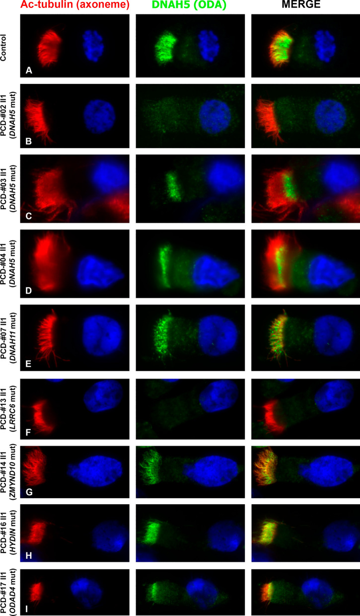
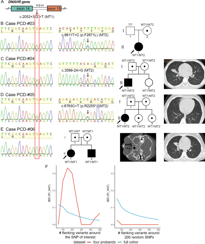
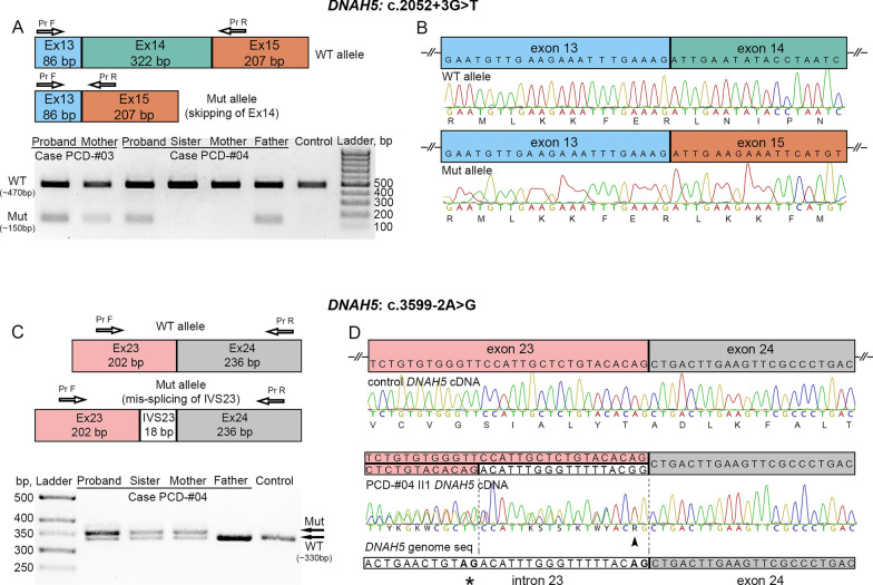
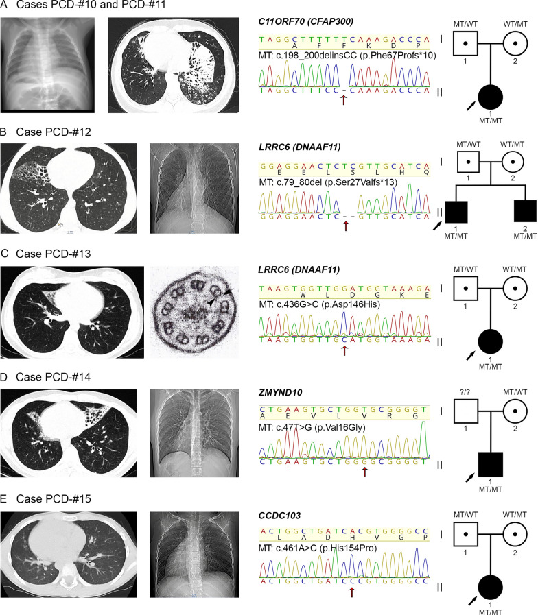
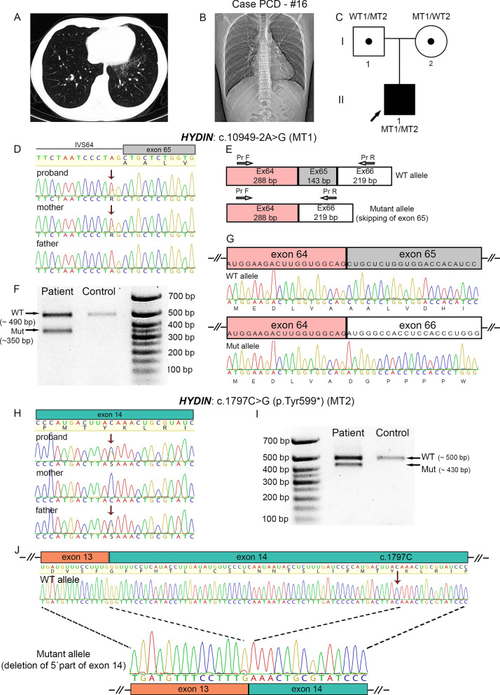
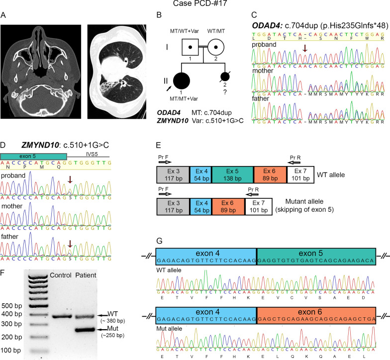
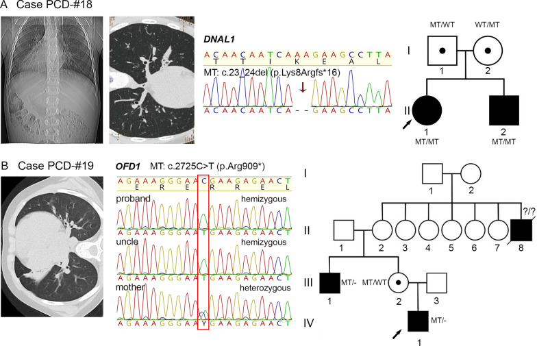
Results: The present study provides a comprehensive clinical and genetic characterization of 21 Russian families with PCD living in various country regions. Anomalies of ciliary beating in patients` respiratory epithelial cells were confirmed by high-speed video microscopy. In the most cases, custom-designed panel sequencing allowed to uncover causative variants in well-known or rarely mentioned PCD-related genes, including DNAH5, DNAH11, CFAP300, LRRC6, ZMYND10, CCDC103, HYDIN, ODAD4, DNAL1, and OFD1. The variations comprised common mutations, as well as novel genetic variants, some of which probably specific for Russian patients. Additional targeted analysis of mRNA transcripts from ciliated cells enabled us to specify functional effects of newly identified genetic variants in DNAH5 (c.2052+3G>T, c.3599-2A>G), HYDIN (c.10949-2A>G, c.1797C>G), and ZMYND10 (c.510+1G>C) on splicing process. In particular, the splice site variant c.2052+3G>T, detected in four unrelated families, resulted in skipping of exon 14 in DNAH5 transcripts and, according to haplotype analysis of affected probands, was proposed as an ancestral founder mutation in Udmurt population.

Conclusions: The reported data provide a vital insight into genetic background of primary ciliary dyskinesia in the Russian population. The findings clearly illustrate the utility of gene panel sequencing coupled with transcriptional analysis in identification and clinical interpretation of novel genetic variants.

Keywords: DNAH5; HYDIN; ZMYND10; Founder mutation; Gene-panel sequencing; Primary ciliary dyskinesia; Russian population; Splice site variants; Transcript analysis.


Conflict of interest statement

The authors declare that they have no competing interests.

Figures

Fig. 1
Fig. 1
Localization of DNAH5 in respiratory epithelial cells from PCD-patients harboring pathogenic variants in different ciliary genes. Immunofluorescence staining of ciliated cells with antibodies against acetylated α-tubulin (marker of axoneme, red) and anti-DNAH5 antibodies (green). In contrast to a healthy control sample (A), complete absence of DNAH5 from ciliary axoneme was noted in patient PCD-#02 II1 with mutated DNAH5 (B) and patient PCD-#13 II1 with mutated LRRC6 (F). Mis-localization of DNAH5 was detected in patients PCD-#03 II1 and PCD-#04 II1 with mutated DNAH5 (C, D), patient PCD-#14 II1 with mutated ZMYND10 (G) and patient PCD-#17 II1 with mutated ODAD4 (I). Seemingly normal pattern of DNAH5 distribution was noted in patient PCD-#07 II1 with mutated DNAH11 (E) and patient PCD-#16 II1 with mutated HYDIN (H)
Fig. 2
Fig. 2
Identification of a novel DNAH5 variant c. 2052+3G>T in four unrelated PCD families from Udmurtia. A Schematic image of a DNAH5 fragment comprising exon 14 with the c.2052+3G>T variant pointed by an arrow and marked by a red frame. B Case PCD-#03. On the left, Sanger sequencing of the proband PCD-#03 II1 demonstrates compound heterozygosity for the c.2052+3G>T (MT1) and c.8611T>C (p.F2871L) (MT2). On the right, there is a family pedigree. C Case PCD-#04. On the left, Sanger sequencing of the proband PCD-#04 II1 demonstrates compound heterozygosity for the c.2052+3G>T (MT1) and c.3599-2A>G (MT2). In the middle, there is a family pedigree. On the right, computed-tomography (CT) scans of the proband with atelectasis and bronchiectatic lung disease. D Case PCD-#05. On the left, Sanger sequencing of the proband PCD-#05 II1demonstrates compound heterozygosity for the c.2052+3G>T (MT1) and c.6763C>T (p.R2255*) (MT2). In the middle, there is a family pedigree. On the right, CT scans of the proband with subpleural fibrous changes and bronchiectasis. E Case PCD-#06. On the left, Sanger sequencing of the proband PCD-#06 II1 demonstrates homozygosity for the c.2052+3G>T (MT1). In the middle, there is a family pedigree. On the right, CT scans of the proband`s lungs showing bronchiectatic disease and paranasal sinuses demonstrating sphenoiditis and ethmoiditis. F Change in identity by descent (IBD) as a function of increasing interval (step equals 50 SNPs downstream and upstream of the locus). In a range from 0 to 1, the p̂ (PI_HAT) metric indicates the proportion of alleles that are identical by descent. A value of 0 signifies that individuals are unrelated in terms of recent common ancestry, while a value of 1 means that individuals are genetically identical by IBD, such as identical twins or clones. The left panel shows the change in p̂ around NC_000005.10:g.13901249C>A (c.2052+3G>T) with increasing number of SNPs included in the analysis, for the four probands and the full cohort. Similarly, the right panel shows the mean change in p̂ around 200 randomly selected non-overlapping (within 500 SNPs) intervals, for the four probands and the full cohort. The analysis revealed a shared haplotype surrounding the DNAH5 gene in four probands PCD-#03–06 II1 carrying the c. 2052+3G>T variant
Fig. 3
Fig. 3
The intronic variants c.2052+3G>T and c.3599-2A>G result in abnormal splicing of DNAH5 transcripts. AD Analysis of DNAH5 transcripts from nasal respiratory epithelial cells of the probands PCD-#03 II1 and PCD-#04 II1 and their relatives. A On the top: schematic results of PCR amplification of the DNAH5 cDNA region covering exons 13–15, that demonstrate a larger WT allele and a smaller mutant allele in carriers of the c.2052+3G>T variant. From below: gel-electrophoresis of RT-PCR samples from the PCD-#03 family (the proband and her mother), the PCD-#04 family (the proband and his unaffected sister, mother and father) and a control sample. B Sanger sequencing confirmed that the 472-bp fragment corresponded to the WT allele (on the top), while a smaller mutant fragment (150 bp) resulted from the skipping of exon 14 (from below). C On the top: schematic results of PCR amplification of the DNAH5 cDNA region comprising exons 23 and 24, that demonstrate a WT allele and a slightly larger mutant allele due to an 18-bp insertion in carriers of the c.3599-2A>G variant. From below: gel-electrophoresis of RT-PCR samples from the PCD-#04 family (the proband and his unaffected sister, mother and father) and a control sample. D Sequencing verified that the control fragment corresponded to the WT allele, while the proband`s sample represented a mix of the WT allele and altered transcript sequences harboring an 18-bp insertion between exon 23 and 24 (dashed lines), which corresponded to the 3’ part of the intron 23 sequence. From below, a DNAH5 genomic region comprising an intron 23/exon 24 junction is shown. A canonical (AG) and a presumed cryptic (AG with an asterisk) splice acceptor sites are in bold
Fig. 4
Fig. 4
Clinical and genetic analysis of PCD families with common mutations in CFAP300, LRRC6, ZMYND10 and CCDC103. A Cases PCD-#10 and PCD-#11. On the left, CT scans of the proband PCD-#10 II1 demonstrating situs inversus totalis and the proband PCD-#11 II1 showing a tree-in-bud pattern and fibrocystic transformation in the lungs. In the middle, Sanger sequencing of the PCD-#11 II1 demonstrates homozygosity for the c.198_200delinsCC variant in CFAP300 (MT); the same variant is harbored by the PCD-#10 II1 (data not shown). B Case PCD-#12. On the left, CT scans of the proband PCD-#12 II1 illustrating bronchiectatic lung disease and his affected brother PCD-#12 II2 showing laterality defects. In the middle, Sanger sequencing of the proband confirms homozygosity for the c.79_80del variant in LRRC6 (MT). C Case PCD-#13. On the left, there is a CT scan of the proband`s lungs showing bronchiectasis and an electron microscopy image of the axoneme cross-section which demonstrates the absence of ODA and IDA. In the middle, Sanger sequencing of the proband shows homozygosity for the c.436G>C (p.Asp146His) variant in LRRC6 (MT). D Case PCD-#14. On the left, CT scans of the proband demonstrating bronchiectasis and dextrocardia. In the middle, Sanger sequencing of the proband verifies homozygosity for the c.47T>G (p.Val16Gly) variant in ZMYND10 (MT). E Case PCD-#15. On the left, CT scans of the proband demonstrating situs anomalies. In the middle, Sanger sequencing of the proband confirms homozygosity for the c.461A>C (p.His154Pro) variant in CCDC103 (MT). AE On the right, family pedigrees are shown
Fig. 5
Fig. 5
Identification of two loss-of-function variants in HYDIN and confirmation of their deleterious impact on splicing. A, B CT scans of the proband PCD-#16 II1 demonstrating situs solitus, atelectasis with bronchiectatic lung disease. C A family pedigree: the proband is a compound heterozygote for the c.10949-2A (MT1) and c.1797C>G (MT2) variants; the mother and the father are heterozygous carriers of MT1 and MT2, respectively. DG Molecular characterization of the c.10949-2A variant. D Sanger sequencing results show that the proband and his mother possess the c.10949-2A as a heterozygote (pointed by arrows). E Schematic results of PCR amplification of the HYDIN cDNA region covering exons 64–66, that detect a larger WT allele and a smaller mutant allele in the proband carrying the c.10949-2A. F Gel-electrophoresis of RT-PCR samples from the patient and the control. G Sanger sequencing of RT-PCR products verified that a larger fragment (~ 490 bp) corresponded to the WT allele (on the top), while a smaller mutant fragment (~ 350 bp) resulted from the skipping of exon 65 (from below). HJ Molecular characterization of the c.1797C>G variant. Sanger sequencing results show that the proband and his father possess the c.1797C>G as a heterozygote (pointed by arrows). I Gel-electrophoresis of RT-PCR samples covering exons 13 and 14 of HYDIN cDNA from the patient and the control. J Sanger sequencing of RT-PCR products confirmed that a larger fragment (~ 500 bp) corresponded to the WT allele (on the top), while a smaller fragment (~ 430 bp) represented a mutant allele where the first 59 nucleotides of exon 14 sequence were skipped (from below)
Fig. 6
Fig. 6
The PCD-#17 family combining a frameshift variant in ODAD4 and a splice-site variant in ZMYND10. A CT scans of the proband PCD-#17 II1 demonstrating bilateral parietal thickening of the mucosa of the paranasal sinuses, polyposis and rhinosinusitis, as well as fibroatelectasis with bronchiectatic lung disease. B A family pedigree: the proband harbors a homozygous c.704dup variant in ODAD4 (MT) and a heterozygous c.510+1G>C variant in ZMYND10 (Var). C Sanger sequencing results demonstrate that the proband is homozygous for the c.704dup (p.His235Glnfs*48) variant in ODAD4, with the parents being heterozygous carriers of the variant. D Sanger sequencing results confirm that the proband and her father carry the c.510+1G>C variant in ZMYND10. EG Analysis of ZMYND10 transcripts from nasal respiratory epithelial cells of the proband. E Schematic results of PCR amplification of the ZMYND10 cDNA region covering exons 3–7 that demonstrate a larger wild-type (WT) allele and a smaller mutant allele. F Gel-electrophoresis of RT-PCR samples from the proband and the control. G Sanger sequencing verified that a larger fragment (~ 380 bp) corresponded to the WT allele (on the top), while a smaller fragment (~ 250 bp) resulted from the skipping of exon 5 (from below)
Fig. 7
Fig. 7
Identification of novel PCD-related loss-of-function variants in DNAL1 and OFD1 genes. A Case PCD-#18. On the left, CT scans of the proband PCD-#18 II1 with situs inversus totalis and a pattern of chronic bronchitis without bronchiectasis. In the middle, Sanger sequencing of the proband shows homozygosity for the c.23_24del (p.Lys8Argfs*16) in DNAL1 (MT). On the right, a family pedigree: the proband and her affected brother are homozygotes; the parents are heterozygous carriers of the variant. B Case PCD-#19. On the left, CT scans of the proband PCD-#19 IV1 demonstrating bronchiectasis. In the middle, Sanger sequencing results show that the proband and his affected uncle PCD-#19 III1 are hemizygous for the c.2725C>T (p.Arg909*) variant in OFD1 gene, with the proband`s mother PCD-#19 III2 being a heterozygous carrier of the variant (marked by a red frame); on the right, a family pedigree for four generations is shown

References

    1. Leigh MW, Ferkol TW, Davis SD, Lee HS, Rosenfeld M, Dell SD, et al. Clinical features and associated likelihood of primary ciliary dyskinesia in children and adolescents. Ann Am Thorac Soc. 2016;13:1305–13. 10.1513/AnnalsATS.201511-748OC - DOI - PMC - PubMed
    1. Shapiro AJ, Zariwala MA, Ferkol T, Davis SD, Sagel SD, Dell SD, et al. Diagnosis, monitoring, and treatment of primary ciliary dyskinesia: PCD foundation consensus recommendations based on state of the art review. Pediatr Pulmonol. 2016;51:115–32. 10.1002/ppul.23304 - DOI - PMC - PubMed
    1. Fliegauf M, Benzing T, Omran H. When cilia go bad: Cilia defects and ciliopathies. Nat Rev Mol Cell Biol. 2007;8:880–93. 10.1038/nrm2278 - DOI - PubMed
    1. Shah AS, Yehuda BS, Moninger TO, Kline JN, Welsh MJ. Motile cilia of human airway epithelia are chemosensory. Science. 1979;2009(325):1131–4. - PMC - PubMed
    1. Cowan MJ, Gladwin MT, Shelhamer JH. Disorders of ciliary motility. Am J Med Sci. 2001;321:3–10. 10.1097/00000441-200101000-00002. 10.1097/00000441-200101000-00002 - DOI - PubMed

Substances

LinkOut - more resources