. 2024 Sep 23;52(17):10705-10716.
doi: 10.1093/nar/gkae717.

Structural insights into RNA cleavage by a novel family of bacterial RNases

Affiliations

Structural insights into RNA cleavage by a novel family of bacterial RNases

Ruoxi Wu et al. Nucleic Acids Res. .

Abstract

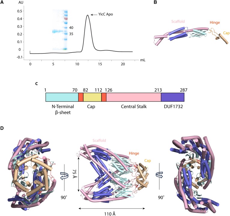
Processing of RNA is a key regulatory mechanism for all living systems. Escherichia coli protein YicC belongs to the well-conserved YicC family and has been identified as a novel ribonuclease. Here, we report a 2.8-Å-resolution crystal structure of the E. coli YicC apo protein and a 3.2-Å-cryo-EM structure of YicC bound to an RNA substrate. The apo YicC forms a dimer of trimers with a large open channel. In the RNA-bound form, the top trimer of YicC rotates nearly 70° and closes the RNA substrate inside the cavity to form a clamshell-pearl conformation that resembles no other known RNases. The structural information combined with mass spectrometry and biochemical data identified cleavage on the upstream side of an RNA hairpin. Mutagenesis studies demonstrated that the previously uncharacterized domain, DUF1732, is critical in both RNA binding and catalysis. These studies shed light on the mechanism of the previously unexplored YicC RNase family.


Figures

Graphical Abstract
Graphical Abstract
Figure 1.
Figure 1.
Crystal structure of apo YicC. (A) Purification of YicC, with chromatogram from a Superdex 200 Increase column and Coomassie gel of peak fractions (35 and 40 kDa molecular weight markers indicated). (B) Crystal structure of YicC monomer. (C) Domain architecture of YicC protein and its homologs. The different domains and regions are colored; the color scheme is used for all panels in this figure. (D) Crystal structure of YicC from three different angles in cartoon representation. The opening was measured from B chain L196/CA to E chain Q195/CA.
Figure 2.
Figure 2.
Cryo-EM structure of YicC bound to an RNA substrate. (A) Electron density of the structure, colored by chain. (B) Overall model of YicC-RNA complex. The structure is shown in cartoon representation, colored by chain. The RNA is shown in stick format, in the protein cavity. (C) surface view of YicC-RNA complex. One trimer has been removed for clarity, and the structure is colored by domain, as in Figure 1. (D) Schematic of RNA structure, with basepairing indicated. The loop that is not observed in the structure is indicated by dotted lines. (E) Structure of RNA shown in stick form, with bases numbered and the missing loop indicated. The central cavity was measured by I212B/CA to Y35B/CA, which is 53.7Å
Figure 3.
Figure 3.
Structural determinants of RNA binding. (A) Overall structure of YicC-RNA complex shown with electrostatic potential, with positive charge shown in blue and negative charge in red. (B) Closeup of protein-RNA interactions, with side chains indicated and potential hydrogen bonds and ionic interactions shown. Three different segments of the RNA, labeled as I, II and III are shown. (C) Schematic of amino acid–RNA interactions, with the residue number and chain indicated after the slash, colored by domain. (D) Axial view of the DUF–RNA interactions, with the RNA shown in spherical representation.
Figure 4.
Figure 4.
RNA cleavage activity of YicC and site identification. (A) Fluorescent cleavage gel assay. Total RNA concentration was 600 nM; YicC protein concentration was as indicated above each set of reactions. Reactions were incubated at 37°C and aliquots were removed at times indicated above each lane. Control lane (–), no YicC protein added. Migration of full-length RNA oligo (A) and cleavage products (B and C) indicated at left. (B) Schematic of cleavage fragments resulting from two cleavages in DHB2051. Predicted RNA fragments are labeled A-F. Asterisk indicates the site of the fluorescent dye modification for gel experiments. For the mass spectrometry experiments they are unmodified hydroxyl 5′ ends. (C) Time course of cleavage activity on 26-mer RNA for E. coli YicC, B. subtilis YloC, and B. burgdorferi (Bb) YicC. RNA concentration was 600 nM; YicC protein concentration indicated above each set of reactions. Aliquots were removed at times (min) indicated above each lane. Control lane (–), no protein added. Migration of full-length RNA oligo (A) and E. coli YicC cleavage products (B and C) indicated at left. (D) High-resolution mass spectrometry analysis of cleavage of DHB2051. The reaction was purified and the sample (∼10 μM) was directly infused to an orbitrap mass spectrometer and analyzed under the negative ionization mode. Upper panel shows the full MS spectrum of the RNA analytes at 400–1150 m/z. Zoom-in of three dominant RNA species including salt adducts (590–680 m/z) is shown in the lower panel. Their precise m/z measurement, respective molecular compositions and mass accuracies in parts per million (ppm) are denoted. (E) Schematic showing cleavage sites of RNA hairpin by YicC, marked by red arrows. We hypothesize that the site 2 cleavage occurs on an alternative hairpin structure (see Discussion). (F) Proposed catalytic site, shown as zoomed-in view. The cluster of three glutamates are shown along with presumed water molecules shown as green spheres. The nucleotides between which site 1 cleavage occurs are shown in yellow.
Figure 5.
Figure 5.
Analysis of catalysis and substrate binding. (A) Mutagenesis and activity measurements. Alanine mutants of the indicated residues were generated. DHB2051 RNA concentration was 600 nM; YicC protein concentration was 20 nM. Reactions were incubated at 37 °C and aliquots were removed at 1 min and 16 min. The YicC residue that was mutated to alanine is indicated for each mutant protein. Control lane (–), no YicC protein added. (B) Analysis of YicC mutant binding to RNA by fluorescence anisotropy. Binding constants were measured using a fixed concentration of 5′-Cy3 labeled RNA 36-mer and varied concentrations of wild-type or mutant YicC protein. Fluorescent polarization was measured on a multimode plate reader. The binding constants are shown in Supplementary Table S4. n = 3 for each data point, shown with error bars for standard deviation.

Update of

References

    1. Bechhofer D.H., Deutscher M.P.. Bacterial ribonucleases and their roles in RNA metabolism. Crit. Rev. Biochem. Mol. Biol. 2019; 54:242–300. - PMC - PubMed
    1. Ingle S., Chhabra S., Chen J., Lazarus M.B., Luo X., Bechhofer D.H.. Discovery and initial characterization of YloC, a novel endoribonuclease in Bacillus subtilis. RNA. 2022; 28:227–238. - PMC - PubMed
    1. Poulsen P., Jensen K.F.. Three genes preceding pyrE on the Escherichia coli chromosome are essential for survival and normal cell morphology in stationary culture and at high temperature. Res. Microbiol. 1991; 142:283–288. - PubMed
    1. Martins D., DiCandia M.A., Mendes A.L., Wetzel D., McBride S.M., Henriques A.O., Serrano M.. CD25890, a conserved protein that modulates sporulation initiation in Clostridioides difficile. Sci. Rep. 2021; 11:7887. - PMC - PubMed
    1. Chen J., To L., de Mets F., Luo X., Majdalani N., Tai C.H.. Gottesman S. (2021) A fluorescence-based genetic screen reveals diverse mechanisms silencing small RNA signaling in E. coli. Proc. Natl. Acad. Sci. U.S.A. 118:e2106964118. - PMC - PubMed

LinkOut - more resources